

Specialized Reagents & Physiological Buffers
Four groups, one page. Find your group in the catalog below and go straight to the product page — the design rationale, history, applications, and selection guidance follow.
| Product | Buffer Base | Primary Application | Product Page |
|---|---|---|---|
| 1 Physiological Buffers — ex vivo tissue & cell viability 3 products | |||
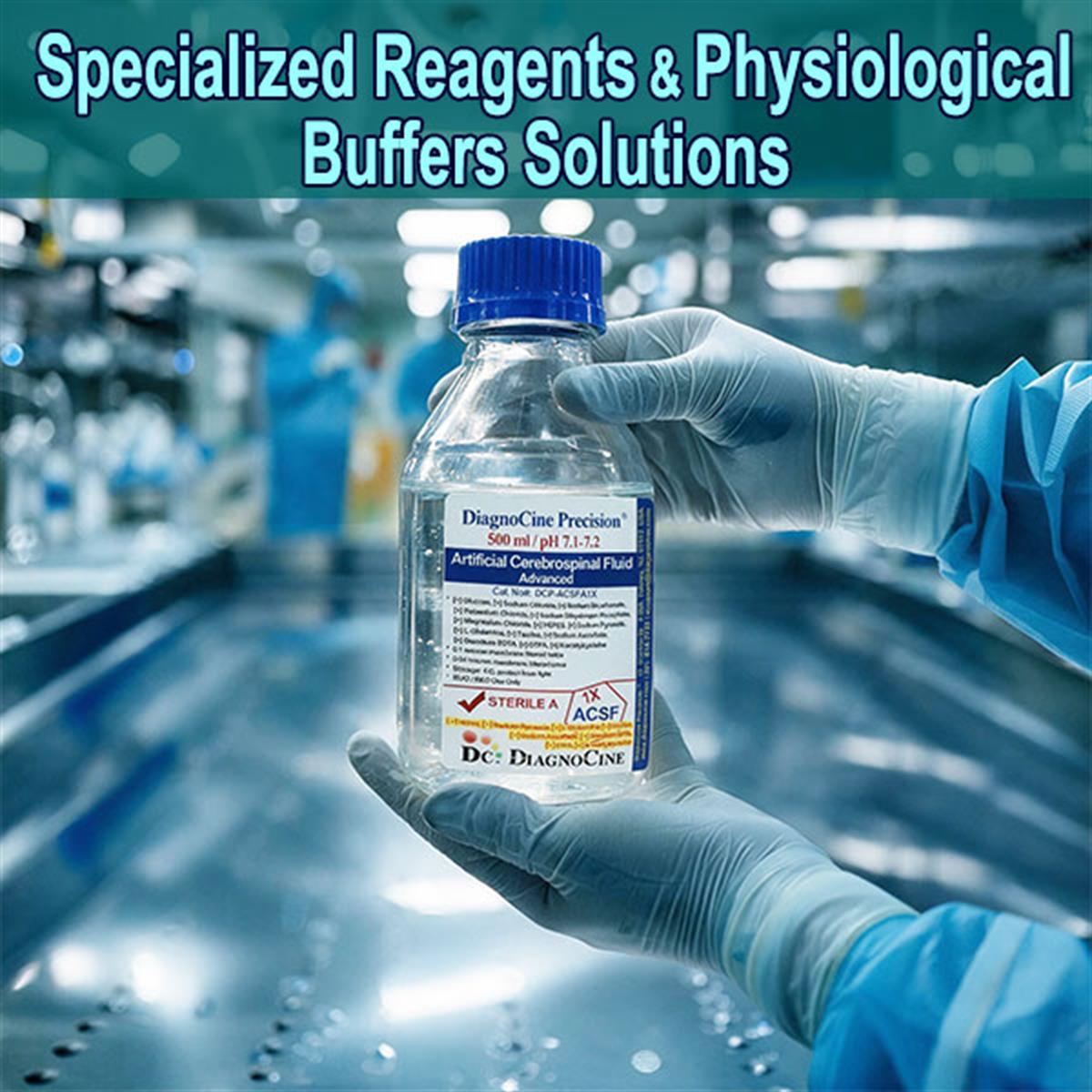
| Artificial Cerebrospinal Fluid (ACSF) | CSF-matched | Neurobiology standard | Viewarrow_forward |
| UltraClean Krebs-Ringer Bicarbonate Buffer with Calcium chloride, 10mM HEPES, 0.2% Bovine Serum Albumin and Sodium bicarbonate, without Glucose, 1X (Liquid), 0.04um sterile filtered | HEPES + NaHCO3 | Metabolic & microfluidic studies | Viewarrow_forward |
| UltraClean Krebs-Ringer Bicarbonate Buffer with Calcium Chloride, 10mM HEPES Buffer, and 0.2% Bovine Serum Albumin, without Glucose and Sodium Bicarbonate, 1X (Liquid) | HEPES | Open-air / custom-gas workflows | Viewarrow_forward |
| 2 Signal Enhancement — SigMax™ Series 8 products · PBS & TBS | |||
| SigMax 1Ab-PBS | PBS | Primary-antibody (1Ab) buffer | Viewarrow_forward |
| SigMax 1Ab-TBS | TBS | Primary-antibody (1Ab) buffer | Viewarrow_forward |
| SigMax 2Ab-PBS | PBS | Secondary-antibody (2Ab) buffer | Viewarrow_forward |
| SigMax 2Ab-TBS | TBS | Secondary-antibody (2Ab) buffer | Viewarrow_forward |
| SigMax HRP-PBS kit | PBS | — | Viewarrow_forward |
| SigMax HRP-TBS kit | TBS | — | Viewarrow_forward |
| SigMax ALP-PBS kit | PBS | — | Viewarrow_forward |
| SigMax ALP-TBS kit | TBS | — | Viewarrow_forward |
| 3 Enzyme Quench Buffers — matched to the detection method 4 products · PBS & TBS | |||
| HRPQuench-PBS | PBS | Exclusively for HRP-based kits | Viewarrow_forward |
| HRPQuench-TBS | TBS | Exclusively for HRP-based kits | Viewarrow_forward |
| ALPQuench-PBS | PBS | Exclusively for ALP-based kits | Viewarrow_forward |
| ALPQuench-TBS | TBS | Exclusively for ALP-based kits | Viewarrow_forward |
| 4 Other Specialized Reagents 3 products | |||
| 2X PCR Buffer for KOD FX, 100ml | 2X PCR | Reaction buffer for KOD FX DNA polymerase | Viewarrow_forward |
| 2X PCR Buffer for KOD FX, 5mL (3X 1.67ml) | 2X PCR | Reaction buffer for KOD FX DNA polymerase | Viewarrow_forward |
| AZ PRESERVER™, AZP-A1 | — | — | Viewarrow_forward |
Not sure which group? Compare the four subcategories · See applications by group · Read the FAQ
What a specialized reagent has to do that PBS does not
Standard buffers like PBS or Tris are fine for basic biochemistry, but modeling living systems in vitro demands more. Physiological buffers and specialized reagents are multi-component formulations engineered to mimic the electrolyte, nutrient, and buffering profiles of real tissue — keeping primary cells, tissue slices, and microfluidic organ models alive and functioning outside the body.
The same principle carries through the rest of the category: every reagent here is a formulation tuned to one job, not a general-purpose salt solution.
- Group 1 — Physiological Buffers (3): Artificial Cerebrospinal Fluid (ACSF), and two UltraClean Krebs-Ringer Bicarbonate Buffers — one with sodium bicarbonate, one without.
- Group 2 — SigMax™ Series (8): primary- and secondary-antibody buffers plus HRP and ALP kits, each stocked in a PBS and a TBS base.
- Group 3 — Enzyme Quench Buffers (4): HRPQuench and ALPQuench, each in PBS and TBS, matched to the kit's detection method.
- Group 4 — Other Specialized Reagents (3): 2X PCR Buffer for KOD FX in two sizes, and AZ PRESERVER™, AZP-A1.
- Complex electrolyte balance — precise mixtures of monovalent (Na+, K+) and divalent (Ca2+, Mg2+) cations needed for membrane potentials, signaling, and contraction.
- Dual buffering — many formulations combine an organic buffer (HEPES) with a gas-regulated physiological buffer (sodium bicarbonate) to hold pH ~7.4 across different setups.
- Metabolic & structural support — glucose as an energy source; proteins like BSA to prevent surface adhesion and stabilize fragile membrane proteins.
- Microfluidic readiness — 0.04 µm filtration prevents microchannel clogging in organ-on-a-chip platforms.
- Products in this category18
- Subcategories4
- Physiological buffer configurations3
- SigMax™ series products8
- Enzyme quench buffers4
- Buffer bases offered (SigMax / Quench)PBS and TBS
- HEPES concentration (Krebs-Ringer)10 mM
- Bovine serum albumin (Krebs-Ringer)0.2%
- Sterile filtration (bicarbonate Krebs-Ringer)0.04 µm
- Target physiological pH~7.4
Recreating a dynamic, nutrient-rich fluid on the bench
In vivo, cells are bathed in dynamic, nutrient-rich fluid that manages osmotic pressure, supplies energy, and clears waste. Specialized reagents recreate that in the lab — and each component below answers a specific way that a simple saline fails.
Complex electrolyte balance
Precise mixtures of monovalent (Na+, K+) and divalent (Ca2+, Mg2+) cations, needed for membrane potentials, signaling, and contraction.
Dual buffering
Many formulations combine an organic buffer (HEPES) with a gas-regulated physiological buffer (sodium bicarbonate) to hold pH ~7.4 across different experimental setups.
Metabolic support
Glucose serves as the energy source. Removing it deliberately — a glucose-free formulation — is what makes a controlled baseline for measuring cellular respiration and metabolism possible.
Structural support (BSA)
Proteins like BSA prevent surface adhesion and stabilize fragile membrane proteins — the reason 0.2% BSA appears in both Krebs-Ringer configurations.
Microchannel-safe filtration
Organ-on-a-chip microfluidics demands ultra-filtered, high-purity versions of these classic solutions to avoid nanoscale clogging; 0.04 µm filtration prevents microchannel clogging.
Atmosphere compatibility
A bicarbonate-free, glucose-free version retains HEPES buffering and protein stabilization while giving stable pH in normal atmosphere — for open-air and custom-gas workflows.
The specification that decides the workflow
Within the physiological buffers, two variables separate the products more than anything else: whether sodium bicarbonate is present, and whether glucose is present. Bicarbonate sets what atmosphere the experiment has to run under; glucose sets whether the medium is a support solution or a controlled metabolic baseline. Read both before matching a formulation to a protocol.
From a frog heart in tap water to organ-on-a-chip
Every number on a modern physiological buffer datasheet traces back to one of four developments. Each one explains a component you still see in the formulation today.
-
1
1882–1883 Ringer and the calcium requirement
British physiologist Sydney Ringer found that an isolated frog heart keeps beating only when the bathing solution contains the right ratios of sodium, potassium, and calcium — famously discovering the calcium requirement after an assistant used mineral-rich tap water. Significance: founded the field of physiological saline design; “Ringer's solution” is the ancestor of modern cell-culture and tissue media.
-
2
1932 Krebs & Henseleit formalize bicarbonate buffering
Hans Krebs & Kurt Henseleit, in their urea-cycle work, formalized a bicarbonate-buffered, electrolyte-balanced solution (Krebs–Henseleit / Krebs-Ringer bicarbonate buffer) for maintaining tissue slices. Significance: established the bicarbonate-buffered, substrate-supplemented media used for ex vivo metabolic studies.
-
3
1957 The first brain-slice recordings
Choh-Luh Li & Henry McIlwain made the first electrical recordings from mammalian brain slices, showing (among other things) the need for glucose in the bathing medium. Significance: launched brain-slice electrophysiology and the artificial cerebrospinal fluid (ACSF) formulations that support it.
-
4
Late 20th century–present Refinement and miniaturization
Neuroscience drove refinement of ACSF (including sucrose / NMDG variants to reduce excitotoxicity during slicing), and organ-on-a-chip microfluidics now demands ultra-filtered, high-purity versions of these classic solutions to avoid nanoscale clogging. Significance: extended physiological media from tissue baths to microfluidic and translational platforms.
How this category is organized
Four groups, split by what the reagent does rather than by what it is made of. Use this table to find the right group before drilling into a product.
| Group | What it is | Buffer bases | Products |
|---|---|---|---|
| 1 · Physiological Buffers | Multi-component formulations that mimic the electrolyte, nutrient, and buffering profiles of real tissue, keeping primary cells, tissue slices, and microfluidic organ models alive outside the body | CSF-matched; HEPES ± NaHCO3 | 3 |
| 2 · Signal Enhancement — SigMax™ | Immunoassay antibody buffers. The TBS primary-antibody buffer is a PEG 6000 / PVP-40 / BSA blend that raises signal intensity while reducing background noise for low-abundance targets; SigMax 1Ab and 2Ab ship with every Diagnocine ELISA kit order | PBS and TBS | 8 |
| 3 · Enzyme Quench Buffers | The component of the ELISA buffer set matched to the kit's detection method: HRPQuench exclusively for HRP-based kits, ALPQuench exclusively for ALP-based kits | PBS and TBS | 4 |
| 4 · Other Specialized Reagents | Reagents that do not fall into the three groups above: 2X PCR Buffer for KOD FX (reaction buffer for KOD FX DNA polymerase) in two sizes, and AZ PRESERVER™, AZP-A1 | — | 3 |
Which workflow, which group
Select a group to see the workflows it is intended to support.
- Neurophysiology / brain-slice electrophysiology — incubating and recording from living brain or spinal-cord slices; ACSF matches the CNS electrolyte profile and maintains acute brain-slice viability for electrophysiology and patch-clamp recording
- Isolated organ & tissue-bath experiments — sustaining muscle contraction or vascular tone in isolated cardiovascular tissue
- Microfluidics / organ-on-a-chip — circulating physiological media through microscopic cellular channels; 0.04 µm filtration prevents microchannel clogging and BSA limits surface adhesion
- Metabolic flux studies — controlled-substrate media (e.g., glucose-free) for measuring cellular respiration and metabolism
- Sucrose / NMDG ACSF variants were developed to reduce excitotoxicity during slicing
- Immunoassay antibody incubation steps, in either a phosphate-buffered saline or a Tris-buffered saline environment
- A proprietary PEG 6000, PVP-40, and BSA blend that substantially increases signal intensity while reducing background noise
- Detection of low-abundance targets, by optimizing the microenvironment for antibody–antigen interactions
- Preserves native molecular conformations for improved epitope recognition and minimal non-specific binding
- SigMax 1Ab and SigMax 2Ab are included with every Diagnocine ELISA kit order
- The component of the ELISA buffer set matched to the kit's detection method
- HRPQuench — exclusively for HRP-based kits
- ALPQuench — exclusively for ALP-based kits
- Supplied in the same PBS and TBS bases as the SigMax series, so the whole set stays on one buffer chemistry
- 2X PCR Buffer for KOD FX — reaction buffer for KOD FX DNA polymerase; stocked in a 100 mL bottle and a 5 mL pack (3 × 1.67 mL)
- AZ PRESERVER™, AZP-A1 — specification not yet published on this page; contact support for the current datasheet
Physiological buffers — how the three formulations compare
Core attributes and the workflow each product is intended for.
| Product | Core Attributes | Best Use |
|---|---|---|
| Artificial Cerebrospinal Fluid (ACSF) | Matches CNS electrolyte profile | Neurobiology standard — maintains acute brain-slice viability for electrophysiology and patch-clamp recording |
| UltraClean Krebs-Ringer Bicarbonate Buffer + CaCl2, 10 mM HEPES, 0.2% BSA, + sodium bicarbonate, glucose-free, 0.04 µm filtered | Glucose-free, HEPES/bicarbonate double-buffered | Metabolic & microfluidic studies — glucose-free baseline for controlled metabolism; BSA limits surface adhesion; 0.04 µm filtration prevents microchannel clogging |
| UltraClean Krebs-Ringer Bicarbonate Buffer + CaCl2, 10 mM HEPES, 0.2% BSA, glucose-free & bicarbonate-free | Bicarbonate-free, glucose-free | Open-air / custom-gas workflows — stable pH in normal atmosphere without a 5% CO2 incubator, while retaining HEPES buffering and protein stabilization |
Frequently asked questions
The questions that come up most often when a purchasing spec meets a bench protocol.
The primary literature behind the physiological buffers
The three papers that define the lineage of every physiological buffer in Group 1.
- Ringer, S. (1883). A further contribution regarding the influence of the different constituents of the blood on the contraction of the heart. The Journal of Physiology, 4(1), 29–42. — The landmark paper establishing that calcium (and potassium) are essential to sustain contraction; the origin of physiological saline design.
- Krebs, H. A., & Henseleit, K. (1932). Untersuchungen über die Harnstoffbildung im Tierkörper. Hoppe-Seyler's Zeitschrift für physiologische Chemie, 210(1–2), 33–66. — The work that established the Krebs–Henseleit (Krebs-Ringer bicarbonate) buffer for sustaining tissue ex vivo.
- Li, C.-L., & McIlwain, H. (1957). Maintenance of resting membrane potentials in slices of mammalian cerebral cortex and other tissues in vitro. The Journal of Physiology, 139(2), 178–190. — The founding study of brain-slice electrophysiology and the ACSF-type media that support it.