SigMax 2Ab-TBS
FluxMPS™ SigMax 2Ab-TBS
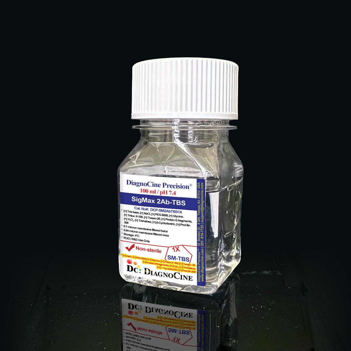
FluxMPS™ SigMax 2Ab-TBS is an MPS-grade, ultra-filtered Tris-buffered saline (TBS) solution engineered to enhance secondary antibody binding efficiency across Western blotting, immunocytochemistry, and multiplex immunoassay workflows, including microfluidic and organ-on-a-chip (OoC) based immunodetection platforms. Dual-stage 0.1 µm and 0.04 µm membrane filtration delivers a low-particulate, low-background buffer base for high-clarity signal detection of low-abundance targets.
- Dual-stage filtration: 0.1 µm membrane once and 0.04 µm membrane once for an ultra-clean, low-particulate TBS base
- Tris-buffered saline core: 50 mM Tris Base and 150 mM Sodium Chloride at pH 7.4
- Proprietary PEG 6000, Glycine, Trehalose, and β-Cyclodextrin blend enhances secondary antibody binding and signal-to-noise ratio
- Manufactured with Ultrapure Type 1 water (18.2 MΩ·cm)
- Non-Sterile, research-use formulation optimized for Western blot, ICC, and multiplex immunoassays
- Compatible with microfluidic and organ-on-a-chip (OoC) immunodetection workflows
- Customizable pH, additive, and concentration formulations available on request
- pH
- 7.4
- Tris Base
- 50 mM
- Sodium Chloride
- 150 mM
- Appearance
- Clear, colorless liquid
- Sterility
- Non-Sterile
- Filtration
- 0.1 µm membrane once + 0.04 µm membrane once
- Water Quality
- Ultrapure Type 1 (18.2 MΩ·cm)
- Storage
- 4°C
- Shelf Life
- 1 year
- Format
- 100 mL
Engineered where standard antibody diluents fail
Conventional single-pass, 0.22 µm-filtered TBS carries subvisible particulates and lacks a defined binding-enhancement chemistry, allowing non-specific binding, background noise, and inconsistent antibody performance to creep into sensitive immunoassays. FluxMPS™ SigMax 2Ab-TBS is formulated and filtered to remove these failure points at the source.
Microchannel-safe purity
Sequential 0.1 µm and then 0.04 µm membrane filtration produces a low-particulate TBS base suited to fine microfluidic channels and sensitive optical detection surfaces.
Precise, stable pH
A defined 50 mM Tris / 150 mM NaCl formulation holds pH 7.4, giving consistent antigen-antibody interaction conditions from lot to lot.
Ultrapure-grade water
Prepared with Ultrapure Type 1 water (18.2 MΩ·cm) to minimize trace contaminants that can elevate assay background.
Low background for imaging & assays
The PEG 6000, Glycine, Trehalose, and β-Cyclodextrin blend reduces non-specific binding, supporting clean signal detection in Western blot, ICC, and multiplex immunoassays.
Defined, traceable composition
Every component is disclosed with its concentration and verified against lot-specific appearance and pH quality control checks.
Customization on demand
Alternate concentrations, additional chemicals, compounds, proteins, supplements, or a different pH can be formulated on request.
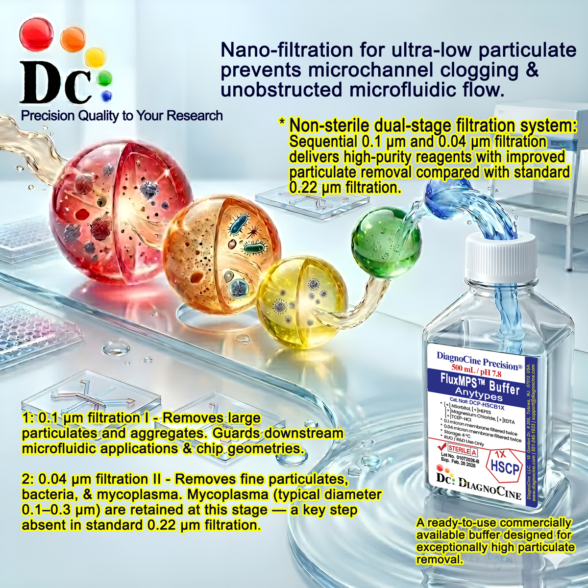
Dual-stage filtration system
SigMax 2Ab-TBS is filtered through a 0.1 µm membrane once and a 0.04 µm membrane once, giving a low-particulate buffer base appropriate for sensitive immunoassay and microfluidic immunodetection work.
- 1
0.1 µm Pre-filtration I
Removes large particulates and aggregates, protecting the buffer base and extending the working life of the finishing filter.
- 2
0.04 µm Pre-filtration II
Retains fine particulates for a final polish, delivering an ultra-clean TBS base suitable for low-background immunodetection.
Performance vs. conventional buffer
Sequential 0.1 µm and 0.04 µm filtration removes finer particulates than a single 0.22 µm pass used in conventional secondary antibody diluents.
© Diagnocine® — DCP-SM2AbTBS1X
Where SigMax 2Ab-TBS performs
Formulated to enhance secondary antibody binding efficiency and reduce background across a range of immunodetection workflows, from bench immunoassays to microfluidic and organ-on-a-chip platforms.
Automated Bioreactors & Robotics
For automated liquid-handling platforms and closed-loop perfusion systems, an optional 0.01 µm (10 nm) ultra-filtered variant of SigMax 2Ab-TBS can be produced on request, aimed at protecting fine-bore valves and sensors from residual particulates.
- Total Particulate Exclusion: finer sub-0.04 µm filtration for automated fluidic paths
- Valve & Sensor Protection: reduces particulate accumulation in precision instrumentation
- Extended Perfusion Stability: supports longer unattended run times in closed systems
Inquiry Required: the 0.01 µm ultra-filtered grade is produced to order — contact support@diagnocine.com to request this configuration.
Micro Physiological System (MPS) & Chip Immunodetection
Low-particulate TBS base suited to on-chip immunostaining and detection workflows.
Antibody Dilution & Reconstitution
Serves as a diluent and rinse buffer during secondary antibody preparation steps.
iPSC-Derived Model Immunostaining
Supports low-background immunolabeling of iPSC-derived cell models.
Endothelial & Primary Cell Immunolabeling
Isotonic Tris-buffered saline base compatible with endothelial and primary cell handling during immunostaining.
Secondary Antibody Signal Enhancement
Improves antigen-antibody binding efficiency and lowers background across ELISA, Western blot, and multiplex platforms.
Microscopy & Optical Detection
Low-background formulation supports clear signal capture in optical and biosensor-based detection.
Full specification sheet
Measured and declared values for SigMax 2Ab-TBS, Catalog No. DCP-SM2AbTBS1X.
| Parameter | Specification |
|---|---|
| Formulation | Tris-buffered saline with PEG 6000, Glycine, Trehalose, and β-Cyclodextrin |
| Appearance | Clear, colorless liquid |
| pH | 7.4 |
| Tris Base / Sodium Chloride | 50 mM Tris Base, 150 mM Sodium Chloride |
| Parameter | Specification |
|---|---|
| Sterility | Non-Sterile |
| Water Quality | Ultrapure Type 1 water (18.2 MΩ·cm) |
| Manufacturing Standard ISO 13485 | ISO 13485-certified, CE-approved facility |
| Filtration System | 0.1 µm membrane once, 0.04 µm membrane once |
| Parameter | Specification |
|---|---|
| Storage Temperature | 4°C |
| Shelf Life | 1 year |
| Parameter | Specification |
|---|---|
| Manufacturing QMS ISO 13485 | ISO 13485-certified, CE-approved facility (Suppliers of DiagnoCine Precision) |
| Production & Customization | Final packaging, QA, and testing at the DiagnoCine R&D and Quality Testing Center; customization and assembly at DiagnoCine Precision, Totowa, New Jersey, USA |
| Intended Use | Research Use Only (RUO) |
Full composition
Complete component listing for SigMax 2Ab-TBS, expressed exactly as declared in molarity or percent w/v / v/v.
| Component | CAS Number | Concentration |
|---|---|---|
| Tris Base | 77-86-1 | 50 mM |
| Sodium Chloride | 7647-14-5 | 150 mM |
| Polyethylene Glycol (PEG 6000) | 25322-68-3 | 5.0% w/v |
| Glycine | 56-40-6 | 0.75% w/v |
| Triton X-100 | 9036-19-5 | 0.05% v/v |
| Tween-20 | 9005-64-5 | 0.1% v/v |
| Recombinant Protein G fragments | 0.00005% w/v | |
| Hydrogen peroxide (H2O2) | 7722-84-1 | 0.03% v/v |
| Trehalose | 99-20-7 | 1% w/v |
| β-Cyclodextrin | 7585-39-9 | 0.1% w/v |
| ProClin 300 | 55965-84-9 | 0.05% v/v |
Manufactured under controlled quality systems
SigMax 2Ab-TBS is produced, packaged, and tested under a documented quality management system.
ISO 13485:2016 QMS
Manufactured under ISO 13485-certified, CE-approved facilities (Suppliers of DiagnoCine Precision).
Ultrapure Type 1 Water
Formulated with Ultrapure Type 1 water (18.2 MΩ·cm) to minimize background contamination.
Dual-Stage Filtration
Filtered through a 0.1 µm membrane once and a 0.04 µm membrane once for a low-particulate buffer base.
Micro-Batch Precision
Final packaging, quality assurance, and testing performed at the DiagnoCine R&D and Quality Testing Center; customization and assembly completed at DiagnoCine Precision in Totowa, New Jersey, USA.
Appearance & Clarity QC
Verified as a clear, colorless liquid per lot.
pH Verification
Confirmed at pH 7.4 per lot.
Dual-Stage Filtration Assurance
0.1 µm membrane filtration once, followed by 0.04 µm membrane filtration once.
Documentation / CoA
Certificate of Analysis available on request.
Buffer comparison
SigMax 2Ab-TBS against conventional secondary antibody diluents.
| Parameter | DCP-SM2AbTBS1X (FluxMPS™) | Conventional 0.22 µm-filtered buffer | Standard alternative (0.22 µm-filtered) |
|---|---|---|---|
| PEG 6000 / Glycine / Trehalose / β-Cyclodextrin enhancer blend | check_circle | cancel | cancel |
| Final filtration pore size | 0.04 µm | 0.22 µm | 0.22 µm |
| Number of filtration stages | 2 | 1 | 1 |
| Ultrapure Type 1 water (18.2 MΩ·cm) | check_circle | cancel | cancel |
| ISO 13485-certified manufacturing | check_circle | cancel | cancel |
| Microfluidic / OoC channel compatibility | check_circle | cancel | cancel |
| Formulation customization available | check_circle | cancel | cancel |
| Certificate of Analysis available | check_circle | cancel | cancel |
Frequently asked questions
Common questions about SigMax 2Ab-TBS, Catalog No. DCP-SM2AbTBS1X.
Supporting literature
Curated literature relevant to Tris-buffered saline chemistry, secondary antibody signal enhancement, and microfluidic immunodetection.
- Mahmood, T. & Yang, P.C. Western blot: technique, theory, and trouble shooting. N Am J Med Sci. doi:10.4103/1947-2714.100998
- Kurien, B.T. & Scofield, R.H. Western blotting: an introduction. Methods Mol Biol. doi:10.1007/978-1-4939-2694-7_17
- Hnasko, R.M. & Lin, A. A short review on blocking of non-specific antibody binding. Methods Mol Biol. doi:10.1007/978-1-4939-2742-5_8
- Bittner, M. et al. Comparison of blocking agents in immunoblotting. J Immunol Methods. doi:10.1016/0022-1759(86)90040-1
- Bhattacharya, R. et al. Trehalose as a stabilizer of protein and antibody formulations. Int J Pharm. doi:10.1016/j.ijpharm.2008.06.011
- Loftus, N.J. et al. Cyclodextrins as excipients in protein and antibody formulations. J Pharm Sci. doi:10.1002/jps.21750
- Bhatia, S.N. & Ingber, D.E. Microfluidic organs-on-chips. Nat Biotechnol. doi:10.1038/nbt.2989
- Zhang, B. et al. Organ-on-a-chip devices for research on the effects of drugs. Nat Rev Drug Discov. doi:10.1038/s41573-018-0005-0