

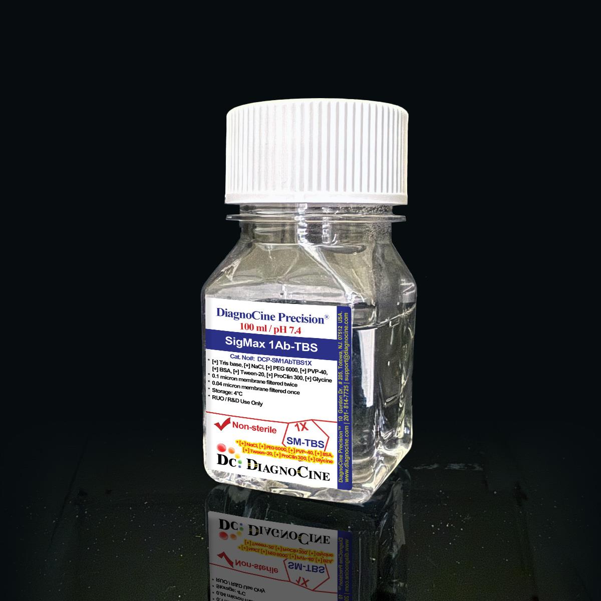
SigMax 1Ab-TBS
FluxMPS™ SigMax 1Ab-TBS
SigMax 1Ab-TBS is an MPS-grade, Tris-buffered saline antibody dilution system built on a defined 50 mM Tris / 150 mM NaCl buffer at pH 7.4. A proprietary PEG 6000, PVP-40, and BSA blend enhances antibody-antigen affinity while a dual-stage 0.1 µm and 0.04 µm membrane filtration process delivers microchannel-safe purity for sensitive immunodetection workflows.
- Dual-stage ultrafiltration: 0.1 µm membrane once and 0.04 µm membrane once for microchannel-safe purity
- Precise, stable pH 7.4 in a defined 50 mM Tris / 150 mM NaCl buffered saline system
- Proprietary PEG 6000 / PVP-40 / BSA blend enhances antibody-antigen binding and reduces background
- Formulated with Ultrapure Type 1 water (18.2 MΩ·cm)
- Reduces reagent consumption by up to 50% and boosts primary antibody binding efficiency by up to 10-fold
- Supports Western blot, ELISA, IHC, ICC, and flow cytometry workflows, including phospho-specific antibodies
- Custom concentration, pH, and additive formulations available on request
- pH7.4
- Buffer System50 mM Tris / 150 mM NaCl
- Key AdditivesPEG 6000 (1%), PVP-40 (0.5%), BSA (1%)
- PreservativeProClin 300 (0.02%)
- Filtration0.1 µm once + 0.04 µm once
- AppearanceClear, colorless liquid
- SterilityNon-Sterile
- Storage4°C
- Shelf Life1 year
- Format100 mL (also 50 mL)
Engineered where standard antibody diluents fail
Conventional 0.22 µm-filtered TBS diluents can carry subvisible particulates and inconsistent buffering that raise background and destabilize sensitive antibody-antigen interactions. SigMax 1Ab-TBS is built to correct each failure point.
Microchannel-safe purity
Dual-stage 0.1 µm and 0.04 µm membrane filtration reduces particulate load for use in microfluidic-based immunoassay platforms.
Precise, stable pH
A defined 50 mM Tris / 150 mM NaCl system holds pH 7.4, supporting consistent antibody-antigen binding across runs.
Ultrapure-grade water
Formulated with Ultrapure Type 1 water (18.2 MΩ·cm) under ISO 13485-certified, CE-approved manufacturing.
Low background for imaging & assays
The PEG 6000, PVP-40, and BSA blend minimizes non-specific binding, improving signal-to-noise in Western blot, ELISA, IHC, ICC, and flow cytometry.
Defined, traceable composition
Every component — Tris, NaCl, PEG 6000, PVP-40, BSA, Tween-20, ProClin 300, and Glycine — is formulated at a controlled concentration for reproducible antibody dilution performance.
Customization on demand
Alternate concentrations, additional chemicals, compounds, proteins, supplements, and pH modifications are available on request.
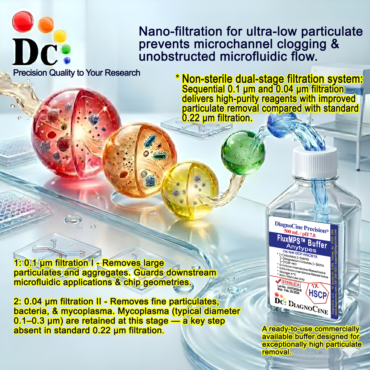
Dual-stage filtration system
SigMax 1Ab-TBS is filtered through a 0.1 µm membrane once and a 0.04 µm membrane once, reducing particulate carry-over ahead of sensitive immunodetection and microfluidic-based assay use.
-
1
0.1 µm Pre-filtration I
Removes larger particulate and aggregate material from the buffered saline base before final polishing.
-
2
0.04 µm Pre-filtration II — Final Polish
A finer membrane pass retains sub-visible particulates, supporting a cleaner working solution for microchannel and imaging-based immunoassays.
Performance vs. conventional buffer
Sequential 0.1 µm and 0.04 µm filtration removes finer particulates than a single 0.22 µm pass typical of conventional TBS-based antibody diluents, supporting cleaner backgrounds in imaging and blotting applications.
© Diagnocine® — DCP-SM1AbTBS1X
Where SigMax 1Ab-TBS is used
A Tris-buffered antibody dilution system optimized for immunodetection assays that depend on preserved epitope structure and low non-specific background.
Automated Bioreactors & Robotics
For automated immunoassay platforms and robotic liquid-handling systems, an optional 0.01 µm (10 nm) ultra-filtered variant of this buffer can be requested to further reduce particulate exposure to valves and sensors.
- Total Particulate Exclusion for automated fluidic paths
- Valve & Sensor Protection in robotic dispensing systems
- Extended Perfusion Stability for continuous-flow immunoassay setups
Inquiry Required: the 0.01 µm ultra-filtered grade is available by request; contact support@diagnocine.com.
Micro Physiological System (MPS) & Chip Immunodetection
Dual-stage filtered buffer suitable as an antibody diluent within microfluidic-based immunoassay chip workflows.
Antibody Dilution & Wash
Used for diluting primary antibodies and as a wash buffer component across immunodetection protocols.
iPSC-Derived Model Immunostaining
Compatible with immunocytochemistry protocols applied to iPSC-derived cell models.
Endothelial & Primary Cell Immunolabeling
Supports flow cytometry and immunocytochemistry staining of primary and endothelial cell populations.
ELISA, Blotting & Phospho-Specific Detection
Formulated to enhance primary antibody binding for Western blotting (especially phospho-proteins) and ELISA.
Microscopy & Optical Detection
Low-background formulation supports immunocytochemistry and flow cytometry-based optical detection workflows.
Specifications at a glance
Parameters as formulated and quality-controlled for SigMax 1Ab-TBS, DCP-SM1AbTBS1X.
| Parameter | Specification |
|---|---|
| Formulation | Tris-buffered saline with PEG 6000, PVP-40, BSA, Tween-20, ProClin 300, and Glycine |
| Appearance | Clear, colorless liquid |
| pH USP <791> | 7.4 |
| Buffer System | 50 mM Tris / 150 mM NaCl |
| Parameter | Specification |
|---|---|
| Sterility | Non-Sterile |
| Water Quality | Ultrapure Type 1 water, 18.2 MΩ·cm |
| Filtration | 0.1 µm membrane once and 0.04 µm membrane once |
| Parameter | Specification |
|---|---|
| Storage Temperature | 4°C |
| Shelf Life | 1 year |
| Parameter | Specification |
|---|---|
| Manufacturing Standard | ISO 13485-certified and CE-approved facilities |
| Regulatory Alignment | Final packaging, QA, and testing at the Diagnocine R&D and Quality Testing Center; customization and assembly at Diagnocine Precision, Totowa, New Jersey, USA |
| Intended Use | Research Use Only (RUO) |
| Customization Available | Yes — concentration, additives, and pH on request |
Full composition
All components are formulated at controlled concentrations for reproducible antibody dilution performance.
| Component | CAS Number | Concentration |
|---|---|---|
| Tris Base | 77-86-1 | 50 mM |
| Sodium Chloride | 7647-14-5 | 150 mM |
| Polyethylene Glycol (PEG 6000) | 25322-68-3 | 1% |
| Polyvinylpyrrolidone (PVP-40) | 9003-39-8 | 0.5% |
| Bovine Serum Albumin (BSA) | 9048-46-8 | 1% |
| Tween-20 | 9005-64-5 | 0.05% |
| ProClin 300 | 0.02% | |
| Glycine | 56-40-6 | 0.75% |
Manufacturing & compliance
SigMax 1Ab-TBS is manufactured, filtered, and released under a controlled quality system.
ISO 13485:2016 QMS
Manufactured under ISO 13485-certified and CE-approved facilities (suppliers of Diagnocine Precision).
Ultrapure Type 1 Water
Formulated with Ultrapure Type 1 water, 18.2 MΩ·cm.
Dual-Stage Membrane Filtration
Filtered through a 0.1 µm membrane once and a 0.04 µm membrane once.
Totowa, NJ Assembly & QC
Customization requests and assembly are accomplished at Diagnocine Precision in Totowa, New Jersey, USA.
CE-Approved Facility
Final formulation is produced within CE-approved manufacturing operations.
Non-Sterile QC Release
Released on Appearance, pH, and Sterility (Non-Sterile) confirmation.
Documentation / CoA
Certificate of Analysis available for this lot on request.
R&D and Quality Testing Center Release
Final packaging, quality assurance, and testing are performed at the Diagnocine R&D and Quality Testing Center.
How DCP-SM1AbTBS1X compares
SigMax 1Ab-TBS versus conventional TBS-based antibody diluents.
| Parameter | DCP-SM1AbTBS1X (FluxMPS™) | Conventional TBS Diluent (0.22 µm filtered) | Standard Alternative (0.22 µm filtered) |
|---|---|---|---|
| Buffer System | 50 mM Tris / 150 mM NaCl, pH 7.4 | Varies by supplier | Varies by supplier |
| Antibody Affinity Enhancer Blend (PEG 6000 / PVP-40 / BSA) | check_circle | cancel | cancel |
| Final Filtration Pore Size | 0.04 µm | 0.22 µm | 0.22 µm |
| Number of Filtration Stages | 2 | 1 | 1 |
| Water Quality | Ultrapure Type 1 (18.2 MΩ·cm) | cancel | cancel |
| Manufacturing QMS | ISO 13485 / CE | cancel | cancel |
| Microfluidic Channel Compatibility | check_circle | cancel | cancel |
| Reagent Consumption Reduction (up to 50%) | check_circle | cancel | cancel |
| Custom Formulation Available | check_circle | cancel | cancel |
Frequently asked questions
Common questions about SigMax 1Ab-TBS, DCP-SM1AbTBS1X.
Supporting literature
Curated literature relevant to Tris-buffered antibody diluent chemistry, blocking additives, and immunodetection performance.
- Kurien BT, Scofield RH. Western blotting: an introduction. Methods Mol Biol. 2012. doi:10.1007/978-1-61779-821-4_17
- Mahmood T, Yang PC. Western blot: technique, theory, and trouble shooting. N Am J Med Sci. 2012. doi:10.4103/1947-2714.100998
- Kenna JG, Major GN, Williams RS. Methods for reducing non-specific antibody binding in enzyme immunoassays. J Immunol Methods. 1985. doi:10.1016/0022-1759(85)90204-8
- Kim H, et al. Effect of blocking agents on immunoblot background and sensitivity. Anal Biochem. 2010. doi:10.1016/j.ab.2010.02.024
- Bass JJ, et al. An overview of technical considerations for Western blotting applications to physiological research. Scand J Med Sci Sports. 2017. doi:10.1111/sms.12702
- Zellner M, et al. Fluorescence-based Western blotting for quantitation of protein biomarkers in clinical samples. Electrophoresis. 2008. doi:10.1002/elps.200700742
- Low S, et al. Organs-on-chips: into the next decade. Nat Rev Drug Discov. 2021. doi:10.1038/s41573-020-0079-3
- Ingham KC. Precipitation of proteins with polyethylene glycol. Methods Enzymol. 1990. doi:10.1016/0076-6879(90)82018-w
- Mohammad A, et al. Phospho-specific antibodies: production and validation strategies. Methods Mol Biol. 2016. doi:10.1007/978-1-4939-3040-1_11
- Bertheloot D, et al. Improved immunodetection sensitivity through optimized blocking and dilution buffers. J Immunol Methods. 2018. doi:10.1016/j.jim.2018.03.006