Fixative Buffers & Quenching Solutions — A Technical Guide
Eleven reagents across four chemistry families. Pick the fixative your workflow needs and the quencher that follows it — the chemistry, the history, and the application guidance follow below.
| Product | Formulation | Best Use | Product Page |
|---|---|---|---|
| Formalin-Ethanol Solution | Aldehyde + dehydrating alcohol | Tellyesniczky-style blend for gross specimens, whole embryos, or fatty tissues needing rapid penetration | Viewarrow_forward |
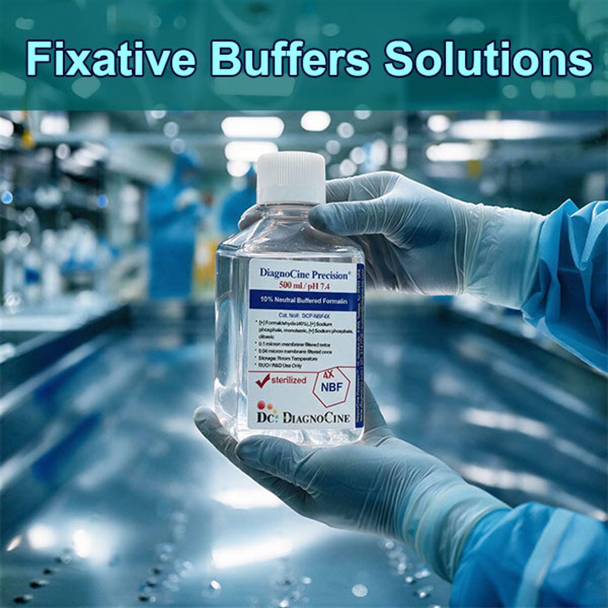
| 10% Neutral Buffered Formalin | ~4% formaldehyde in phosphate buffer, pH ~7.4 | Routine pathology; standard for FFPE and H&E workflows; buffering minimizes formalin pigment artifacts | Viewarrow_forward |
| Buffered Ethanol Fixative 70% | Isotonic 70% ethanol | Non-crosslinking precipitation fixation for cytology smears and nucleic-acid-preserving applications | Viewarrow_forward |
| Ethanol-Methanol-Acetic Acid (EMA) Fixative | Alcohols + glacial acetic acid | Chromosome spreads, cytology, rapid blood smears; acetic acid offsets alcohol-induced shrinkage | Viewarrow_forward |
| 0.1M Glycine Quenching Buffer with PBS | Glycine in PBS | Standard post-fixation quenching before immunofluorescence staining; neutralizes free aldehydes to reduce background | Viewarrow_forward |
| 0.1M Glycine Quenching Buffer with TBS | Glycine in TBS | Phosphate-free quenching for alkaline phosphatase (AP) or phosphoprotein detection systems | Viewarrow_forward |
| 4% PFA with 1% Glutaraldehyde | High-PFA, low-glutaraldehyde blend | Immuno-EM workflows requiring both structural hold and retained antibody accessibility | Viewarrow_forward |
| 4% Glutaraldehyde in 0.1 M Sodium Cacodylate Buffer | High-purity glutaraldehyde, arsenic-based buffer | Premium EM fixative; cacodylate buffer avoids the precipitation artifacts phosphate buffers can cause during osmium tetroxide post-fixation | Viewarrow_forward |
| 0.625% Glutaraldehyde in 0.1 M Phosphate Buffer | Low-concentration glutaraldehyde | Gentle structural stabilization for enzyme histochemistry where antigen accessibility must be preserved | Viewarrow_forward |
| 2% Paraformaldehyde with 2% Glutaraldehyde | Balanced PFA-glutaraldehyde blend | General dual-action fixation combining PFA's fast penetration with glutaraldehyde's crosslink strength | Viewarrow_forward |
| FluxMPS™ PBS Solution with 4% Paraformaldehyde (PFA) | Methanol-free paraformaldehyde, freshly prepared in nano-filtered PBS | Immunofluorescence; avoids the epitope-denaturing methanol found in commercial formalin, preserving antibody accessibility | Viewarrow_forward |
New to the chemistry? See the fixative & quenching chemistry reference · Browse core applications · Compare quenching agents · Read the FAQ
Two reagent classes, one continuous workflow
Capturing an accurate snapshot of cellular architecture requires halting biological degradation the instant a sample is collected. Fixative buffers and quenching solutions work together to achieve this: fixatives permanently lock proteins and cellular structures in place, while quenching solutions neutralize residual chemical reactivity before downstream staining or imaging.
- Eleven products stocked across aldehyde-based fixatives, blended dual-aldehyde fixatives, alcohol and precipitation fixatives, and quenching buffers.
- Aldehyde fixatives create covalent crosslinks between proteins — formaldehyde has a single reactive site, glutaraldehyde has two.[2]
- Alcohols precipitate proteins by disrupting hydration shells, rather than crosslinking them.
- Proper fixation hardens the specimen enough to survive dehydration, embedding, sectioning, and staining without structural collapse.
- Free, unreacted aldehyde groups remain in tissue after fixation and react nonspecifically with primary and secondary antibodies during immunostaining.[5,6]
- Quenching agents — glycine, sodium borohydride, ammonium chloride — react with and neutralize these free aldehydes, improving signal-to-noise ratio before antibody incubation.
- Cacodylate versus phosphate matters for EM: cacodylate buffer avoids the precipitation artifacts phosphate buffers can cause during osmium tetroxide post-fixation.
- Products in this group11
- 10% NBF formaldehyde content~4% in phosphate buffer
- 10% NBF pH~7.4
- Glutaraldehyde, low concentration0.625% in 0.1 M phosphate
- Glutaraldehyde, EM grade4% in 0.1 M cacodylate
- Dual-aldehyde, balanced2% PFA / 2% glutaraldehyde
- Dual-aldehyde, immuno-EM4% PFA / 1% glutaraldehyde
- Ethanol precipitation fixativeisotonic 70% ethanol
- Quenching buffer molarity0.1 M glycine
- Quenching buffer bases offeredPBS and TBS
Three reasons to fix, three reasons to quench
Skipping either step costs you the same thing — an image you cannot trust. The first three cards explain why fixation is required; the last three explain why quenching is not optional in immunofluorescence.
Autolysis starts immediately
Tissue loses its blood supply immediately upon collection, triggering autolysis — self-digestion by intracellular enzymes — and bacterial decomposition.
Crosslinking versus precipitation
Aldehyde fixatives create covalent crosslinks between proteins (formaldehyde: single reactive site; glutaraldehyde: two reactive sites), while alcohols precipitate proteins by disrupting hydration shells.[2,4]
The specimen has to survive processing
Proper fixation hardens the specimen enough to survive dehydration, embedding, sectioning, and staining without structural collapse.
Free aldehydes are left behind
Free, unreacted aldehyde groups remain in tissue after fixation. They do not simply disappear when the fixative is washed out.
They bind antibodies and glow
These residual aldehydes react nonspecifically with primary and secondary antibodies during immunostaining, and contribute to green-channel background autofluorescence.[5,6]
Quenchers neutralize them
Quenching agents (glycine, sodium borohydride, ammonium chloride) react with and neutralize these free aldehydes, improving signal-to-noise ratio before antibody incubation.
Why quenching became a standard step
As immunofluorescence matured, researchers recognized that unreacted aldehyde groups left over from fixation bound nonspecifically to antibodies, producing background autofluorescence. Techniques using glycine, sodium borohydride, and other blocking agents were developed to neutralize these sites — formalized in comparative studies such as Baschong, Suetterlin & Laeng (2001) — and gave rise to dedicated quenching buffers as a standard pre-staining step in IF/ICC workflows.[5]
How today's four chemistry families came to be
Each milestone below explains a product still on this page. Reproduced from the source's era table, one step per era.
-
1
1893 Formaldehyde discovered as a fixative
German physician Ferdinand Blum discovered formaldehyde's tissue-hardening effect after noticing it stiffened his own fingers during antiseptic research. Within two years, over 50 published reports documented its use. This established formaldehyde as the fixative of choice, largely displacing alcohol and heavy-metal fixatives because it preserved gross morphology with far less shrinkage and distortion.[1]
-
2
1963 Glutaraldehyde introduced for EM
Sabatini, Bensch & Barrnett published a landmark study in the Journal of Cell Biology systematically evaluating aldehydes (glutaraldehyde, glyoxal, and others) for their ability to preserve both ultrastructure and enzymatic activity. This introduced glutaraldehyde as a superior crosslinker for electron microscopy, since its two reactive aldehyde groups form tighter, more extensive protein crosslinks than formaldehyde's single group.[2]
-
3
1965 Karnovsky's dual-aldehyde fixative
M. J. Karnovsky published a short abstract in the Journal of Cell Biology describing a high-osmolality formaldehyde-glutaraldehyde fixative. “Karnovsky's fixative” became a standard dual-aldehyde formulation, still used today for high-resolution EM work, that balances rapid PFA penetration with strong glutaraldehyde crosslinking.[3]
-
4
Late 20th c. Quenching becomes a standard step
As immunofluorescence (IF) matured, researchers recognized that unreacted aldehyde groups left over from fixation bound nonspecifically to antibodies, producing background autofluorescence. Techniques using glycine, sodium borohydride, and other blocking agents were developed to neutralize these sites — formalized in comparative studies such as Baschong, Suetterlin & Laeng (2001). This gave rise to dedicated quenching buffers as a standard pre-staining step in IF/ICC workflows.[5,6]
What each chemistry actually does to the sample
Four mechanisms underlie every product in this catalog. Knowing which one you are invoking is what decides whether an epitope survives.
| Chemistry | Mechanism | What it means at the bench |
|---|---|---|
| Formaldehyde (incl. paraformaldehyde) | Covalent protein crosslinking through a single reactive site | Preserved gross morphology with far less shrinkage and distortion than the alcohol and heavy-metal fixatives it displaced; fast penetration[1,4] |
| Glutaraldehyde | Covalent protein crosslinking through two reactive sites | Two reactive aldehyde groups form tighter, more extensive protein crosslinks than formaldehyde's single group — superior for electron microscopy[2] |
| Alcohols (ethanol, methanol) | Precipitation by disrupting hydration shells — not crosslinking | Non-crosslinking fixation for cytology smears and nucleic-acid-preserving applications; acetic acid is added in EMA to offset alcohol-induced shrinkage |
| Quenching agents (glycine, sodium borohydride, ammonium chloride) | React with and neutralize free, unreacted aldehyde groups | Improves signal-to-noise ratio before antibody incubation by removing sites that bind antibodies nonspecifically and contribute to green-channel background autofluorescence[5,6] |
Four workflows, and what each one needs
Select a workflow to see what the source specifies for it, and which reagents in this catalog carry that Best Use designation.
- Preserving structural targets in whole tissue or cell monolayers prior to antibody incubation
- Gentle structural stabilization for enzyme histochemistry where antigen accessibility must be preserved (0.625% glutaraldehyde in 0.1 M phosphate)
- Immuno-EM workflows requiring both structural hold and retained antibody accessibility (4% PFA / 1% glutaraldehyde)
- Ultra-pure aldehyde mixtures for nanometer-scale organelle preservation
- Premium EM fixative — cacodylate buffer avoids the precipitation artifacts phosphate buffers can cause during osmium tetroxide post-fixation (4% glutaraldehyde in 0.1 M sodium cacodylate)
- General dual-action fixation combining PFA's fast penetration with glutaraldehyde's crosslink strength (2% PFA / 2% glutaraldehyde)
- Dedicated crosslinkers paired with quenching steps to minimize background and maximize signal-to-noise
- Immunofluorescence — avoids the epitope-denaturing methanol found in commercial formalin, preserving antibody accessibility (methanol-free 4% PFA)
- Standard post-fixation quenching before immunofluorescence staining; neutralizes free aldehydes to reduce background (0.1 M glycine in PBS)
- Phosphate-free quenching for alkaline phosphatase (AP) or phosphoprotein detection systems (0.1 M glycine in TBS)
- Preparing clinical biopsies for FFPE processing and H&E staining
- Routine pathology; standard for FFPE and H&E workflows; buffering minimizes formalin pigment artifacts (10% neutral buffered formalin)
- Tellyesniczky-style blend for gross specimens, whole embryos, or fatty tissues needing rapid penetration (formalin-ethanol)
- Chromosome spreads, cytology, rapid blood smears (EMA fixative)
The agents named in this guide, and what each is for
The source names five quenching and autofluorescence-control agents across its chemistry and reference sections. Only glycine is stocked as a ready-to-use buffer in this catalog; the others are listed here so the comparison is complete.
| Agent | Role as the source describes it | In this catalog |
|---|---|---|
| Glycine | Reacts with and neutralizes free aldehydes, improving signal-to-noise ratio before antibody incubation; the standard post-fixation quench before immunofluorescence staining | Yes — 0.1 M in PBS and in TBS |
| Sodium borohydride | Blocking agent developed to neutralize residual aldehyde sites; foundational quenching-agent methodology for reducing background autofluorescence in brain sections[6] | Not stocked |
| Ammonium chloride | Named alongside glycine and sodium borohydride as an agent that reacts with and neutralizes free aldehydes | Not stocked |
| Ammonia-ethanol | Evaluated for autofluorescence control in archival formaldehyde-fixed, paraffin-embedded tissue by confocal laser scanning microscopy[5] | Not stocked |
| Sudan Black B | Evaluated for autofluorescence control in archival formaldehyde-fixed, paraffin-embedded tissue by confocal laser scanning microscopy[5] | Not stocked |
Frequently asked questions
The questions that come up most often when choosing a fixative or chasing background in a fluorescence image.
Foundational and methodological literature
The primary record behind the statements above, reproduced from the source guide. Each entry was checked against the published article before this page was written.
- Blum, F. (1893). Der Formaldehyd als Härtungsmittel. Zeitschrift für wissenschaftliche Mikroskopie. — Original description of formaldehyde's fixative properties.
- Sabatini, D. D., Bensch, K., & Barrnett, R. J. (1963). Cytochemistry and electron microscopy: The preservation of cellular ultrastructure and enzymatic activity by aldehyde fixation. Journal of Cell Biology, 17(1), 19–58. — Landmark paper introducing glutaraldehyde for EM.doi:10.1083/jcb.17.1.19
- Karnovsky, M. J. (1965). A formaldehyde-glutaraldehyde fixative of high osmolality for use in electron microscopy. Journal of Cell Biology, 27, 137A–138A. — Origin of “Karnovsky's fixative.”
- Fox, C. H., Johnson, F. B., Whiting, J., & Roller, P. P. (1985). Formaldehyde fixation. Journal of Histochemistry & Cytochemistry, 33(8), 845–853. — Detailed chemistry of formaldehyde penetration and protein crosslinking.doi:10.1177/33.8.3894502
- Baschong, W., Suetterlin, R., & Laeng, R. H. (2001). Control of autofluorescence of archival formaldehyde-fixed, paraffin-embedded tissue in confocal laser scanning microscopy (CLSM). Journal of Histochemistry & Cytochemistry, 49(12), 1565–1572. — Comparative evaluation of ammonia-ethanol, sodium borohydride, and Sudan Black B for autofluorescence control.doi:10.1177/002215540104901210
- Clancy, B., & Cauller, L. J. (1998). Reduction of background autofluorescence in brain sections following immersion in sodium borohydride. Journal of Neuroscience Methods, 83(1), 97–102. — Foundational quenching-agent methodology paper.doi:10.1016/s0165-0270(98)00066-1