4% PFA with 1% Glutaraldehyde
FluxMPS™ 4% PFA with 1% Glutaraldehyde Fixative Buffer
DCP-PFAGA1X is an MPS-grade, dual-aldehyde fixative buffer combining 4% paraformaldehyde with 1% glutaraldehyde in 0.1M phosphate buffer at pH 7.4 for rapid tissue penetration paired with superior ultrastructural preservation. Sequentially filtered through 0.1 µm and 0.04 µm membranes and manufactured under ISO 13485-certified, CE-approved facilities, this methanol-free, ready-to-use formulation is suited to electron microscopy, immunohistochemistry, developmental biology, and organ-on-a-chip (OoC) tissue-model fixation.
- Non-sterile, ultrapure, sequentially filtered through a 0.1 µm membrane once and a 0.04 µm membrane once
- Dual-aldehyde formulation: 4% paraformaldehyde plus 1% glutaraldehyde
- Prepared in 0.1M phosphate buffer at pH 7.4 for physiological compatibility
- Methanol-free — eliminates membrane permeabilization artifacts of methanol-stabilized formalin
- Ready-to-use, pre-mixed format for consistent results
- Manufactured under ISO 13485-certified, CE-approved facilities
- Custom concentrations, additives, and pH available on request
- pH7.4
- Formulation4% PFA / 1% Glutaraldehyde
- Buffer System0.1M Phosphate Buffer
- Filtration0.1 µm once + 0.04 µm once
- SterilityNon-Sterile
- AppearanceClear solution
- Storage4°C, protected from light
- Shelf Life6 months
- ShippingTypically within 48 hours (up to 1 week)
- Size500 mL
Engineered where standard fixatives fall short
Single-aldehyde formalin and unfiltered fixative solutions carry subvisible particulates, inconsistent aldehyde ratios, and variable pH that compromise ultrastructure and antigen accessibility. DCP-PFAGA1X is formulated and filtered to remove these failure modes.
Microchannel-safe purity
Sequential 0.1 µm and 0.04 µm membrane filtration reduces particulate load for clean fixation of tissue models in confined channels.
Precise, stable pH
Prepared in 0.1M phosphate buffer at pH 7.4 to maintain osmotic balance and minimize cellular distortion during fixation.
Ultrapure-grade water
Formulated with ultrapure Type 1 water (18.2 MΩ·cm) to minimize trace contaminants that can interfere with downstream staining.
Low background for imaging & assays
Compatible with fluorescent proteins such as hyperfolder YFP, retaining approximately 70% initial fluorescence intensity compared with methanol-free 4% PFA alone.
Defined, traceable composition
A controlled, methanol-free dual-aldehyde formulation containing paraformaldehyde and glutaraldehyde in a fixed phosphate buffer background.
Customization on demand
Other concentrations, additions of chemicals, compounds, proteins, or supplements, different pH, and formulation modifications are available on inquiry.
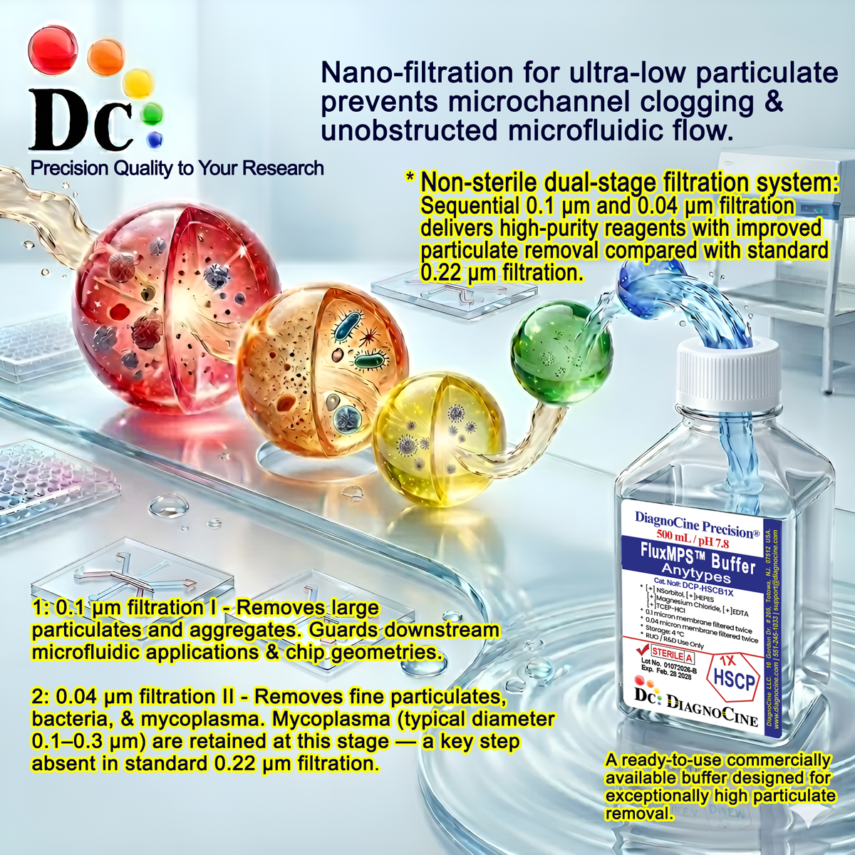
Dual-stage filtration system
DCP-PFAGA1X is filtered through a 0.1 µm membrane once and a 0.04 µm membrane once, reducing particulate load beyond a conventional single-pass filtration step while the fixative remains non-sterile as formulated.
-
1
0.1 µm Pre-filtration I
Large particulate and aggregate removal, extending the service life of downstream filtration.
-
2
0.04 µm Pre-filtration II
Fine particulate retention for a cleaner finished fixative solution.
Performance vs. conventional buffer
A sequential 0.1 µm and 0.04 µm filtration pass removes finer particulates than a single conventional filtration step, supporting cleaner backgrounds for downstream imaging and staining without altering the fixative chemistry.
© Diagnocine® — DCP-PFAGA1X
Where DCP-PFAGA1X is used
A balanced dual-aldehyde ratio makes this fixative suited to workflows that demand both rapid tissue stabilization and preserved ultrastructure, antigenicity, and fluorescence.
Automated Bioreactors & Robotics
An optional 0.01 µm (10 nm) ultra-filtered variant is available on inquiry for automated tissue processors and robotic liquid-handling platforms used in high-throughput histology and imaging workflows.
- Total Particulate Exclusion for particulate-sensitive automated staining lines
- Valve & Sensor Protection in robotic liquid-handling systems
- Extended Stability during automated, unattended processing runs
Inquiry Required: contact support@diagnocine.com to request the 0.01 µm ultra-filtered grade.
Organ-on-Chip & MPS Tissue Fixation
In-situ dual-aldehyde fixation of tissue models within microfluidic channels prior to imaging or immunostaining.
Ultrastructural Preservation for EM
The 1% glutaraldehyde concentration provides sufficient cross-linking for membrane preservation without excessive hardening, facilitating easier sectioning after osmium tetroxide staining.
Fluorescent Protein & Live-Cell Marker Preservation
Compatible with fluorescent proteins like hyperfolder YFP, retaining approximately 70% initial fluorescence intensity compared with methanol-free 4% PFA alone; retained fluorescence demonstrated for mitochondrial markers like MitoTracker Red.
Whole-Embryo & Craniofacial Tissue Fixation
Validated for embryonic tissue fixation, showing 23% less craniofacial distortion versus Karnovsky’s original 5% glutaraldehyde formula.
IHC & IF Sample Preparation
Lower glutaraldehyde content preserves epitope accessibility, enabling reliable immunohistochemistry and immunofluorescence without extensive antigen retrieval steps.
Standard Histology & Cytology Fixation
Ready-to-use, pre-mixed format ensures consistency and reduces preparation time compared with traditional ampoule-based fixation kits.
Product parameters
All parameters below reflect DCP-PFAGA1X as formulated, filtered, and released by Diagnocine.
| Parameter | Specification |
|---|---|
| Formulation | 4% PFA + 1% Glutaraldehyde |
| Buffer System | 0.1M Phosphate Buffer |
| Appearance | Clear solution |
| pH USP <791> | 7.4 |
| Paraformaldehyde Penetration Rate | 1–2 mm/hour |
| Parameter | Specification |
|---|---|
| Sterility | Non-Sterile |
| Filtration | 0.1 µm membrane once + 0.04 µm membrane once |
| Water Quality | Ultrapure Type 1 water (18.2 MΩ·cm) |
| Manufacturing Standard ISO 13485 | ISO 13485:2016-certified facility |
| Parameter | Specification |
|---|---|
| Storage Temperature | 4°C, protected from light (amber) |
| Shelf Life | 6 months |
| Shipping Condition | Typically within 48 hours; up to 1 week during unforeseen events |
| Fixation Immersion Time | 4–24 hours at 4°C |
| Rinse Protocol | 3x with PBS or buffer |
| Parameter | Specification |
|---|---|
| Raw Material / Facility Grade | ISO 13485-certified, CE-approved supplier facilities |
| Manufacturing QMS | ISO 13485:2016 |
| Regulatory Alignment | CE-approved |
| Production & QA Location | DiagnoCine R&D and Quality Testing Center, Totowa, New Jersey, USA |
| Intended Use | Research Use Only (RUO) |
Full composition
Dual-aldehyde fixative composition, prepared in 0.1M phosphate buffer.
| Component | CAS Number | Concentration |
|---|---|---|
| Paraformaldehyde (PFA) | 30525-89-4 | 4% |
| Glutaraldehyde | 111-30-8 | 1% |
| Phosphate Buffer | 0.1M |
Manufacturing & compliance
DCP-PFAGA1X is manufactured under ISO 13485-certified and CE-approved facilities, with final packaging, quality assurance, and testing completed at the DiagnoCine R&D and Quality Testing Center.
ISO 13485:2016 QMS
Manufactured under ISO 13485-certified and CE-approved supplier facilities.
Ultrapure Type 1 Water
Formulated with ultrapure Type 1 water (18.2 MΩ·cm).
Sequential Membrane Filtration
0.1 µm membrane filtration once and 0.04 µm membrane filtration once for particulate control.
Micro-Batch Traceability
Final packaging, QA, and testing performed at the DiagnoCine R&D and Quality Testing Center, Totowa, New Jersey, USA.
pH Verification USP <791>
Each lot is verified to a pH of 7.4.
Appearance QC
Each lot is verified as a clear solution.
Sterility Status
Non-Sterile; sequentially filtered through a 0.1 µm membrane once and a 0.04 µm membrane once.
Documentation / CoA
A Certificate of Analysis is available on request.
How DCP-PFAGA1X compares
A qualitative comparison against conventional and standard-alternative fixative solutions.
| Parameter | DCP-PFAGA1X (FluxMPS™) | Conventional Fixative (Unfiltered) | Standard Alternative (0.22 µm Filtered) |
|---|---|---|---|
| Dual-aldehyde formulation (4% PFA + 1% GA) | check_circle | cancel | cancel |
| Final filtration pore size | 0.04 µm | Unfiltered | 0.22 µm |
| Number of filtration stages | 2 | 0 | 1 |
| Methanol-free formulation | check_circle | cancel | check_circle |
| Physiological phosphate buffering (0.1M, pH 7.4) | check_circle | cancel | check_circle |
| ISO 13485 manufacturing QMS | check_circle | cancel | cancel |
| Fluorescent protein compatibility | check_circle | cancel | cancel |
| Custom formulation available | check_circle | cancel | cancel |
Frequently asked questions
Common questions about DCP-PFAGA1X.
Supporting literature
Curated literature relevant to dual-aldehyde fixation chemistry and its research applications.
- Karnovsky MJ. A formaldehyde-glutaraldehyde fixative of high osmolality for use in electron microscopy. J Cell Biol. 1965. doi:10.1083/jcb.27.2.137A
- Hopwood D. Fixatives and fixation: a review. Histochem J. 1969. doi:10.1007/BF01003278
- Hayat MA. Principles and Techniques of Electron Microscopy: Biological Applications. Cambridge University Press. doi:10.1017/CBO9780511605377
- Kiernan JA. Formaldehyde, formalin, paraformaldehyde and glutaraldehyde: what they are and what they do. Microsc Today. 2000. doi:10.1017/S1551929500057060
- Melan MA, Sluder G. Redistribution and differential extraction of soluble proteins in permeabilized cultured cells. J Cell Sci. 1992. doi:10.1242/jcs.101.4.731
- Schnell U, Dijk F, Sjollema KA, Giepmans BN. Immunolabeling artifacts and the need for live-cell imaging. Nat Methods. 2012. doi:10.1038/nmeth.1855
- Low LA, Mummery C, Berridge BR, Austin CP, Tagle DA. Organs-on-chips: into the next decade. Nat Rev Drug Discov. 2021. doi:10.1038/s41573-020-0079-3
- Bhatia SN, Ingber DE. Microfluidic organs-on-chips. Nat Biotechnol. 2014. doi:10.1038/nbt.2989
- Richter KN, Revelo NH, Seitz KJ, et al. Glyoxal as an alternative fixative to formaldehyde in immunostaining. EMBO J. 2018. doi:10.15252/embj.201695709