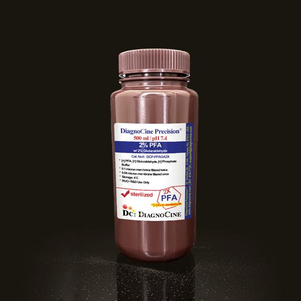
0.1M Glycine Quenching Buffer with PBS
FluxMPS™ 0.1M Glycine Quenching Buffer with PBS
An MPS-grade, ready-to-use quenching buffer combining 0.1 M glycine with physiological PBS to rapidly neutralize residual aldehyde groups after formaldehyde or paraformaldehyde fixation. Dual-stage 0.1 µm and 0.04 µm membrane filtration keeps particulate background low, helping preserve epitope integrity and image quality in downstream immunodetection.
- Formulated with 0.1 M glycine in physiological PBS to rapidly quench residual aldehyde groups after formaldehyde/paraformaldehyde fixation
- Dual-stage membrane filtration (0.1 µm then 0.04 µm) for reduced particulate background
- Precise pH 7.4 for consistent, reproducible quenching kinetics
- Non-sterile, ready-to-use liquid format for research workflows
- Compatible with IF, ICC, IHC, flow cytometry, and select ChIP protocols
- Helps reduce autofluorescence and background signal in confocal and widefield microscopy
- Manufactured under ISO 13485:2016-certified, CE-approved facilities
- Custom pH, molarity, and formulation options available on request
- pH7.4
- Glycine Concentration0.1 M
- Buffer BasePBS (NaCl, Na2HPO4, KCl, KH2PO4)
- AppearanceClear Solution
- Filtration0.1 µm once + 0.04 µm once
- SterilityNon-sterile (membrane filtered)
- FormatReady-to-use liquid, 500 mL
- Storage4°C, protect from light and excessive heat
- Shelf Life1 year
- Intended UseResearch Use Only (RUO)
Engineered where standard quenching buffers fall short
Conventional single-pass 0.22 µm-filtered glycine buffers can carry subvisible particulates and inconsistent pH from lot to lot, adding background noise and variability to sensitive immunofluorescence, ICC, and ChIP workflows. FluxMPS™ DCP-GQPBS0.1X is filtered through a dual-stage membrane process and formulated to a defined, reproducible pH and glycine concentration for cleaner, more consistent quenching.
Microchannel-safe purity
Sequential 0.1 µm and 0.04 µm membrane filtration reduces particulate load prior to non-sterile fill, supporting cleaner downstream microscopy and immunodetection.
Precise, stable pH
Formulated to pH 7.4 with 0.1 M glycine in physiological PBS to deliver consistent, reproducible aldehyde-quenching kinetics batch to batch.
Ultrapure-grade water
Prepared with Ultrapure Type 1 water (18.2 MΩ·cm) as a manufacturing input to minimize trace contaminants in the finished buffer.
Low background for imaging & assays
Rapid, controlled quenching of unreacted aldehyde groups helps reduce background and autofluorescence in confocal and widefield microscopy.
Defined, traceable composition
Every buffer salt and the glycine quenching agent are declared with concentration values so protocols can be reproduced exactly.
Customization on demand
pH, molarity, and formulation can be adjusted to your protocol — contact support@diagnocine.com.
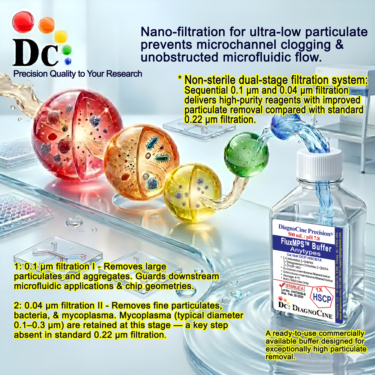
Dual-stage filtration system
DCP-GQPBS0.1X is processed through two sequential membrane stages — a 0.1 µm pass followed by a 0.04 µm pass — before non-sterile fill, reducing particulate carryover relative to a single conventional 0.22 µm pass.
-
1
0.1 µm Pre-filtration I
Removes larger particulates and aggregates from the glycine/PBS solution, protecting the finer downstream membrane and extending its service life.
-
2
0.04 µm Pre-filtration II
A second, finer membrane pass retains fine particulates approaching the smallest known mycoplasma size (about 0.2 micron), further reducing bioburden ahead of fill. This buffer is filtered through both stages but is supplied non-sterile.
Performance vs. conventional buffer
Sequential 0.1 µm and 0.04 µm membrane filtration removes finer particulates than a single 0.22 µm pass typical of conventional glycine quenching buffers, supporting lower background in downstream imaging and immunodetection.
© Diagnocine® — DCP-GQPBS0.1X
Where DCP-GQPBS0.1X fits your workflow
Glycine's amino group (-NH2) reacts with unreacted formaldehyde or paraformaldehyde molecules, halting further cross-linking at a controlled time point and preventing over-fixation that can compromise antibody epitope recognition.
Automated Bioreactors & Robotics
For automated fixation/staining platforms and liquid-handling robotics, an optional 0.01 µm (10 nm) ultra-filtered variant of this quenching buffer can be produced on request to further minimize particulate load in fluidic pathways.
- Total Particulate Exclusion: Finer filtration for closed fluidic systems
- Valve & Sensor Protection: Reduced particulate risk to precision components
- Extended Perfusion Stability: Lower fouling potential in automated lines
Inquiry Required: the 0.01 µm ultra-filtered grade is produced to order — contact support@diagnocine.com to request this configuration.
Post-Fixation Quenching for MPS & Chip Assays
Quenches residual aldehyde groups on fixed cells within microfluidic chips prior to permeabilization, blocking, or antibody incubation.
Wash & Reconstitution After Fixation
Used directly after fixative removal and a PBS rinse to stop crosslinking at the desired time point in cell and tissue samples.
iPSC-Derived Model Handling
Reduces fixative-derived background in immunostaining of fixed iPSC-derived neuronal and cardiac models.
Endothelial & Primary Cell Immunostaining
Stabilizes fixed endothelial and primary cell samples ahead of permeabilization and antibody staining steps.
ELISA, Blotting & Blocking
Improves signal-to-noise ratio in immunocytochemistry and immunohistochemistry by removing fixative-derived background prior to blocking.
Microscopy & Confocal Background Reduction
Reduces fixative-derived autofluorescence, supporting cleaner confocal and widefield imaging of fixed samples.
Detailed product specifications
Values below reflect the formulation and quality parameters specific to DCP-GQPBS0.1X.
| Parameter | Specification |
|---|---|
| Formulation / Composition | 0.1 M Glycine in PBS (NaCl, Na2HPO4, KCl, KH2PO4) |
| Appearance | Clear Solution |
| pH USP <791> | 7.4 |
| Glycine Molarity / Concentration | 0.1 M |
| Parameter | Specification |
|---|---|
| Sterility | Non-sterile; filtered 0.1 µm once and 0.04 µm once |
| Water Quality | Ultrapure Type 1 water (18.2 MΩ·cm) |
| Manufacturing Standard ISO 13485 | ISO 13485:2016-certified facility |
| Parameter | Specification |
|---|---|
| Storage Temperature | 4°C, protect from light and excessive heat |
| Shelf Life | 1 year |
| Parameter | Specification |
|---|---|
| Manufacturing QMS | ISO 13485-certified facility |
| Regulatory Alignment | CE-approved facilities (Suppliers of DiagnoCine Precision) |
| Production Method | Final packaging, QA, and customization at DiagnoCine Precision, Totowa, New Jersey, USA |
| Intended Use | Research Use Only (RUO) |
Full composition
Glycine and PBS salt concentrations are declared below exactly as formulated.
| Component | CAS Number | Concentration |
|---|---|---|
| Glycine | 56-40-6 | 0.1 M |
| Sodium Chloride | 7647-14-5 | 137 mM |
| Disodium Hydrogen Phosphate | 7558-79-4 | 10 mM |
| Potassium Chloride | 7447-40-7 | 2.7 mM |
| Monopotassium Phosphate | 7778-77-0 | 1.8 mM |
Manufacturing & compliance
DCP-GQPBS0.1X is produced, packaged, and quality-tested under a controlled quality system.
ISO 13485:2016 QMS
Manufactured under ISO 13485-certified and CE-approved facilities (Suppliers of DiagnoCine Precision).
Ultrapure Type 1 Water
Prepared using Ultrapure Type 1 water (18.2 MΩ·cm) as a manufacturing input.
Dual-Stage Membrane Filtration
Filtered through 0.1 µm and 0.04 µm membranes prior to non-sterile fill.
Micro-Batch Precision
Final packaging, quality assurance, and testing are completed at the DiagnoCine R&D and Quality Testing Center.
Facility Certification
ISO 13485-certified, CE-approved manufacturing facilities.
Custom Assembly Site
Specific customization requests and assembly completed at DiagnoCine Precision in Totowa, New Jersey, USA.
pH Control
Formulated and released at pH 7.4.
Documentation
Certificate of Analysis available on request.
How DCP-GQPBS0.1X compares
A qualitative comparison against conventional 0.22 µm-filtered glycine quenching buffers.
| Parameter | DCP-GQPBS0.1X (FluxMPS™) | Conventional 0.22 µm-Filtered Buffer | Standard Alternative (0.22 µm Filtered) |
|---|---|---|---|
| Glycine-Based Aldehyde Quenching | check_circle | cancel | cancel |
| Final Filtration Pore Size | 0.04 µm | 0.22 µm | 0.22 µm |
| Number of Filtration Stages | 2 | 1 | 1 |
| Formulated in Physiological PBS | check_circle | cancel | cancel |
| ISO 13485:2016 Manufacturing | check_circle | cancel | cancel |
| Ultrapure Type 1 Water Input | check_circle | cancel | cancel |
| Custom Formulation Available | check_circle | cancel | cancel |
| Optimized for IF, ICC, IHC, Flow, ChIP | check_circle | cancel | cancel |
Frequently asked questions
Common questions about DCP-GQPBS0.1X 0.1M Glycine Quenching Buffer with PBS.
Supporting literature
Curated references on formaldehyde/paraformaldehyde fixation chemistry, glycine quenching, and background reduction in immunodetection.
- Fox CH, Johnson FB, Whiting J, Roller PP. Formaldehyde fixation. J Histochem Cytochem. 1985;33(8):845-853. doi:10.1177/33.8.3894502
- Thavarajah R, Mudimbaimannar VK, Elizabeth J, Rao UK, Ranganathan K. Chemical and physical basics of routine formaldehyde fixation. J Oral Maxillofac Pathol. 2012;16(3):400-405. doi:10.4103/0973-029X.102496
- Hoffman EA, Frey BL, Smith LM, Auble DT. Formaldehyde crosslinking: a tool for the study of chromatin complexes. J Biol Chem. 2015;290(44):26404-26411. doi:10.1074/jbc.R115.651679
- Metz B, Kersten GF, Hoogerhout P, et al. Identification of formaldehyde-induced modifications in proteins. J Biol Chem. 2004;279(8):6235-6243. doi:10.1074/jbc.M310752200
- Solomon MJ, Varshavsky A. Formaldehyde-mediated DNA-protein crosslinking: a probe for in vivo chromatin structures. Proc Natl Acad Sci USA. 1985;82(19):6470-6474. doi:10.1073/pnas.82.19.6470
- Jamur MC, Oliver C. Cell fixatives for immunostaining. Methods Mol Biol. 2010;588:55-61. doi:10.1007/978-1-59745-324-0_8
- Kim SO, Kim J, Okajima T, Cho NJ. Mechanical properties of paraformaldehyde-treated individual cells. Nano Converg. 2017;4(1):5. doi:10.1186/s40580-017-0099-9
- Im K, Mareninov S, Diaz MFP, Yong WH. An introduction to performing immunofluorescence staining. Methods Mol Biol. 2019;1897:299-311. doi:10.1007/978-1-4939-8935-5_26
- Robinson JM, Vandre DD. Efficient immunocytochemical labeling of leukocyte microtubules with FluoroNanogold. J Histochem Cytochem. 1997;45(5):631-642. doi:10.1177/002215549704500813