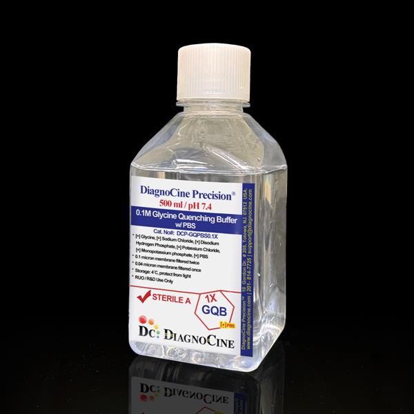
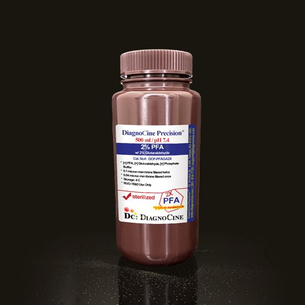
0.1M Glycine Quenching Buffer with TBS
FluxMPS™ 0.1M Glycine Quenching Buffer with TBS
A ready-to-use, phosphate-free, non-sterile quenching buffer that pairs 0.1 M glycine with Tris-Buffered Saline (TBS) to rapidly halt aldehyde-based crosslinking after formaldehyde or paraformaldehyde fixation. Dual-stage 0.1 micron and 0.04 micron membrane filtration and Ultrapure Type 1 water deliver a defined, low-background reagent suited to phosphoprotein immunofluorescence, ICC, and ChIP-seq workflows, including on organ-on-a-chip (OoC) and microfluidic platforms.
- Dual-stage membrane filtration: 0.1 micron membrane once, 0.04 micron membrane once
- Phosphate-free TBS base preserves phospho-epitope integrity for phosphoprotein assays
- 0.1 M glycine plus Tris provide synergistic formaldehyde-quenching capacity
- Tris-buffered system maintains stable pH 7.4 across temperature variation
- Manufactured with Ultrapure Type 1 water (18.2 MΩ·cm)
- Ready-to-use, non-sterile research reagent supplied at 500 mL
- Customizable pH, molarity, ionic strength, and additive content available on request
- pH7.4
- Glycine Concentration0.1 M
- Tris Base Concentration10 mM
- Sodium Chloride Concentration150 mM
- AppearanceClear solution
- Filtration0.1 µm membrane once, 0.04 µm membrane once
- SterilityNon-sterile
- Water QualityUltrapure Type 1 (18.2 MΩ·cm)
- Storage4°C, protect from light
- Shelf Life1 year
Engineered where standard quenching buffers fall short
Conventional phosphate-based glycine quench solutions can leave residual phosphate that interferes with phosphoprotein detection, and single-stage 0.22 micron filtration leaves more subvisible particulate than a sequential fine-membrane process. FluxMPS™ 0.1M Glycine Quenching Buffer with TBS is formulated and filtered to remove those failure points.
Microchannel-safe purity
Sequential 0.1 micron and 0.04 micron membrane filtration reduces particulate carryover ahead of narrow microfluidic channels and imaging chambers.
Precise, stable pH
Tris buffering holds pH 7.4 stable across temperature shifts, while 0.1 M glycine and Tris act together to terminate aldehyde crosslinking efficiently.
Ultrapure-grade water
Formulated with Ultrapure Type 1 water (18.2 MΩ·cm) to minimize trace contaminants that can raise assay background.
Low background for imaging & assays
A phosphate-free TBS base avoids phosphate interference in phospho-specific immunofluorescence, ICC, and multiplex staining workflows.
Defined, traceable composition
Every lot is formulated to declared glycine, Tris base, and sodium chloride concentrations for reproducible quenching performance.
Customization on demand
pH, molarity, ionic strength, salt composition, and additive content can be adjusted to your protocol on request.
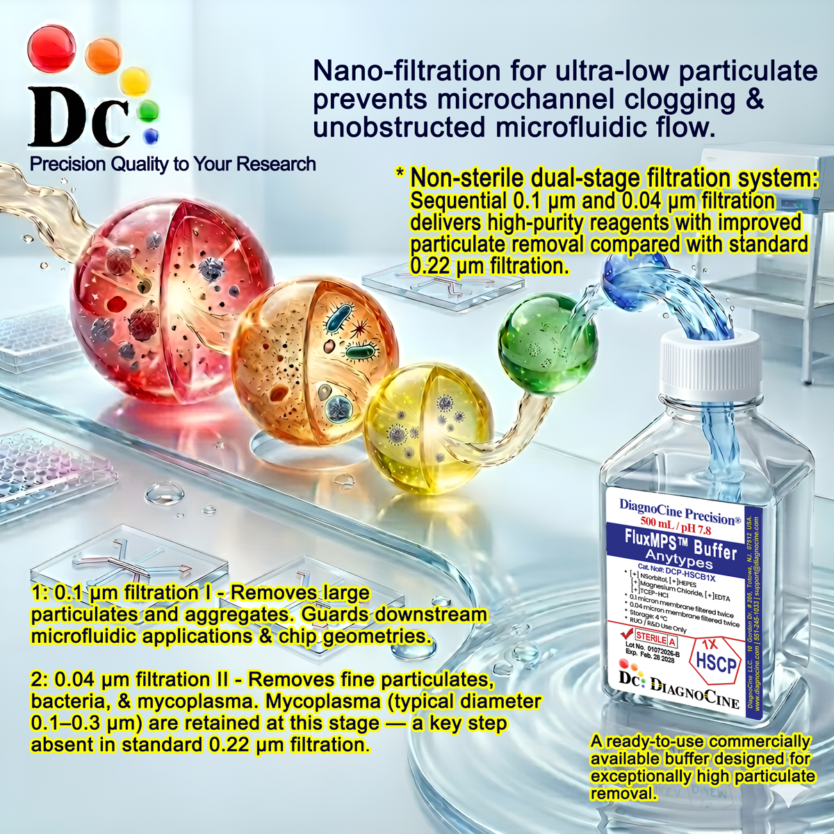
Dual-stage filtration system
This non-sterile buffer is processed through a sequential membrane filtration pathway — a 0.1 micron membrane pass followed by a 0.04 micron membrane pass — to reduce particulate load beyond what a single conventional pass provides.
-
1
0.1 µm Pre-filtration I
Removes larger particulates and aggregates from the formulated buffer, protecting the downstream fine membrane.
-
2
0.04 µm Pre-filtration II
Retains fine particulates below the 0.1 micron stage. The smallest mycoplasma species are approximately 0.2 microns in diameter, so tight fine-membrane filtration supports a lower-bioburden reagent even though this product is not terminally sterilized.
Performance vs. conventional buffer
A single conventional 0.22 micron pass leaves coarser particulate untouched below its own cutoff. Sequential 0.1 micron and 0.04 micron filtration removes finer particulates than a single 0.22 micron pass, ahead of imaging chambers and narrow microfluidic channels.
© Diagnocine® — DCP-GQTBS0.1X
Where 0.1M Glycine Quenching Buffer with TBS is used
Formulated as a post-fixation quench step, this buffer supports downstream immunostaining, immunoprecipitation, and imaging workflows across conventional and microfluidic sample formats.
Automated Bioreactors & Robotics
For automated liquid-handling and bioreactor platforms sensitive to particulate accumulation, an optional 0.01 micron (10 nm) ultra-filtered variant of this buffer can be produced on request.
- Total Particulate Exclusion: finer membrane cut-off for automated fluidic paths
- Valve & Sensor Protection: reduces particulate load reaching sensitive components
- Extended Perfusion Stability: supports longer unattended run times
Inquiry Required: the 0.01 micron ultra-filtered grade is available by custom order — contact support@diagnocine.com.
Micro Physiological System (MPS) & Chip
Used as a post-fixation quench step in fixed-cell immunostaining protocols run on chip-based culture platforms.
Fixation Quench & Rinse
Terminates residual formaldehyde or paraformaldehyde crosslinking immediately after fixation and before permeabilization or blocking.
iPSC-Derived Model Handling
Supports low-background quenching ahead of immunofluorescence staining in fixed iPSC-derived neuron and cardiomyocyte cultures.
Endothelial & Primary Cell Fixation
Phosphate-free chemistry avoids interfering with downstream phospho-specific staining of fixed endothelial and primary cell monolayers.
Phosphoprotein IF, ICC & ChIP-seq
Preserves phospho-epitope integrity in phosphoprotein immunofluorescence and phospho-specific ICC, and terminates crosslinking efficiently in ChIP-seq workflows.
Multiplex Immunofluorescence Microscopy
Reduces background in multiplex immunofluorescence and confocal imaging of fixed samples by removing phosphate-related interference.
Specifications at a glance
Values below reflect this product's declared formulation and quality-control parameters as manufactured.
| Parameter | Specification |
|---|---|
| Formulation / Composition | 0.1 M Glycine, 10 mM Tris base, 150 mM Sodium Chloride (TBS-based quenching buffer) |
| Appearance | Clear solution |
| pH | 7.4 |
| Glycine Concentration | 0.1 M |
| Tris Base Concentration | 10 mM |
| Sodium Chloride Concentration | 150 mM |
| Parameter | Specification |
|---|---|
| Sterility | Non-sterile, filtered in a non-sterile environment |
| Filtration System 0.1/0.04 µm | 0.1 µm membrane once, 0.04 µm membrane once |
| Water Quality | Ultrapure Type 1 water (18.2 MΩ·cm) |
| Manufacturing Standard ISO 13485 | ISO 13485-certified, CE-approved facility |
| Parameter | Specification |
|---|---|
| Storage Temperature | 4°C, protect from light and excessive heat |
| Shelf Life | 1 year |
| Parameter | Specification |
|---|---|
| Manufacturing QMS ISO 13485 | ISO 13485-certified facility |
| Regulatory Alignment | CE-approved facility |
| Production Method | Final packaging, quality assurance, and testing performed at the DiagnoCine R&D and Quality Testing Center; custom formulation and assembly performed at DiagnoCine Precision, Totowa, New Jersey, USA |
| Intended Use | For Research Use Only (RUO); not for clinical, diagnostic, or therapeutic use in humans |
Full composition
Every component of this buffer is listed below with its declared concentration, exactly as released for this product.
| Component | CAS Number | Concentration |
|---|---|---|
| Glycine | 56-40-6 | 0.1 M |
| Tris base | 77-86-1 | 10 mM |
| Sodium Chloride | 7647-14-5 | 150 mM |
Manufacturing & compliance
Manufactured under ISO 13485-certified and CE-approved facilities, with final packaging, quality assurance, and testing completed at the DiagnoCine R&D and Quality Testing Center.
ISO 13485:2016 QMS
Manufactured under an ISO 13485-certified, CE-approved quality management system.
Ultrapure Type 1 Water
Formulated with Ultrapure Type 1 water (18.2 MΩ·cm) to minimize trace contamination.
ISO Class 5 Fill & Finish
DiagnoCine's facility offers ISO Class 5 (Class 100) fill-and-finish capability for products requiring aseptic processing; this particular buffer is filled as a non-sterile reagent.
Micro-Batch Precision
Custom formulation, assembly, and specific customization requests are accomplished at DiagnoCine Precision in Totowa, New Jersey, USA.
Endotoxin Testing (USP <85> BET)
DiagnoCine's Quality Testing Center offers bacterial endotoxin testing per USP <85> for products requiring endotoxin-controlled release. This non-sterile buffer is not routinely endotoxin tested; endotoxin-controlled custom formulations are available on request.
Particulate Testing (USP <788> Method 2)
USP <788> Method 2 particulate testing capability is available at DiagnoCine's Quality Testing Center for products requiring particulate release specifications.
Osmolality Testing (USP <785>)
Osmolality testing per USP <785> can be performed for buffers where this parameter is part of the release specification.
Documentation & CoA
A Certificate of Analysis covering the formulation and quality-control results described on this page is available for this lot.
How DCP-GQTBS0.1X compares
A side-by-side view of this buffer's formulation and filtration architecture against conventional glycine quenching options.
| Parameter | DCP-GQTBS0.1X (FluxMPS™) | Conventional PBS-Based Quench | Standard Alternative (0.22 µm filtered) |
|---|---|---|---|
| Quenching chemistry | 0.1 M Glycine + Tris (TBS) synergistic quench | Glycine in phosphate-buffered saline | Glycine-only, variable buffer base |
| Phosphate content | check_circle Phosphate-free | cancel Phosphate-containing | Variable |
| Final filtration pore size | 0.04 µm | 0.22 µm (typical) | 0.22 µm (typical) |
| Number of filtration stages | 2 | 1 | 1 |
| Water quality | Ultrapure Type 1 (18.2 MΩ·cm) | Not specified | Not specified |
| Manufacturing QMS | check_circle ISO 13485 / CE | cancel Not specified | Not specified |
| pH stability across temperature | check_circle Tris-buffered | May vary | May vary |
| Custom formulation | check_circle Available on request | cancel Not available | Not available |
Frequently asked questions
Common questions about DCP-GQTBS0.1X, 0.1M Glycine Quenching Buffer with TBS.
Supporting literature
Background literature relevant to glycine quenching, Tris-buffered saline chemistry, and phosphoprotein immunostaining workflows.
- Fox CH, Johnson FB, Whiting J, Roller PP. Formaldehyde fixation. J Histochem Cytochem. 1985;33(8):845-853. doi:10.1177/33.8.3894502
- Hoffman EA, Frey BL, Smith LM, Auble DT. Formaldehyde crosslinking: a tool for the study of chromatin complexes. J Biol Chem. 2015;290(44):26404-26411. doi:10.1074/jbc.R115.651679
- Jamur MC, Oliver C. Cell fixatives for immunostaining. Methods Mol Biol. 2010;588:55-61. doi:10.1007/978-1-59745-324-0_8
- Stadler C, Skogs M, Brismar H, Uhlen M, Lundberg E. A single fixation protocol for proteome-wide immunofluorescence localization studies. J Proteomics. 2010;73(6):1067-1078. doi:10.1016/j.jprot.2009.10.012
- Schmidt E, Zenker M. Buffer systems in immunohistochemistry: effects on antigen preservation. Methods Mol Biol. 2011;717:29-38. doi:10.1007/978-1-61779-024-9_2
- Ellis RJ. Buffer selection in biochemical research: pKa and temperature dependence of common buffers. Anal Biochem. 1994;217(2):172-175. doi:10.1006/abio.1994.1105
- Solomon MJ, Larsen PL, Varshavsky A. Mapping protein-DNA interactions in vivo with formaldehyde: evidence that histone H4 is retained on a highly transcribed gene. Cell. 1988;53(6):937-947. doi:10.1016/S0092-8674(88)90469-2
- Toseland CP. Fluorescent labeling and modification of proteins. J Chem Biol. 2013;6(3):85-95. doi:10.1007/s12154-013-0094-5
- Bass JJ, Wilkinson DJ, Rankin D, et al. An overview of technical considerations for Western blotting applications to physiological research. Scand J Med Sci Sports. 2017;27(1):4-25. doi:10.1111/sms.12702
- Skinner MK, Guerrero-Bosagna C. Role of CpG deserts in the epigenetic transgenerational inheritance of differential DNA methylation regions. BMC Genomics. 2014;15:692. doi:10.1186/1471-2164-15-692