Fluorescent Protein/Antibody
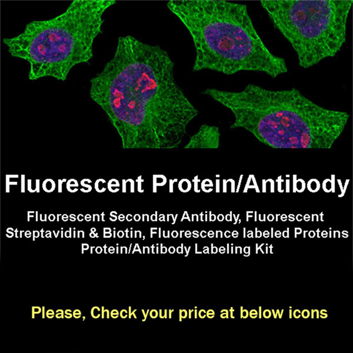
Fluorescence labeled proteins and antibodies might provide great advantages in the identification of their binding partners or in the observation of biological processes. They allow to visualize cellular environment, to define their function, regulation and interactions. In advanced assays, fluorescent proteins can be utilized in measuring of protein–protein interactions or conformational changes, by measuring intensity changes or FRET energy transfer. An ideal fluorescent label should be small, bright, stable, and without any perturbation to the biological system. Additionally, the fluorescence labeling should be performed at non-active site with the possibility for multiplexing and avoided to the tendency to aggregate.
Organic fluorescent dye-labeled proteins have superior properties over fluorescent protein–conjugated counterparts including, wide spectral range, smaller size, greater photostability, and higher brightness. However, dye–labeled proteins also have some limitations that their sensitivity is low due to lack of the amplification step associated with the secondary antibody, and the protein should be free of contaminants and stabilizing agents. BioActs provides a wide range of fluorophore-conjugated proteins and antibodies for a variety of biological applications.
Fluorescent Dye conjugated Secondary Antibody
|
Indirect or secondary immunofluorescence method employs specific interactions between primary and secondary antibody. It is more commonly used than the direct immunofluorescence method due to strong signal amplification and cost-effectiveness. Non-labeled primary antibody in indirect immunofluorescence specifically binds to target molecule, and fluorescence conjugated secondary antibody acts as an antigen of the primary antibody. In addition, the polyclonal nature of secondary antibody features multiplex binding of secondary antibodies per a primary antibody resulting in amplified fluorescence signal. However, since indirect immunofluorescence method utilizes complicated antibody-antibody interaction, the experiment process is complicated and the nonspecific fluorescence signals along with cross reaction might occur. BioActs provides a wide spectrum of fluorescent dye conjugated secondary antibodies directed against IgG from mouse, rabbit, rat, and goat.
In spite of competing approaches such as peptide tagging or mass spectrometry, antibody-based detection is the most broadly used application in analysis of specific protein in complex samples. Fluorescent secondary antibodies are raised against IgG heavy and light chains of the target IgG and minimized cross reactivity by affinity purification and by adsorption against the sera of a number of species. For multiple labeling experiments where cross reactivity is the critical issue, we prepare highly cross-adsorbed goat anti–mouse IgG, goat anti–rabbit IgG, and goat anti–rat IgG fluorescent antibodies. Our antibody probes are conjugated with Flamma® Fluor series, which displays strong fluorescence and photostability, along with ICG and horseradish peroxidase (HRP). Due to their outstanding optical and biological properties, Flamma® Fluor conjugates are superior to most conventional fluorescent secondary antibodies and being optimal molecular probes in many applications. BioActs offers series of fluorescent secondary antibodies as suitable molecular probes for many biological experiments such as fluorescence microscopy, flow cytometry, microplate assays, as protein and nucleic acid blots, in situ hybridization, etc. |
Goat anti-mouse IgG
|
 |
 |
 |
 |
 |
 |
|
| 495/519 |
554/565 |
593/618 |
651/667 |
679/696 |
751/774 |
774/790 |
 |
| HRP |
| Quick link (Cat.#) |
Series |
Quick link (Cat.#) |
Series |
| RSA1145 |
Goat anti-mouse IgG, FSD™ 488 |
RSA1141 |
Goat anti-mouse IgG, Flamma® 488 |
| RSA1155 |
Goat anti-mouse IgG, FSD™ 555 |
RSA1151 |
Goat anti-mouse IgG, Flamma® 552 |
| RSA1195 |
Goat anti-mouse IgG, FSD™ 594 |
RSA1191 |
Goat anti-mouse IgG, Flamma® 594 |
| RSA1165 |
Goat anti-mouse IgG, FSD™ 647 |
RSA1161 |
Goat anti-mouse IgG, Flamma® 648 |
| RSA1175 |
Goat anti-mouse IgG, FSD™ 680 |
RSA1171 |
Goat anti-mouse IgG, Flamma® 675 |
| RSA1105 |
Goat anti-mouse IgG, FSD™ 750 |
RSA1101 |
Goat anti-mouse IgG, Flamma® 749 |
| RSA1185 |
Goat anti-mouse IgG, FSD™ 800 |
RSA1122 |
Goat anti-mouse IgG, HRP |
Goat anti-rabbit IgG
|
 |
 |
 |
 |
 |
 |
|
| 495/519 |
554/565 |
593/618 |
651/667 |
679/696 |
751/774 |
774/790 |
|
| HRP |
| Quick link (Cat.#) |
Series |
Quick link (Cat.#) |
Series |
| RSA1245 |
Goat anti-rabbit IgG, FSD™ 488 |
RSA1241 |
Goat anti-rabbit IgG, Flamma® 488 |
| RSA1255 |
Goat anti-rabbit IgG, FSD™ 555 |
RSA1251 |
Goat anti-rabbit IgG, Flamma® 552 |
| RSA1295 |
Goat anti-rabbit IgG, FSD™ 594 |
RSA1291 |
Goat anti-rabbit IgG, Flamma® 594 |
| RSA1265 |
Goat anti-rabbit IgG, FSD™ 647 |
RSA1261 |
Goat anti-rabbit IgG, Flamma® 648 |
| RSA1275 |
Goat anti-rabbit IgG, FSD™ 680 |
RSA1271 |
Goat anti-rabbit IgG, Flamma® 675 |
| RSA1205 |
Goat anti-rabbit IgG, FSD™ 750 |
RSA1201 |
Goat anti-rabbit IgG, Flamma® 749 |
| RSA1285 |
Goat anti-rabbit IgG, FSD™ 800 |
RSA1221 |
Goat anti-rabbit IgG, HRP |
Goat anti-rat IgG
|
 |
 |
 |
 |
 |
 |
|
| 495/519 |
554/565 |
593/618 |
651/667 |
679/696 |
751/774 |
774/790 |
|
| HRP |
| Quick link (Cat.#) |
Series |
Quick link (Cat.#) |
Series |
| RSA1545 |
Goat anti-rat IgG, FSD™ 488 |
RSA1541 |
Goat anti-rat IgG, Flamma® 488 |
| RSA1555 |
Goat anti-rat IgG, FSD™ 555 |
RSA1551 |
Goat anti-rat IgG, Flamma® 552 |
| RSA1595 |
Goat anti-rat IgG, FSD™ 594 |
RSA1591 |
Goat anti-rat IgG, Flamma® 594 |
| RSA1565 |
Goat anti-rat IgG, FSD™ 647 |
RSA1561 |
Goat anti-rat IgG, Flamma® 648 |
| RSA1575 |
Goat anti-rat IgG, FSD™ 680 |
RSA1571 |
Goat anti-rat IgG, Flamma® 675 |
| RSA1505 |
Goat anti-rat IgG, FSD™ 750 |
RSA1501 |
Goat anti-rat IgG, Flamma® 749 |
| RSA1585 |
Goat anti-rat IgG, FSD™ 800 |
RSA1521 |
Goat anti-rat IgG, HRP |
Rabbit anti-goat IgG
|
 |
 |
 |
 |
 |
 |
|
| 495/519 |
554/565 |
593/618 |
651/667 |
679/696 |
751/774 |
774/790 |
|
| HRP |
| Quick link (Cat.#) |
Series |
Quick link (Cat.#) |
Series |
| RSA4445 |
Rabbit anti-goat IgG, FSD™ 488 |
RSA4441 |
Rabbit anti-goat IgG, Flamma® 488 |
| RSA4455 |
Rabbit anti-goat IgG, FSD™ 555 |
RSA4451 |
Rabbit anti-goat IgG, Flamma® 552 |
| RSA4495 |
Rabbit anti-goat IgG, FSD™ 594 |
RSA4491 |
Rabbit anti-goat IgG, Flamma® 594 |
| RSA4465 |
Rabbit anti-goat IgG, FSD™ 647 |
RSA4461 |
Rabbit anti-goat IgG, Flamma® 648 |
| RSA4475 |
Rabbit anti-goat IgG, FSD™ 680 |
RSA4471 |
Rabbit anti-goat IgG, Flamma® 675 |
| RSA4405 |
Rabbit anti-goat IgG, FSD™ 750 |
RSA4401 |
Rabbit anti-goat IgG, Flamma® 749 |
| RSA4485 |
Rabbit anti-goat IgG, FSD™ 800 |
RSA4421 |
Rabbit anti-goat IgG, HRP |
Fluorescent Streptavidin & Biotin
|
Avidin and streptavidin are both tetrameric proteins composed of four identical subunits, each bind four biotins (vitamin H) per molecule with high binding affinity and specificity (Kd ~ 1015 M for avidin and ~ 1014 M for streptavidin). Although the primary sequence homogeneity of both proteins are 30%, their tertiary and quaternary structure are almost identical, and anti-avidin and anti-streptavidin antibodies are not immunologically cross reactive. Avidin, a 67 KDa glycoprotein with an isoelectric point of about 10.5, has the higher affinity than streptavidin, however it also displays more nonspecific binding and aggregation due to its oligosaccharide component (mannose and N-acetylglucosamine) and positive charge. Streptavidin is smaller (53 KDa) and has a little lower affinity than avidin yet displays less non-specific binding due to near-neutral pI value and lack of carbohydrates. Biotin, a 244 dalton vitamin found in all living cells, binds with high affinity to avidin and streptavidin. In biotechnology, biotin is conjugated to antibodies, enzymes, reporter to form the tetravalent binding nature of biotin-avidin/streptavidin complex. The valeric acid side of biotin can be incorporated with various functional groups, reporters and fluorophores that can be utilized in a wide range of biological structures and processes.
Biotin-avidin/streptavidin binding has high affinity, which has been utilized in diverse applications such as ELISA, immunohistochemistry, cell surface labeling, affinity purification, FACS, EMSA, etc. The bond formation between biotin and avidin/streptavidin is very rapid, and once formed, is stable at high temperature and in a wide range of pH, organic solvents and denaturing agents. The system is a simple yet elegant and can be incorporated into virtually every immunoassay where an antibody is conjugated with biotin and then detected with avidin or streptavidin conjugated to various commercially available fluorophores and reporters. These features of biotin and avidin/streptavidin are useful for purifying or detecting proteins conjugated to either component of the interaction. Although biotin-avidin/streptavidin system is simple and easy to use, it also has some limitations: biotinylated compounds might non-selectively bind to any biotin-binding protein, endogenous biotin can cause background noise, and harsh conditions are needed to break their interaction that might limit its application. BioActs offers a variety of fluorescent dye conjugated avidin, streptavidin and biotin as effective detecting and analytic probes for diverse applications in biochemical and biological research fields. |
Fluorescent Streptavidin
|
 |
 |
 |
 |
 |
|
| 496/516 |
550/565 |
648/663 |
675/691 |
749/774 |
774/806 |
| Quick link (Cat.#) |
Series |
Ex * (nm) |
Em* (nm) |
| RFP0716 |
Streptavidin, Flamma® 496 |
496 |
516 |
| RFP0705 |
Streptavidin, Flamma® 552 |
550 |
565 |
| RFP0711 |
Streptavidin, Flamma® 648 |
648 |
663 |
| RFP0712 |
Streptavidin, Flamma® 675 |
675 |
691 |
| RFP0713 |
Streptavidin, Flamma® 749 |
749 |
774 |
| RFP0714 |
Streptavidin, Flamma® 774 |
774 |
806 |
Fluorescent Biotin
|
 |
 |
 |
 |
 |
|
| 496/516 |
550/565 |
648/663 |
675/691 |
749/774 |
774/806 |
| Quick link (Cat.#) |
Series |
Ex * (nm) |
Em* (nm) |
Molar mass (g/mol) |
| RFP0616 |
Biotin, Flamma® 496 |
496 |
516 |
654.64 |
| RFP0605 |
Biotin, Flamma® 552 |
550 |
565 |
913.18 |
| RFP0611 |
Biotin, Flamma® 648 |
648 |
663 |
927.20 |
| RFP0612 |
Biotin, Flamma® 675 |
675 |
691 |
1185.43 |
| RFP0613 |
Biotin, Flamma® 749 |
749 |
774 |
951.22 |
| RFP0614 |
Biotin, Flamma® 774 |
774 |
806 |
1169.25 |
Other Fluorescence labeled Proteins
|
Lectins are carbohydrate-binding proteins or glycoproteins with a high, specific affinity to sugar residues. Most lectins do not possess enzymatic activity, however they play important roles in biological recognition phenomena involving cells, carbohydrates, and proteins. Lectins enable sensitive detection of cellular carbohydrates allowing to distinguish subtle alteration in glycosylation otherwise indistinguishable cells. They also mediate attachment and binding of bacteria and viruses to their intended targets. Oligosaccharide residues are most abundant on the cell surface, yet they also covalently attached to constituents inside the cell. Fluorescence-labeled lectins can bind to specific configurations of sugar molecules, thus they enable to identify cell types or cellular components, making them versatile detecting agents in microscopy, histochemistry and flow cytometry. In addition, fluorescent lectins might be useful markers of certain cancers because those cells often display altered surface glycoproteins. BioActs offers variety of fluorophore–conjugated lectins for identification of various cellular carbohydrates.
Albumin, making up 55 to 62% of the serum protein, is one of the few carbohydrate-free proteins in blood plasma that plays an important role in maintaining the colloidal osmotic pressure. Albumin also acts as a plasma carrier of several hydrophobic steroid hormones and as a transport protein for hemin and fatty acids. Bovine serum albumin (BSA) is a small (~66.5 kDa), stable, moderately non-reactive protein that has been utilized in numerous biochemical applications including ELISA, Western blot, immunohistochemistry, etc. BSA enable to increase antibody functionality and longevity and serves as both a standard in protein quantitation and a stabilizing component in DNA blunt end and replacement assays. Bovine serum albumin (BSA) conjugates are commonly used as tracers in applications where physical dimensions are important. Specifically, fluorescent BSA conjugates have been used in many applications such as quantitative studies of electroporation, measurement of plasma volume, intra cellular protein processing. BioActs provides a wide range of fluorophore-conjugated BSA analogs as well-defined molecular weight tracers for a variety of applications. |
Fluorescent BSA
|
 |
 |
 |
|
 |
 |
| 495/519 |
550/565 |
560/589 |
581/596 |
648/663 |
675/691 |
 |
|
|
|
|
|
| 749/774 |
774/806 |
|
|
|
|
| Quick link (Cat.#) |
Series |
Ex * (nm) |
Em* (nm) |
| RSC0102 |
BSA, Flamma® 488 |
495 |
519 |
| RSC0105 |
BSA, Flamma® 552 |
550 |
565 |
| RSC0106 |
BSA, Flamma® 560 |
560 |
589 |
| RSC0110 |
BSA, Flamma® 581 |
581 |
596 |
| RSC0111 |
BSA, Flamma® 648 |
648 |
663 |
| RSC0112 |
BSA, Flamma® 675 |
675 |
691 |
| RSC0113 |
BSA, Flamma® 749 |
749 |
774 |
| RSC0114 |
BSA, Flamma® 774 |
774 |
806 |
Protein/Antibody Labeling Kit
|
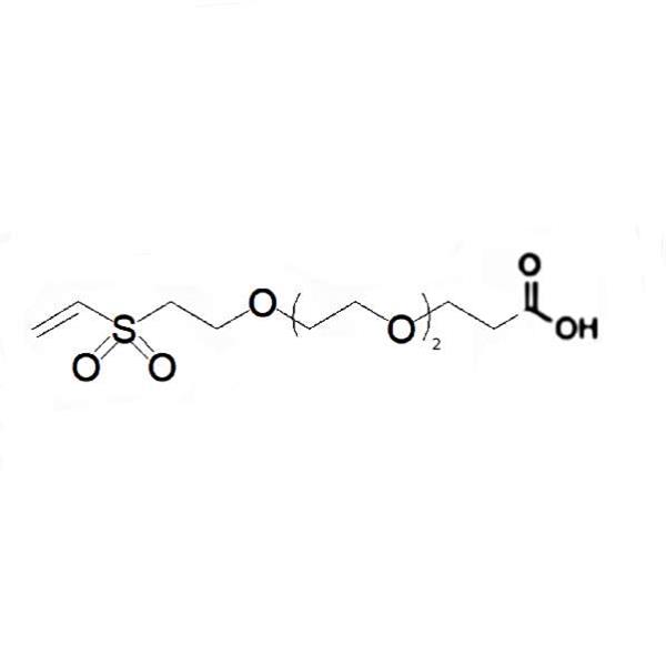
Flamma® Fluor Protein Labeling Kits from BioActs are designed for efficient labeling of antibody or protein with fluorescent materials. Due to strong absorption, high fluorescence quantum yield, and high photostability, Flamma® Fluor Vinylsulfones are selected as reactive dyes, and they maintain good fluorescence activity and stability after conjugated to biomolecules, allowing detection of low-abundance biological structures with great sensitivity. Vinylsulfone reactive group is selectively bind to the primary amines of proteins to create efficient dye-protein conjugates. The Labeling Kits are optimized for labeling 1 mg of antibody per reaction, and contain everything needed to perform the conjugation. Flamma® Fluors Protein Labeling Kits enable to perform the entire step from labeling to purification. |
Protein Labeling Kit
|
 |
 |
 |
 |
|
|
|
| 550/565 |
648/663 |
675/691 |
749/774 |
774/806 |
|
|
| Quick link (Cat.#) |
Series |
Ex * (nm) |
Em* (nm) |
Emission Color |
| XPL1104 |
Flamma® 552 Protein Labeling Kit |
550 |
565 |
Orange |
| XPL2104 |
Flamma® 648 Protein Labeling Kit |
648 |
663 |
Far-red |
| XPL3104 |
Flamma® 675 Protein Labeling Kit |
675 |
691 |
Near-IR |
| XPL4104 |
Flamma® 749 Protein Labeling Kit |
749 |
774 |
Near-IR |
| XPL5104 |
Flamma® 774 Protein Labeling Kit |
774 |
806 |
Near-IR |