Flamma® 749 protein labeling kit
Flamma® 749 protein labeling kit
Cat. No. List below
Description
Key Features:
A. Superior Fluorescence Properties
B. Strong absorption and high fluorescence quantum yield
C. Exceptional photostability for extended imaging sessions
D. Excitation/Emission maxima at 749/774 nm, ideal for far-red/near-infrared detection
E. Maintains fluorescence activity and stability after conjugation to biomolecules
The reactive group is vinylsulfone and it has selective binding to primary amines on proteins. It creates stable & efficient dye-protein conjugates and is optimized for labeling 1 mg of antibody per reaction. Flamma® 749 Protein Labeling Kit is suitable for labeling large amounts of protein or antibody. It is compatible with various proteins, not limited to antibodies. The Flamma® 749 Protein Labeling Kit includes all the necessary components for labeling and purifying proteins or antibodies. The complete labeling process can be performed in 3-6 hours.
Kit contains:
1. Flamma® Fluors 749 vinylsulfone dye
2. Reaction buffer (10×)
3. Purification column
4. PBS buffer (1×)
5. Disposable pipette
6. Detailed protocol for labeling and purification
The Flamma® 749 Protein Labeling Kit is ideal for various far-red/near-infrared fluorescence-based techniques including flow cytometry, in vivo and ex vivo imaging, deep tissue fluorescence microscopy, fluorescence guided surgery, western blotting, immunohistochemistry, and high-content screening.
The Flamma® 749 Protein Labeling Kit provides researchers with a state-of-the-art method for creating high-quality far-red/near-infrared fluorescent protein conjugates. Its unique spectral properties make it particularly suitable for applications requiring deep tissue penetration, reduced background fluorescence, or multi-color imaging. This kit enables sensitive detection of biological structures in complex samples and advances a wide range of fluorescence-based research applications, from basic science to preclinical studies and potential clinical applications.
What are the advantages?
1. High labeling efficiency and excellent conjugate recovery
2. Streamlined workflow with an all-in-one kit design
3. Flexible labeling scale, optimized for 1 mg of protein but adaptable to different amounts
4. Superior brightness and photostability compared to many conventional far-red/near-infrared dyes
5. Minimal autofluorescence and light scattering in the far-red/near-infrared region
6. Excellent for multiplexing with visible light fluorophores
7. Compatible with standard far-red/near-infrared detection systems
Specifications
- Fluorophore: Flamma® Fluors 749
- Reactive group: Vinylsulfone
- Reacting with: Primary amine
- Excitation/Emission Max.(nm): 749/774
- Appearance: Red Liquid
- Storage conditions: 4 ℃, protect from light
Table 1. List of Flamma® Fluors Protein Labeling Kits
| Quick link (Cat.#) | Series | Ex * (nm) | Em* (nm) | Emission Color | Scale |
| XPL1104 | Flamma® 552 Protein Labeling Kit | 550 | 565 | Orange | 1 mg × 3 labeling reaction |
| XPL2104 | Flamma® 648 Protein Labeling Kit | 648 | 663 | Far-red | |
| XPL3104 | Flamma® 675 Protein Labeling Kit | 675 | 691 | Near-IR | |
| XPL4104 | Flamma® 749 Protein Labeling Kit | 749 | 774 | Near-IR | |
| XPL5104 | Flamma® 774 Protein Labeling Kit | 774 | 806 | Near-IR |
Overview
Flamma® Fluor Protein Labeling Kits from BioActs are designed for efficient labeling of antibody or protein with fluorescent materials. Due to strong absorption, high fluorescence quantum yield, and high photostability, Flamma® Fluor Vinylsulfones are selected as reactive dyes, and they maintain good fluorescence activity and stability after conjugated to biomolecules, allowing detection of low-abundance biological structures with great sensitivity. Vinylsulfone reactive group is selectively bind to the primary amines of proteins to create efficient dye-protein conjugates. The Labeling Kits are optimized for labeling 1 mg of antibody per reaction, and contain everything needed to perform the conjugation. Flamma® Fluors Protein Labeling Kits enable to perform the entire step from labeling to purification.
General Information
Flamma® Fluors protein labeling kits are optimized for the fluorescence labeling of IgG antibody. The optimal amount of antibody per reaction is 1 mg (~140 kDa) in 2 mg/mL solution. If the protein preparation condition does not match with above, please refer the "Tips and Troubleshooting" section of this document. However, protein labeling kits can be used for fluorescence labeling of a variety of other proteins. Please check "Tips and Troubleshooting" for efficient labeling. Prior to labeling, the antibody/protein should be purified and dissolved in ammonium ions or amine-free buffer. If the being labeled antibody/protein is dissolved in an unsuitable buffer such as Tris or glycine, replace the buffer with phosphate buffered saline (PBS) by dialysis.
Protocol
Labeling
1. Prepare 0.5 mL of antibody/protein solution in 2 mg/mL concentration at room temperature prior to the reaction.
* If the amount of antibody/protein is less than above, refer "Tips and Troubleshooting" in this document.
2. Add 50 μL of "Component C, 10 × Reaction buffer" to above antibody/protein solution and vortex the mixture.
3. Transfer the [antibody/protein solution in step 2] to "Component A, Flamma® Fluors vinylsulfone dye" vial, cover it with cap, and vortex reaction mixture.
4. The step 3 reaction mixture is stand for 2 hours at room temperature under dark.
* Note: in general antibody labeling condition, Vinylsulfone dyes proceed about 65% of labeling at 1 h, 85% at 2 h, 90% at 4h, and the maximum labeling can be achieved at 6 h.
Purification of conjugates
| 1. As shown in Figure 1, Connect “Component E, Column Adapter” to a 50 mL conical tube,and stand the assembly in a tube rack. 2. Open the upper lid of “Component D, Purification column”, cut along the perimeter of the lower faucet cap, amount it onto the conical tube assembly, and drain internal stock liquid through the bottom faucet. *be careful not to spill resin inside Component D. 3. While the stock liquid is drained, dilute 5 mL of “Component B, 10 × PBS buffer” by 10 fold to make 1 × PBS buffer in a separate 50 mL conical tube. *For dilution, use distilled water of room temperature. 4. After all of the stock liquid is drained, dismount 50 mL conical tube, discard the liquid inside the tube, and reinstall it to complete the construction of the purification system. 5. Treat 5 mL of 1 × PBS buffer to “Component D, Purification column” by using “Component F, Disposable pipette” and drain the buffer, repeat the process several times to make the column to equilibrate with 1 × PBS buffer. 6. When labeling reaction (Labeling, step 4) is complete, the reaction mixture is carefully loaded to “Component D, Purification column”, and fluorescent dye conjugate is separated with resin column by eluting with 1 × PBS buffer. 7. Usually, fluorescence conjugated antibody/protein will come out first, and unreacted antibody/protein will come out later. *Be sure not to be contaminated by unreacted or free dyes. |
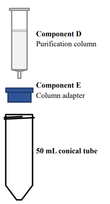
Figure 1. Purification system
|
Calculation of Dyes/Protein ratio
Absorbance measurement
Measure the absorbance of collected fluorescence conjugated antibody/protein solution from purification step 7 and obtain the absorbance value at the maximum Abs. wavelength, which stated in Table 3.
Table 3. Optical Properties of Flamma® Fluors
| Labeling Kit | Abs.max (nm) | Extinction (cm-1M-1 ) | CF280 |
| Flamma® 552 Protein Labeling Kit | 551 | 150,000 | 0.08 |
| Flamma® 648 Protein Labeling Kit | 648 | 250,000 | 0.05 |
| Flamma® 675 Protein Labeling Kit | 675 | 220,000 | 0.18 |
| Flamma® 749 Protein Labeling Kit | 750 | 220,000 | 0.04 |
| Flamma® 774 Protein Labeling Kit | 774 | 200,000 | 0.1 |
Calculation of antibody/protein concentration

*The typical molar extinction coefficient of IgG antibody is around 203,000 cm-1M-1 , and that of other protein may be vary.

*The optimal d/p ratio for IgG antibody is 3 ~ 7.
Storage and handling of dye-conjugates
Dye-conjugated antibody/protein solution shall be stored under refrigerated condition in a dark. The storage period can extended to several months when 0.5 ~ 2.0 mM sodium azide was added. For longer period of storage, store the conjugate solution at -20 ℃, but avoid repeated freezing and melting. When the concentration of conjugated antibody/protein is 1 mg/mL, add 0.1 to 1.0% stabilizing protein such as BSA and store. Do not use aggregated or sediment part, and always centrifuge the stored solution before using and use supernatant only.
Citation & Reference
1. Lim, Chang-Keun. Phthalocyanine-aggregated polymeric nanoparticles as tumor-homing near-infrared absorbers for photothermal therapy of cancer. Theranostics 2.9, 2012, 871-879.
2. Park, Jin Woo. Novel cyanine dyes with vinylsulfone group for labeling biomolecules. Bioconjugate chemistry 23.3, 2012, 350-362.
3. Lim, Chang-Keun. Gadolinium-coordinated elastic nanogels for in vivo tumor targeting and imaging. Biomaterials 34.28, 2013, 6846-6852.
4. Keunsoo Jeong. Poly (oxyethylene sugaramide) s: unprecedented multihydroxyl building blocks for tumor-homing nanoassembly. Journal of Materials Chemistry B 1.28, 2013, 3437-344
5. Lee, So Jin. TNF-α gene silencing using polymerized siRNA/thiolated glycol chitosan nanoparticles for rheumatoid arthritis. Molecular Therapy 22.2, 2014, 397-40
6. Yoon, Hong Yeol. Photo-crosslinked hyaluronic acid nanoparticles with improved stability for in vivo tumor-targeted drug delivery. Biomaterials 34.21, 2013 5273-5280.
7. Park, Solji. Amphiphilized poly (ethyleneimine) nanoparticles: a versatile multi-cargo carrier with enhanced tumor-homing efficiency and biocompatibility. Journal of Materials Chemistry B 3.2, 2015, 198-206.
8. Lee, Eunjung. Co-delivery of chemosensitizing siRNA and an anticancer agent via multiple monocomplexation-induced hydrophobic association. Journal of Controlled Release 210, 2015, 105-114.
9. Xu, Peisheng. Zwitterionic chitosan derivatives for pH-sensitive stealth coating. Biomacromolecules 11.9, 2010, 2352-2358.
10. Ibrahim, Basma M. A strategy to deliver genes to cystic fibrosis lungs: a battle with environment. Journal of controlled release 155.2, 2011, 289-295.
11. Oh, Keun Sang. Accurate sequential detection of primary tumor and metastatic lymphatics using a temperature-induced phase transition nanoparticulate system. International journal of nanomedicine 9, 2014, 2955
12. Yhee, Ji Young. Tumor-targeting transferrin nanoparticles for systemic polymerized siRNA delivery in tumor-bearing mice. Bioconjugate chemistry 24.11, 2013, 1850-1860.
13. Yoon, Hong Yeol. Glycol chitosan nanoparticles as specialized cancer therapeutic vehicles: Sequential delivery of doxorubicin and Bcl-2 siRNA. Scientific reports 4, 2014.
14. Ryu, Ju Hee. Early diagnosis of arthritis in mice with collagen?induced arthritis, using a fluorogenic matrix metalloproteinase 3–specific polymeric probe. Arthritis & Rheumatism 63.12, 2011, 3824-3832.
15. Hollis, Christin P. In vivo investigation of hybrid paclitaxel nanocrystals with dual fluorescent probes for cancer theranostics. Pharmaceutical research 31.6, 2014, 1450-1459.
16. Koo, Heebeom. The movement of self-assembled amphiphilic polymeric nanoparticles in the vitreous and retina after intravitreal injection. Biomaterials 33.12, 2012, 3485-3493.
17. Zhu, Lei. Real-time monitoring of caspase cascade activation in living cells. Journal of controlled release 163.1, 2012, 55-62.
18. Yoon, Hong Yeol. Bioreducible hyaluronic acid conjugates as siRNA carrier for tumor targeting. Journal of Controlled Release 172.3, 2013, 653-661.
19. Yhee, Ji Young. Cancer-targeted MDR-1 siRNA delivery using self-cross-linked glycol chitosan nanoparticles to overcome drug resistance. Journal of Controlled Release 198, 2015, 1-9.
20. Park, Jin Woo. Wide-Ranged Fluorescent Molecular Weight Size Markers for Electrophoresis. Bulletin of the Korean Chemical Society 34.1, 2013, 29-30.
21. Huang, Xinglu. Multiplex Imaging of an Intracellular Proteolytic Cascade by using a Broad?Spectrum Nanoquencher. Angewandte Chemie International Edition 51.7, 2012, 1625-1630