


Anti-Nup62 antibody, rat monoclonal (2A11)
Product#: FNK-70-305
$346.79
Availability:
Ships in 24 hours
Anti-Nup62 antibody, rat monoclonal (2A11)
General information
| Cat. No. | :FNK-70-305 |
| Size | :200 µg |
| Host Species | :Rat |
| Immunogen | :Recombinant human Nup62 (aa 1-300) (GST-Nup62-His) |
| Isotype | :Rat IgG1 kappa |
| Epitope | :aa 1-179 (FG-repeat region) |
| Form | :1mg/ml in PBS, 50% glycerol, filter-sterilized. Carrier protein and sodium azide.free. |
| Specificity | :Specific to human (HeLa cells) and simian (Cos cells). The antibody did not react with mouse. |
| Product | :Purified from serum-free culture medium of the hybridoma by proprietary chromatography under mild conditions. |
| Application | :Western blotting (1/500 ~1/2,000 dilution) :Immunoprecipitation (assay dependent) :Immunofluorescece / Immunocytochemistry (1/400) :ELISA (assay dependent) :When this antibody was micro-injected into the cytoplasm of the HeLa cells, it accumulates into the nuclear pores as examined by immunofluorescence staining. |
| Storage | :Shipped at 4℃ or -20℃, and upon arrival, aliquot and store at -20℃. |
| Data Link | :UniProtKB/Swiss-Prot P37198 (NUP62_HUMAN) |
Description
The nuclear pore complex (NPC) regulates cargo transport between the cytoplasm and the nucleus. Nucleoporins are the main components of the NPC in eukaryotic cells. Nup (Nucleoporin) 62 (522 aa, 53 kDa) is a member of the FG-repeat containing nucleoporins and is localized to the NPC central plug. Nup62 associates with the importin alpha/beta complex which is involved in the import of proteins containing nuclear localization signals. Predicted to contain about 10 N-acetylglucosamine side chain .
 |
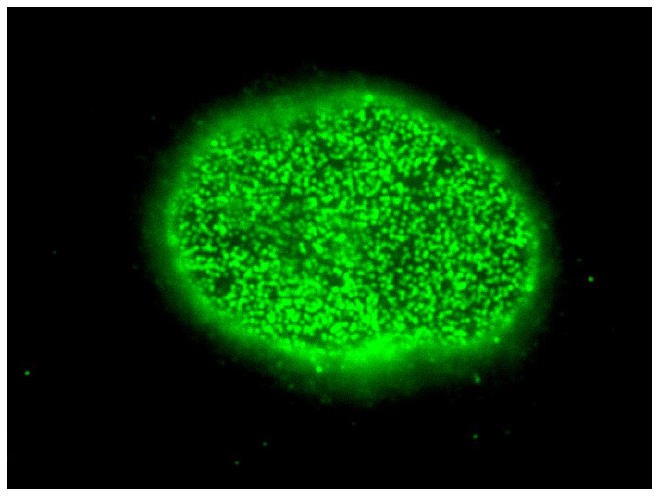 |
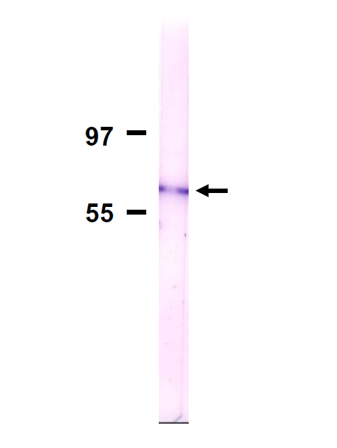
Fig.1 Detection of Nup62 in membrane fraction of HeLa cells by Western blotting with the antibody 2A. Sample is the nuclear membrane fraction of HeLa cells. The antibody was used at 1/500 dilution. As a second antibody, alkaline phosphatase conjugated anti-rat IgG antibody |
Fig.2 Immunofluorescent staining of HeLa cells with the antibody 2A, focused on nuclear surface. HeLa cells were fixed with 3.7% formaldehyde and permeabilized with 0.5% Triton X-100. The anti-Nup62 antibody (2A) was used at 1/400 and as a second antibody, Alexa 488 conjugated goat anti-rat IgG antibody was used at 1/500 dilution. |
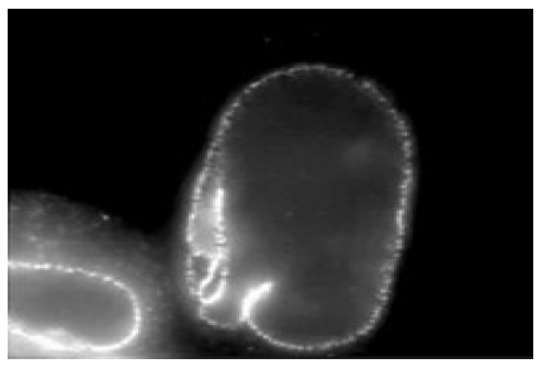 |
Fig.3. Immunofluorescent staining of HeLa cells with the antibody 2A, focused on nuclear rim. Methods are as described in Fig.2. |
References (This antibody was described in Ref.1 and used in Ref.1 and 2.)
- Fukuhara T et al “Functional analysis of nuclear pore complex protein Nup62/p62 using monoclonal antibodies.” Hybridoma 25: 51-59 (2006) PMID 16704304
- Maeshima K et al “Cell-cycle-dependent dynamics of nuclear pores: pore-free islands and lamins.” J Cell Sci 119: 4442-4451 (2006) PMID 17074834
Aliases for NUP62 Gene
