Anti-Nup98 antibody, rat monoclone (2H10)
Product#: FNK-70-310
$346.79
Availability:
Ships in 1-2 Weeks
Anti-Nup98 antibody, rat monoclone (2H10)
General Information
| Cat. No. | :FNK-70-310 |
| Size | :100 ug |
| Antigen Species | :Rabbit |
| Cross Reactivity | :Chicken/Human/Rodents |
| Reactivity | :Human, mouse, rat and S. pombe Nup98 proteins. S. cerevisia nucleoporins, Nup116, Nup100, Nup145N, Nup57, Nup49. This antibody does not react with Tetrahymena. Other species have not been tested. |
| Immunogen | :Recombinant GST-fused human Nup98 (amino acids 1-466) |
| Isotype | :Rat IgG2c (κ) |
| Application | :Western blotting (Fig. 1), Immunocytochemistry (Fig. 2), ELISA, Dot blotting |
| Storage | :Shipped at 4℃ or -20℃, and upon arrival, spin-down and store at -20℃. |
| Form | :Purified IgG (1 mg/ml) in PBS(-), 50% glycerol, filter-sterilized. Azide and carrier free |
| Data Link | :UniProtKB/TrEMBL Q9HDC8 (Q9HDC8_HUMAN) |
Description
Nucleoporin 98 (Nup98) is a component of nuclear pore complex (NPC), which is a large protein assembly embedded in the nuclear envelope. It is localized on both nuclear and cytoplasmic side of NPC. This protein contains glycine-leucine-phenylalanine-glycine (GLFG) amino acid repeats and plays a critical role in nuclear trafficking. Nup98 plays a specific role in the RNA export. Nup98 gene is fused to a variety of partner genes in human myeloid and T-cell malignancies via chromosomal translocation. This hybridoma has been established by Prof. T. Tachibana’s Lab. at Osaka City Univ.
Properties
When injected into the cytoplasm, this antibody accumulates into the nuclear pores of HeLa cells and inhibits nuclear localization of endogenous Ran.
 |
 |
|
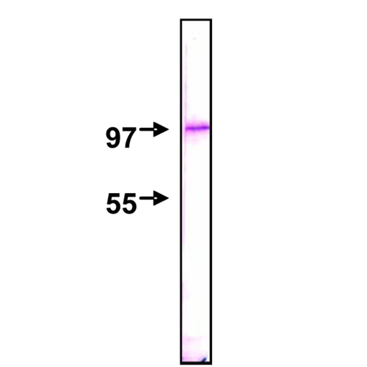
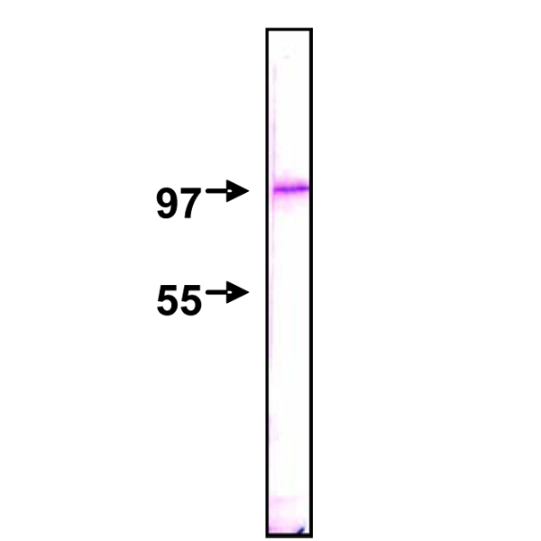
Fig.1 Detection of Nup98 protein by Western blotting using this antibody, 2H10. The sample is HeLa nuclear membrane fraction. The IgG was diluted 2,000 fold before use |
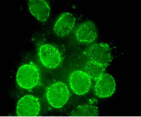
Fig. 2. Immunofluorescent staining of rat neuron with antibody 2H10. The dots are Nup98. |
|
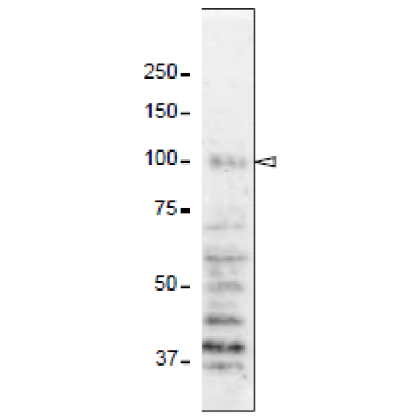 |
Fig. 3. Detection of Np98 protein in whole cell extract of HeLa cells with 2H10 antibody. First antibody was used at 0.4 ug/ml and as second antibody, HRP-conjugated ant-rat IgG was used at 0.4 ug/ml. The arrow head indicates the position of Nup98 protein. The numbers on the left shows the positions of size marker proteins in kDa
|
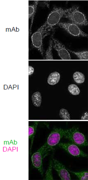 |
Fig.4 Immunofluorescene staining of Nup98 in HeLa cells with 2H10 antibodies. Black-and-white images were obtained with mAb and DAPI. Color images represent merged images of mAb (green) with DAPI (magenta). HeLa cells were cultured in a glass-bottom dish for 2 days before fixation. The cells were fixed with cold methanol (-30°C) for 30 min. After washing with PBS, all fixed samples were blocked with 1% BSA for 2 hr at room temperature. Anti-Nup98 rat mAb 2H10 was diluted to 0.5 µg/ml in PBS. Blocked samples were treated with these primary antibody solutions overnight at 4°C. The secondary antibodies were 4 μg/ml of Alexa488-labelled anti-rat IgG. DNA was stained with 4',6-diamidino-2-phenylindole (DAPI). Samples were washed three times with PBS between treatments.
|
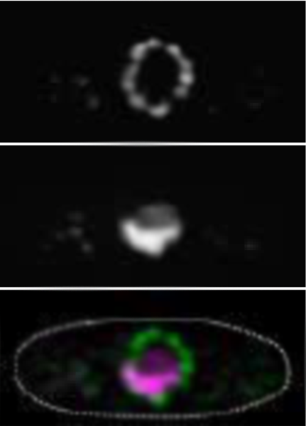 |
Fig. 5. Immunofluorescene staining of Nup98 in S. pombe cells with 2H10 antibody. Black-and-white images were obtained with mAb and DAPI. Color images represent merged images of mAb (green) with DAPI (magenta). Dotted lines represent the outlines of cells. S. pombe cells were fixed with 4% formaldehyde for 10 min, treated with 0.6 mg/ml Zymolyase 100T at 36°C for 70 min, and permeabilized with 1% Triton X-100 for 1 min. 10 µg/ml IgG solution of 2H10 were used. Antibody 2H10 stained the nuclear periphery of S. pombe in a punctate pattern.
|
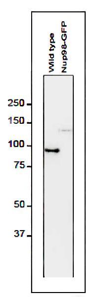 |
Fig. 6. Detection of Nup98 in S. pombe cell extracts by Western blotting with antibodies 2H10. 1 µg/ml IgG solution of 2H10 were used. Left and right lanes represent specimens from a wild type strain and an S. pombe strain in which Nup98 was chromosomally replaced with Nup98-GFP, respectively. S. pombe cells harvested were heated in distilled water at 95°C for 5 min, suspended in 100 mM phosphate buffer (pH 6.8), 4 M urea, 2.5% SDS, 0.05 mM EDTA, and disrupted by glass beads using a Multi Beads Shocker (Yasui Kikai, Osaka,). The sample was clarified by centrifugation, and the supernatant was used as the whole cell extract. First antibody was used at 0.4 ug/ml and as second antibody, HRP-conjugated ant-rat IgG was used at 0.4 ug/ml. Numbers on the left are positions of size marker Proteins in kDa.
|
|
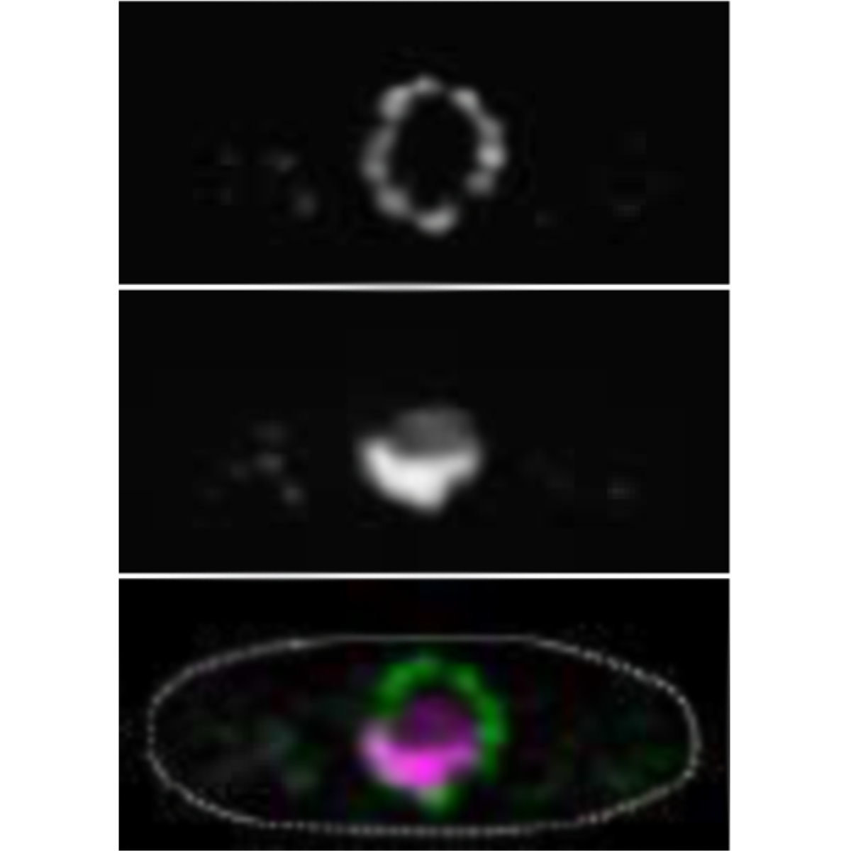
Fig. 7 Immunofluorescence staining of nucleoporin in S. cereviciae cell with 2H10 antibody. 10 µg/ml IgG solution of 2H10 were used. Black-and-white images represent fluorescence images obtained with mAb (top) and DAPI (middle). Color images represent merged images of mAb (green) with DAPI (magenta). Dotted lines represent the outline of cell. Methods were as described for S. pombe.
|
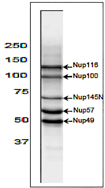 |
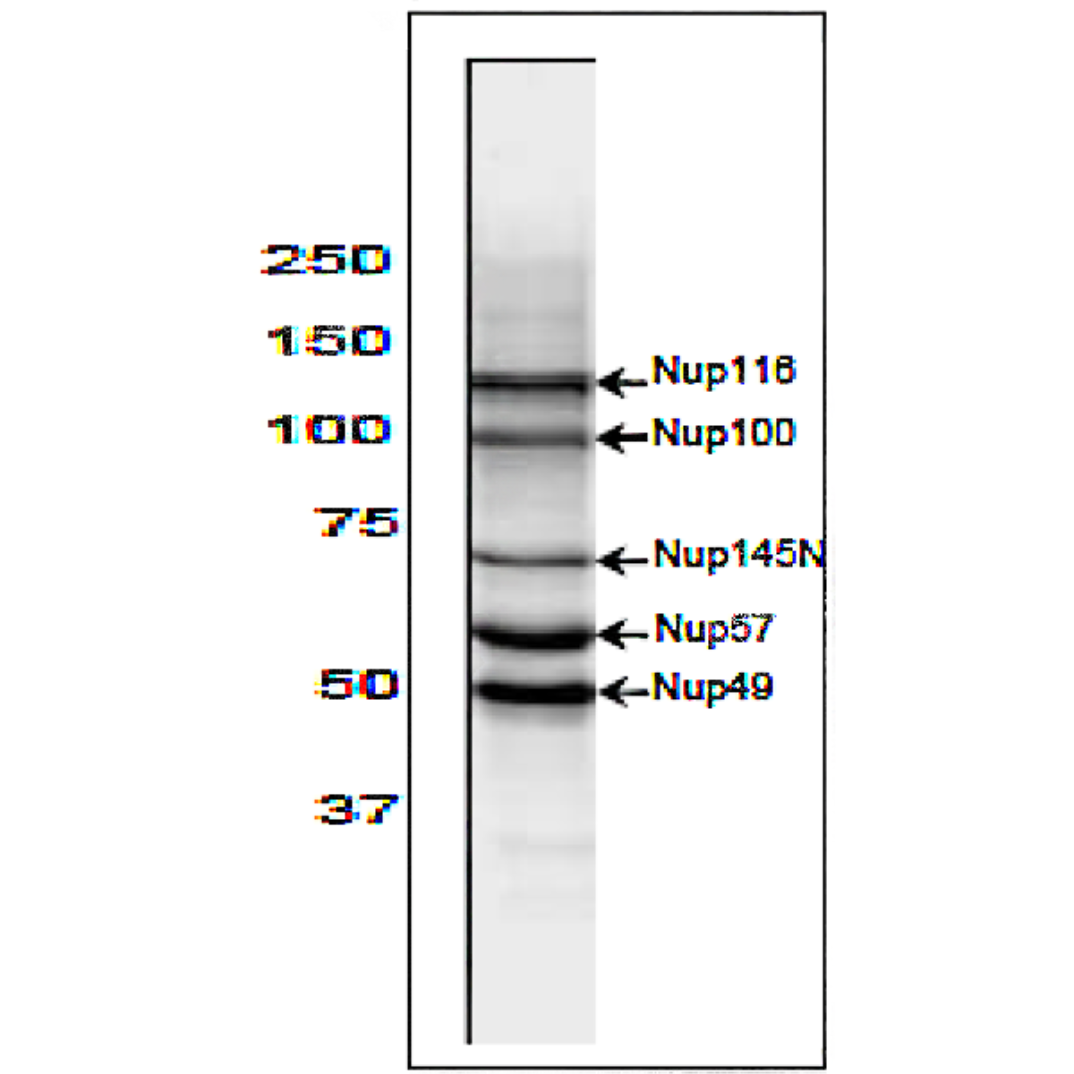
Fig. 8. Western blot analysis of S. cereviciae cell extract with 2H10 antibody. 2 µg/ml IgG solution of 2H10 were used. Arrows represent the positions of the indicated nucleoporins. Methods were as described for S. pombe. Five protein bands identified by this antibody correspond to the sizes of 5 kinds of nucleoporins with multiple GFLG motifs in S. pombe.
|
References
This product has been described and used in reference 1 and 2.
- Fukuhara T et al ”Specific monoclonal antibody against the nuclear pore complex protein, nup98.” Hybridoma 24: 244-247 (2005) PMID: 16225424 WB, IF
- Iwamoto M. et al. (2013) Monoclonal antibodies recognize gly-leu-phe-gly repeat of nucleoporin nup98 of tetrahymena, yeasts, and humans. Monoclon Antib Immunodiagn Immunother. 32: 81-90 PubMed ID: 23607342 WB, IF
Aliases for NUP98 Gene
- Nucleoporin 98 And 96 Precursor 2 3 5
- Nuclear Pore Complex Protein Nup98-Nup96 2 3 4
- Nucleoporin 98kDa 2 3
- Nucleoporin 96 2 3
- Nup98-Nup96 2 3
- Nup98-96 2 3
- NUP96 2 3
- Nuclear Pore Complex Protein Nup98 3
- GLFG-Repeat Containing Nucleoporin 3
- NUP98/PHF23 Fusion 2 Protein 3
- Nucleoporin 98kD 3
- Nucleoporin 98 2
- EC 3.4.21.- 4
- NUP196 3
- ADIR2 3
- NUP98 5
- ADAR2 4