ERseeing (Endoplasmic Reticulum Green)
ERseeing (Endoplasmic Reticulum Green)
Irreversible green-fluorescent ER stain for live-cell imaging (Ex 509 nm / Em 524 nm). Compared with glibenclamide-based ER dyes, ERseeing has a smaller pharmacological effect on cell function and stays in the ER after medium replacement.
For Research Use Only.
Filter compatibility: Commercial FITC filter sets are compatible.
Overview
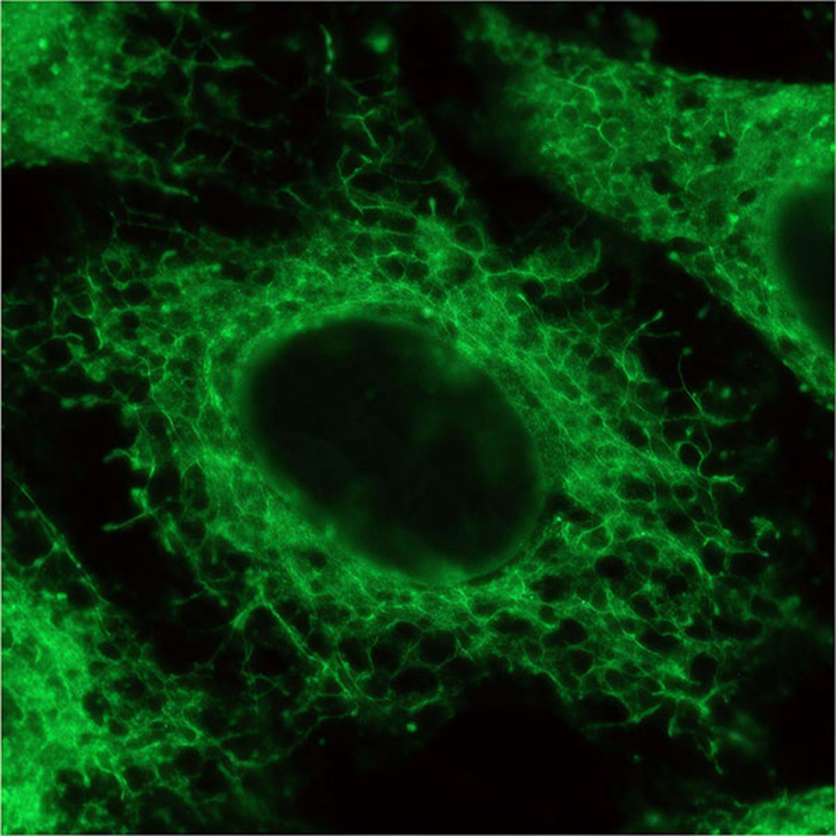
ERseeing is an irreversible endoplasmic reticulum (ER) staining reagent for live-cell imaging. Compared with conventional fluorescently labeled glibenclamide-based ER stains, it has a smaller pharmacological effect on cell function. Because the label is irreversible, the ER can be observed even after medium replacement.
Background: The Endoplasmic Reticulum
The ER is the largest organelle in the cell and has unique, dynamic tubular and sheet structures. It plays essential roles in protein biosynthesis, precise folding and quality control, and it is the origin of the secretory pathway that carries proteins to the Golgi apparatus, exocytosis, the plasma membrane and the extracellular space. Beyond protein synthesis, the ER functions in carbohydrate metabolism, calcium storage, lipid metabolism and lipid droplet synthesis. Visualizing ER structure in live cells is important for understanding ER function and the physiological significance of ER-resident proteins.
Limitations of Glibenclamide-Based ER Dyes
The most common ER-staining dyes are glibenclamide–fluorophore conjugates. Glibenclamide is a potent and specific inhibitor of the sulfonylurea receptors of ATP-sensitive K+ channels, which localize selectively to the ER, so glibenclamide-based dyes can visualize ER structures. These dyes have three drawbacks:
- Their pharmacological activity impairs K+ channel function in the ER.
- Glibenclamide is a reversible inhibitor and is washed out by wash steps and medium changes.
- As a result, they visualize only pharmacologically affected cells and are not suitable for long-term imaging experiments.
Labeling Mechanism
ERseeing has little pharmacological effect on ER function and visualizes the ER structure even after washout or medium change.
ERseeing has two units: a rhodol-type green fluorescent dye, and a thioester-type protein-labeling group carrying a rhodol derivative with high affinity for the ER membrane.


Key Features & Applications
Covalent labeling keeps the ER signal after washout and medium change, enabling long-term imaging.
Smaller effect on cell function than glibenclamide-based ER staining reagents.
Exmax/Emmax = 509/524 nm; commercial FITC filter sets are compatible.
Cells can be fixed with 4% PFA and methanol after staining live cells.
Application
Live-cell ER imaging of cultured cells.
NOTE: Cell fixation is compatible after staining live cells. However, this reagent does not stain the ER specifically in fixed cells, so the staining step must be performed on live cells.Components & Specifications
| Cat No. | FNK-FDV-0038 |
|---|---|
| Size | 10 nmol |
| Molecular formula | C37H28F2N2O4S |
| Molecular weight | 634.6 g/mol |
| Solubility | Soluble in DMSO |
| Ex / Em | 509 nm / 524 nm |
| Filter set | FITC filter sets compatible |
| Storage | -20°C |
Reconstitution & Storage
Protocol: General Procedure for ER Imaging

Application Data
Absorption and Fluorescence Spectra of ERseeing
Excitation (blue) and fluorescence (red) spectra. Exmax/Emmax = 509/524 nm. Commercial FITC filter sets are compatible.

ER Specificity
HeLa cells were stained with ERseeing (100 nM) and organelle markers: a glibenclamide-type ER stain, a lysosomal stain, a mitochondrial stain and a Golgi apparatus stain. ERseeing staining overlapped strongly with the conventional glibenclamide-type ER stain (Pearson’s correlation coefficient >0.9) and did not correlate with the lysosome or mitochondria markers. Only a small portion of ERseeing staining overlapped with the Golgi apparatus stain; this is attributed to vesicle transport of ERseeing or ERseeing-labeled proteins from the ER to the Golgi apparatus. An ER-to-Golgi trafficking inhibitor decreased the overlap between ERseeing staining and Golgi apparatus staining.
Detailed information is described in Ref. 1.
Comparison with a Glibenclamide-Based ER Dye
HeLa cells were treated with ERseeing or a glibenclamide-based dye for 15 min and observed without washout (left); both reagents stained the ER. The cells were then washed with PBS, given fresh medium containing 10% FBS and observed again. The glibenclamide-based dye showed a very weak signal, while ERseeing maintained a good ER signal. ERseeing is suitable for long-term imaging after medium changes.

Organelle Imaging Dyes for Living Cells


References
This product was commercialized and is sold by Funakoshi Co., Ltd. based on the research of Professor Isao Hamachi and Professor Tomonori Tamura, Graduate School of Engineering, Kyoto University. Research use only; not for diagnostic or therapeutic use.
- Fujisawa A., Tamura T., Yasueda Y., Kuwata K., & Hamachi I. (2018). Chemical profiling of the endoplasmic reticulum proteome using designer labeling reagents. J. Am. Chem. Soc., 140, 17060–17070. 10.1021/jacs.8b08606
- Miyamoto H. D., Ikeda M., Ide T., Tadokoro T., Furusawa S., Abe K., Ishimaru K., Enzan N., Sada M., Yamamoto T., Matsushima S., Koumura T., Yamada K. I., Imai H., & Tsutsui H. (2022). Iron overload via heme degradation in the endoplasmic reticulum triggers ferroptosis in myocardial ischemia-reperfusion injury. JACC: Basic to Translational Science, 7(8), 800–819. 10.1016/j.jacbts.2022.03.012
- Lin X. et al. (2026). Four subtypes of disease-causing missense mutations underlie pathogenic protein interactions in neurodegenerative VPS13A disease. J. Clin. Invest., 136(10), e200890. 10.1172/JCI200890
- Ishimaru K. et al. (2024). Deferasirox targeting ferroptosis synergistically ameliorates myocardial ischemia reperfusion injury in conjunction with cyclosporine A. J. Am. Heart Assoc., 13(1), e031219. 10.1161/JAHA.123.031219
- Shimasaki K. et al. (2024). A high-resolution phase-contrast microscopy system for label-free imaging in living cells. Cell Struct. Funct., 49(1), 21–29. 10.1247/csf.24018