CytoSeeing (Reversible Cytoplasm Blue)
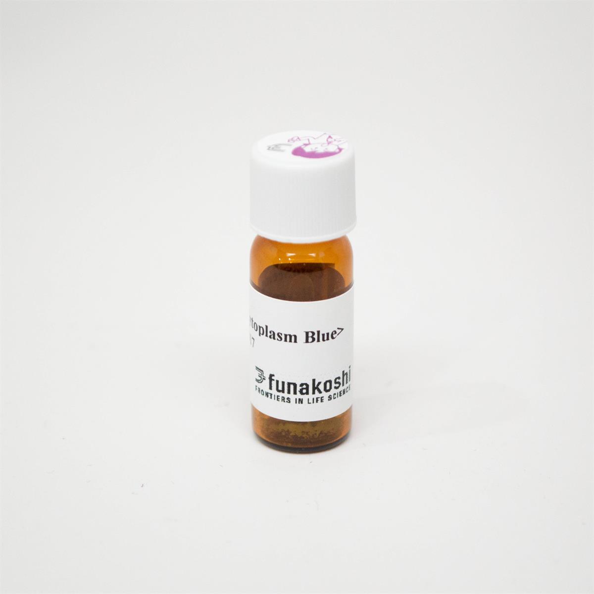
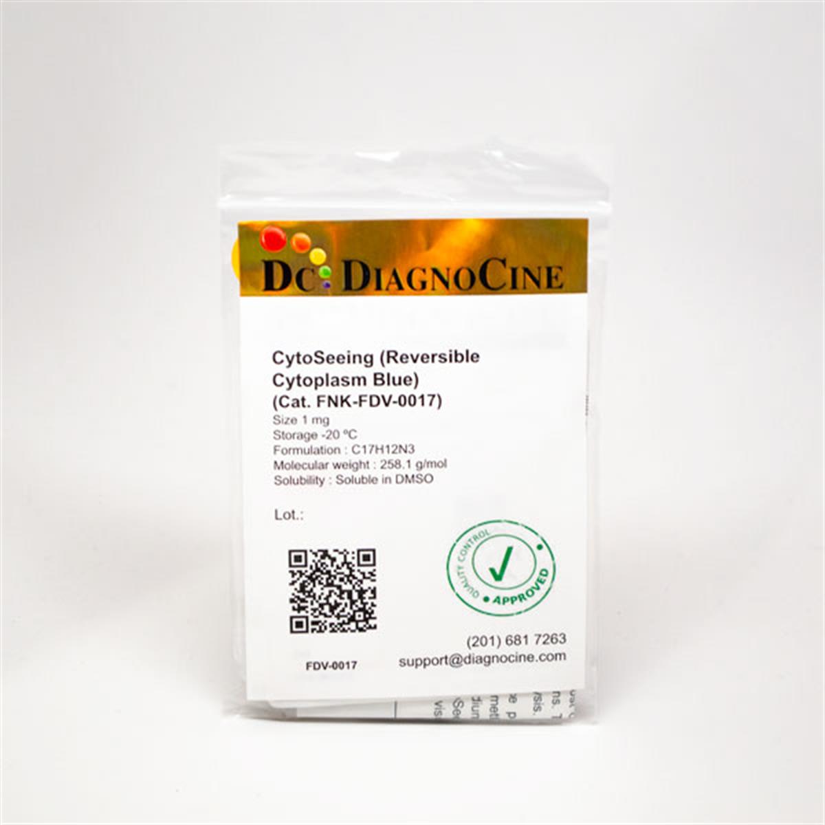
CytoSeeing (Reversible Cytoplasm Blue)
Cat. No. FNK-FDV-0017
Store at -20°C
Size 1mg
Description
Reversible cytoplasm blue fluorescent dye
CytoSeeing is a newly developed fluorescent dye, which can stain cytoplasm promptly by just adding to culture medium. After observation, fluorescent dye can be easily removed by replacing medium which does not contain CytoSeeing.
This product is commercialized based on the research result of Hokkaido University Faculty of Science.
Background
It is well known that morphology changes of nuclear and cytoplasm are related to differentiation, function and signal response of cells. Conventional cytoplasm staining dyes are used for cell tracking.
Therefore, once dyes are incorporated into cells, the dyes remain in cytoplasm even after medium change. The dyes are diluted in the course of cell division, and disappear after 3 to 6 population doubling. If dyes remain in the cells, it is difficult to observe the cell with other probes after live cell imaging.
CytoSeeing overcomes this conventional problem. CytoSeeing can be washed out by just replacing medium without containing CytoSeeing.
Features
- Easy protocol
- Just add CytoSeeing to the culture medium. - No staining on nucleus
- Compatible with green and red fluorescent dyes
- CytoSeeing can be detected by fluorescence filters for DAPI. - Little effect to cell function
- Removable
- Just replace it with fresh medium without containing CytoSeeing. Cells can be used for further assays. - Compatible with both adhesive and suspension cells.
|
Reagent |
Staining Cytoplasm |
Check nuclear morphology |
Time of introduction |
Wash-out |
|
CytoSeeing |
Yes |
Yes |
3 - 9 minutes |
Yes |
|
Company A |
Yes |
No |
15 - 60 minutes |
No |
|
Company B |
Yes |
No |
15 - 60 minutes |
No |
- Molecular Formula : C17H12N3
- Molecular Weight : 258.1
- Purity : >97%
- No effect to cell function
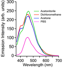
- Ex/Em (*1) :345 nm / 456 nm
- Solubility : DMSO (*2)
*1 : Compatible with DAPI filter.
*2 : 10 mM stock solution is recommended.
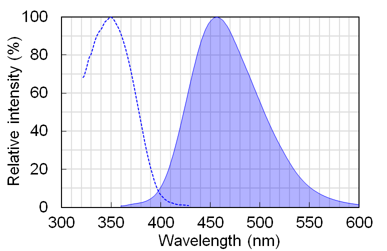
Protocol Overview
- Culture cells
- Add CytoSeeing solution to medium, with the final concentration from 10 μM to 50 μM.
- Incubate for a few minutes.
- Observe cells by fluorescent microscopy.
- If needed, remove CytoSeeing by replacing with fresh medium (without containing CytoSeeing).
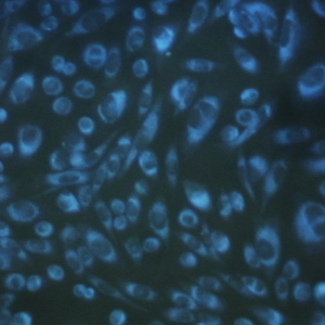
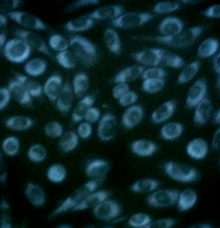
CHO cells were treated with 10μM CytoSeeing.
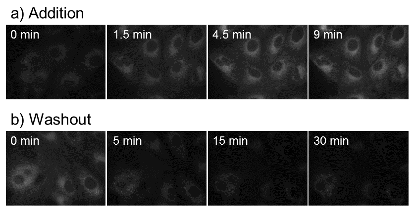
Fig. 2 Incorporation and distribution of CytoSeeing in A549 cells by time course
a) CytoSeeing (10 μM) was added and incubated. CytoSeeing was completely incorporated into cell by 9 minutes.
b) CytoSeeing incorporated cells were washed and incubated in fresh medium (without CytoSeeing).
Almost CytoSeeing was removed in 30 minutes.
Reference
- Kamada, et al., PLOS ONE, 11:e0160625(2016).

<more info.>
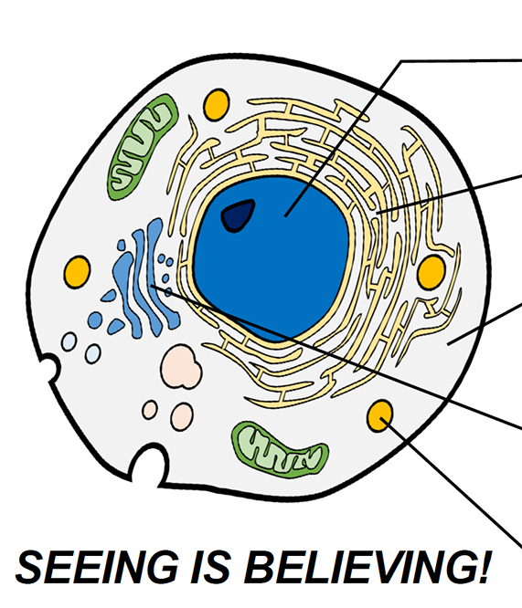
Organelle Imaging Dye in Living Cells
 |
 |
 |
|
 |
|
 |
|
 |
Full PDF Document Download, Click here "Organelle Imaging Dye in Living Cells"