


NucleoSeeing (Live Nucleus Green)
Product#: FNK-FDV-0029
$418.60
Availability:
Ships in 24 hours
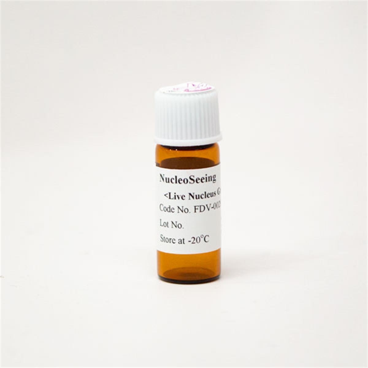
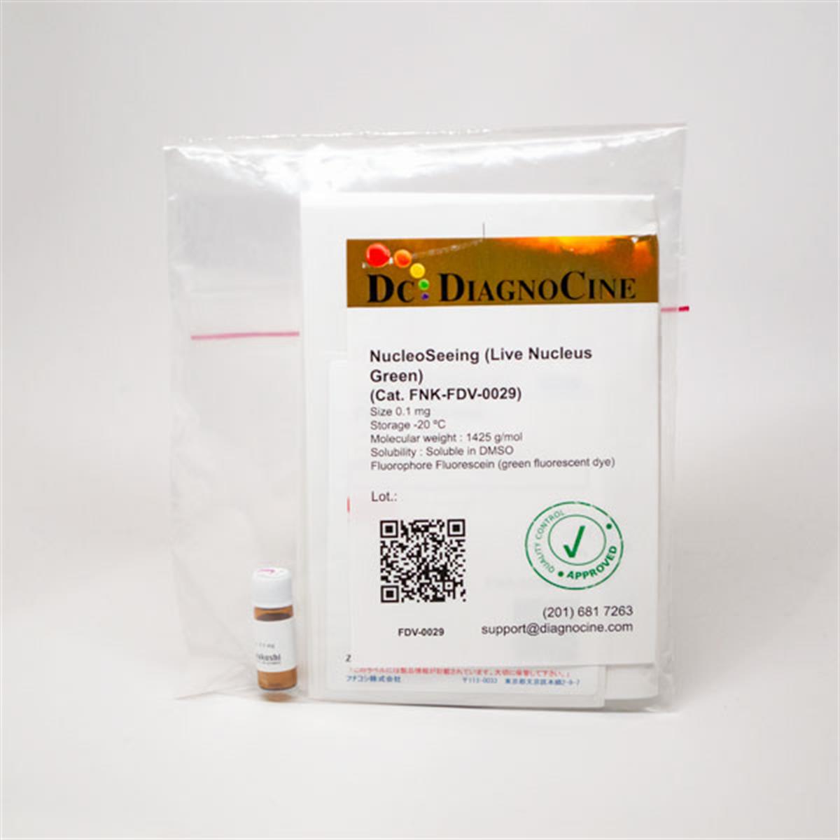
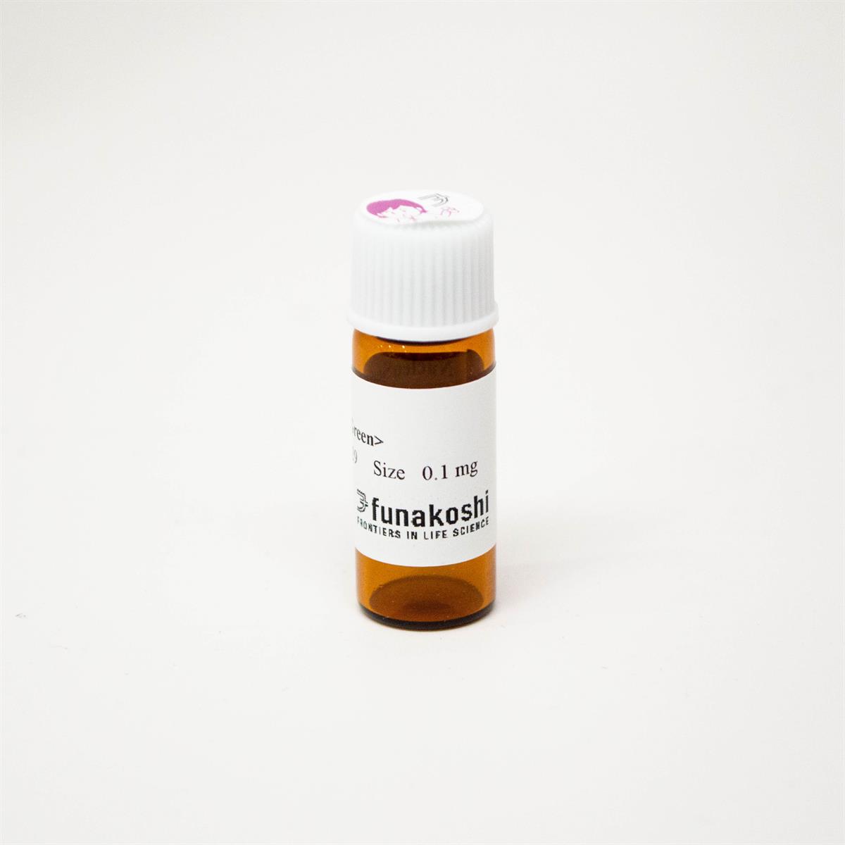
NucleoSeeing (Live Nucleus Green)
Cat. No. FNK-FDV-0029
Size 0.1 mg
Molecular weight 1425 g/mol as tri-TFA salt
Solubility Soluble in DMSO
Fluorophore Fluorescein (green fluorescent dye)
Ex/Em 488 nm/520 nm
Description
Cell nucleus is one of the most important organelle which stores DNA and chromatin complex in eukaryotic cells and plays important roles in various biological functions such as gene expression and epigenetic modulation. To monitor dynamics of cell nucleus in live cells, many cell nucleus-specific dyes have been developed. Hoechst33342 dye is one of the most famous dyes and is used for various applications because of its membrane permeable property.
However, Hoechst33342 requires UV excitation (~350 nm) and has blue fluorescent properties (~450 nm). As UV excitation shows strong photo-toxicity for cells, Hoechst33342 is not suitable for monitoring nucleus dynamics on live cells. Several visible-light excitable dyes for nucleus imaging on live cells had been commercially available, however, they have some disadvantages such as DNA/RNA specificity, or cell toxicity.
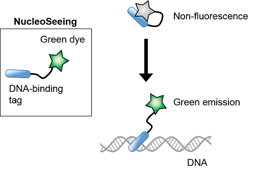
NucleoSeeing is a novel probe which was originally developed by Dr. Shinya Tsukiji and co-workers (Ref.1). This probe has a unique chemical structure containing diacetyl-fluorescein and Hoechst33342 as a DNA-binding tag linked by the optimal PEG linker. Although NucleoSeeing has two fluorophores, fluorescence of this probe is strongly quenched under no DNA conditions. Only in the presence of double stranded DNAs, it binds to DNA and emits fluorescence excited at 488 nm and detected at 520 nm (Figure 1).
Based on this photoproperty, NucleoSeeing allows to monitor cell nucleus with highly S/N value against cytosol or other organelles. Furthermore, this probe specifically interacts with DNA same as Hoechst dyes. Compared with Hoechst33342, NucleoSeeing has advantages which are not only lower photo-toxicity, but also lower compound’s cellular toxicity. Staining of various mammalian cultured cell lines and cultured mouse slice brain tissue were validated. Not only mammalian cells, but also some plant cells were tested, such as Arabidopsis thaliana Guard cells and epidermal cells (Ref.2). Although plant cells show autofluorescence derived from chloroplasts, NucleoSeeing could separate nucleus signal from autofluorescence signal from chloroplasts.
In addition to nucleus-specific imaging, NucleoSeeing can be applied for nucleus-specific pH sensing. A principle is based on pH-dependent fluorescent properties of fluorescein and Hoechst33342, components of NucleoSeeing. When this probe is excited at 405 nm, values of fluorescent ratio (FFluorescein/FHoechst) depend on pH during pH5.5-8.5. This product is the worldfirst commercially available nucleus-specific pH sensing reagent. Please find the “Appendix” for pH sensing.
However, Hoechst33342 requires UV excitation (~350 nm) and has blue fluorescent properties (~450 nm). As UV excitation shows strong photo-toxicity for cells, Hoechst33342 is not suitable for monitoring nucleus dynamics on live cells. Several visible-light excitable dyes for nucleus imaging on live cells had been commercially available, however, they have some disadvantages such as DNA/RNA specificity, or cell toxicity.
NucleoSeeing is a novel probe which was originally developed by Dr. Shinya Tsukiji and co-workers (Ref.1). This probe has a unique chemical structure containing diacetyl-fluorescein and Hoechst33342 as a DNA-binding tag linked by the optimal PEG linker. Although NucleoSeeing has two fluorophores, fluorescence of this probe is strongly quenched under no DNA conditions. Only in the presence of double stranded DNAs, it binds to DNA and emits fluorescence excited at 488 nm and detected at 520 nm (Figure 1).
Based on this photoproperty, NucleoSeeing allows to monitor cell nucleus with highly S/N value against cytosol or other organelles. Furthermore, this probe specifically interacts with DNA same as Hoechst dyes. Compared with Hoechst33342, NucleoSeeing has advantages which are not only lower photo-toxicity, but also lower compound’s cellular toxicity. Staining of various mammalian cultured cell lines and cultured mouse slice brain tissue were validated. Not only mammalian cells, but also some plant cells were tested, such as Arabidopsis thaliana Guard cells and epidermal cells (Ref.2). Although plant cells show autofluorescence derived from chloroplasts, NucleoSeeing could separate nucleus signal from autofluorescence signal from chloroplasts.
In addition to nucleus-specific imaging, NucleoSeeing can be applied for nucleus-specific pH sensing. A principle is based on pH-dependent fluorescent properties of fluorescein and Hoechst33342, components of NucleoSeeing. When this probe is excited at 405 nm, values of fluorescent ratio (FFluorescein/FHoechst) depend on pH during pH5.5-8.5. This product is the worldfirst commercially available nucleus-specific pH sensing reagent. Please find the “Appendix” for pH sensing.
- Live cell nucleus imaging of cultured cells, cultured tissues
- Live cell nucleus imaging of plant cells
- Cell nucleus staining of fixed cells
- Cell nucleus-specific pH sensing (See “Appendix”)
Reconstitution : stock solution in 100% DMSO.
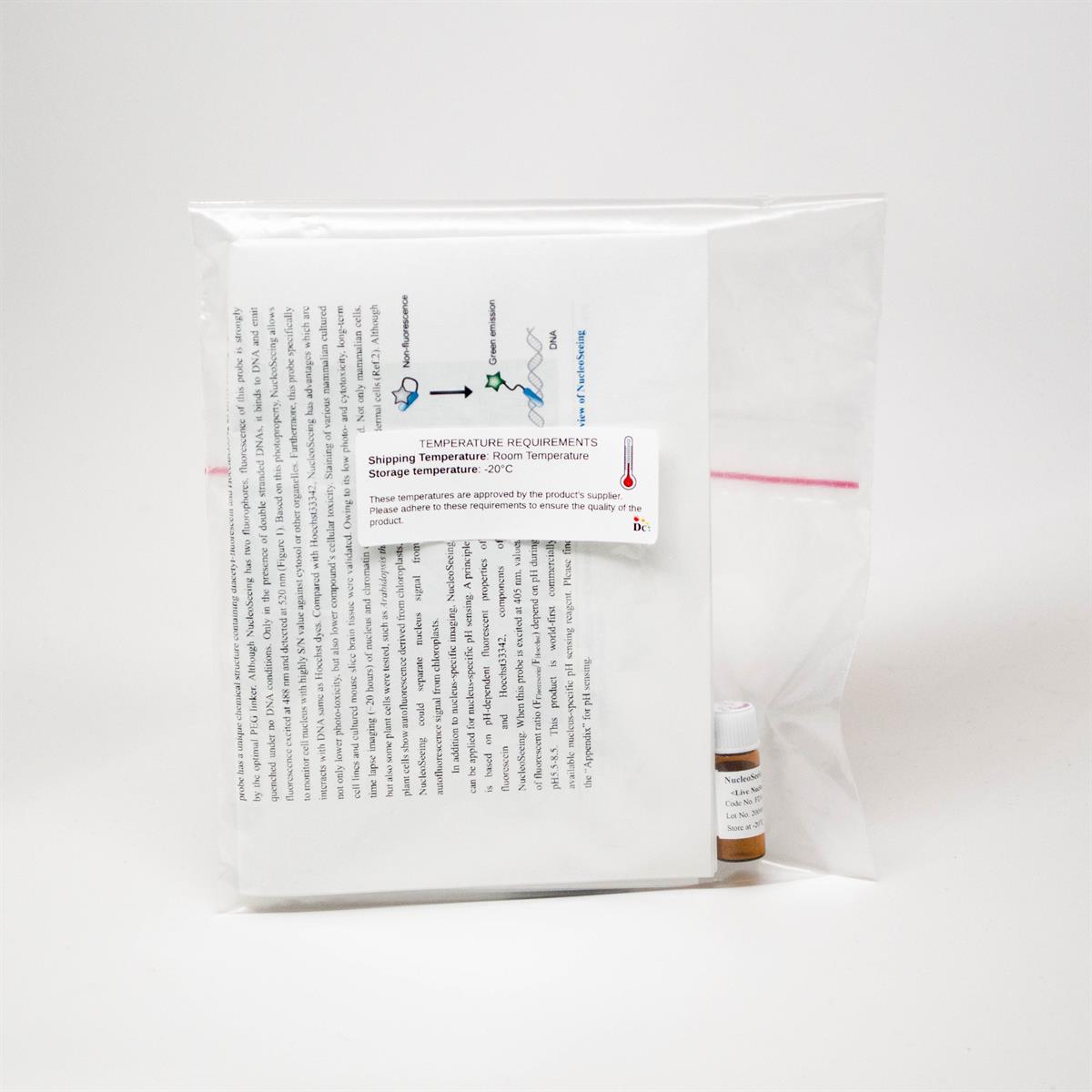
Storage (solution) : Store powder at -20oC. After reconstitution in DMSO, aliquot and store at -20 °C. Avoid repeated freeze-thaw cycles.
Protect from light.
Storage (solution) : Store powder at -20oC. After reconstitution in DMSO, aliquot and store at -20 °C. Avoid repeated freeze-thaw cycles.
Protect from light.
Features
- High S/N ratio
- Excitation / Emission : 488 nm / 520 nm
- Low cytotoxicity
- Can be used for both live and fixed cells
- Can be used for animal samples and plant cells.
- (validated in leaf and guard cell of A. thaliana)
- Reversible : Can be washed out by replacing the medium
- Can be used as nucleus pH sensor
General procedure of nucleus imaging
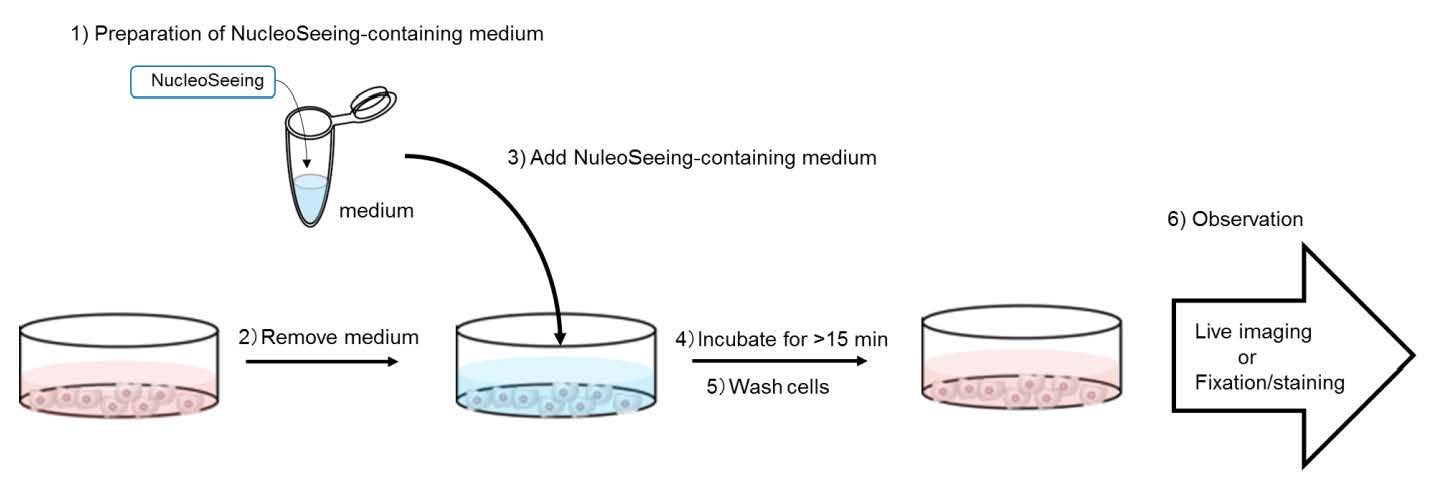
- Prepare 1-20 ?M NucleoSeeing in fresh medium
- Remove culture medium
- Add NucleoSeeing-containing medium to cells
- Incubate cells at 37oC for over 15 min
- Wash cells by PBS or medium **This step is recommended but not essential
- Observe cells under live condition or after fixation by 4% PFA and methanol
Example Data
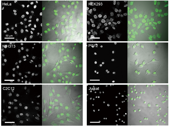
Fig.1 Staining of nucleus in various cultured cells
Six cell lines were treated with 1 μM of NucleoSeeing for 15 min and observed under live condition.
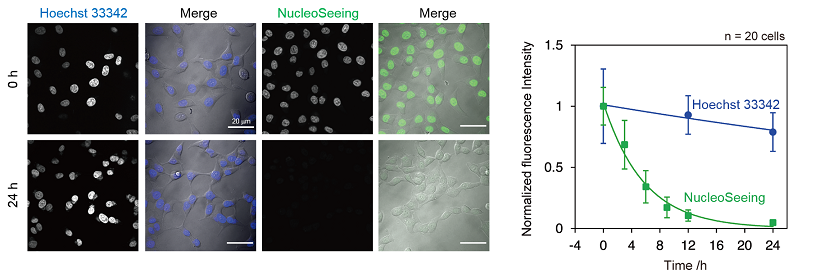
Fig.2 Reversible staining of NucleoSeeing
After HeLa cells were treated 1 μM Hoechst33342 and NucleoSeeing for 15 min, cells were washed by PBS and cultured for 24 hours.
While over 80% of blue signal of Hoechst33342 was still remained, green signals of NucleoSeeing were dramatically reduced within 12 hours.
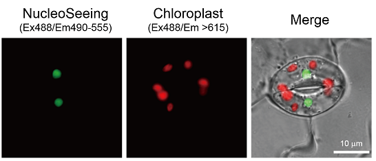
Fig.3 Staining of Arabidopsis thaliana Guard cells
Arabidopsis thaliana leaves were treated with 20 μM NucleoSeeing in 10 mM MES-KOH, 50 mM KCl (pH 6.2) buffer for 60 min and observed under living condition (Ex. 488 nm and Em. 490-555 nm for NucleoSeeing, Em. >615 nm for chloroplast as autofluorescence).
Green signals from NucleoSeeing were clearly separated from plant autofluorescence derived from chloroplast.
Comparison table of dyes for nucleus
|
|
Ex/Em |
Fluorescence |
Photo-toxicity |
Nucleus Specificity |
Cell Mem. Permeability |
Cyto-toxicity |
Live |
Fixed |
|
NucleoSeeing |
488/520 |
Green |
Low |
Yes |
Yes |
Low |
Yes |
Yes |
|
Hoechst |
350/461 |
Blue |
High |
Yes |
Yes |
High |
Yes |
Yes |
|
DAPI |
350/461 |
Blue |
High |
Yes |
No |
N/A |
No |
Yes |
|
Company X |
485/498 |
Green |
Low |
No |
Yes |
N/A |
Yes |
Yes |
|
Company Y |
646/680 |
Red |
Low |
Yes |
Yes |
High |
Yes |
Yes |
Reference
- Nakamura et al., Chem. Commun., 50, 6149-6152 (2014) Hoechst tagging: a modular strategy to design synthetic fluorescent probes for live-cell nucleus imaging.
- Ueda et al., ACS Cent. Sci., 3, 462-472 (2017) Noncanonical function of a small-molecular virulence factor coronatine against plant immunity: an in vivo raman imaging approach.
- Nakamura and Tsukiji, Bioorg. Med. Chem. Lett., 27, 3127-3130 (2017) Ratiometric fluorescence imaging of nuclear pH in living cells using Hoechst-tagged fluorescein.

<more info.>
Organelle Imaging Dye in Living Cells
 |
 |
 |
|
 |
|
 |
|
 |
Full PDF Document Download, Click here "Organelle Imaging Dye in Living Cells"