


Golgiseeing, Golgi Apparatus Green
Product#: FNK-FDV-0053
$538.20
Availability:
Ships in 24 hours
Golgiseeing, Golgi Apparatus Green
Cat. No.: FNK-FDV-0053
Store: -20℃
A green fluorescent dye that can stain the Golgi apparatus of living cells simply by adding it to the culture medium. Compared to conventional ceramide-based Golgi staining reagents, it does not require complicated staining procedures and also suppresses non-specific localization to the endoplasmic reticulum. Can be used under general FITC observation conditions.
*Research use only, not for human or animal therapeutic or diagnostic use.
*This product has been commercialized under a license from the Nagoya Institute of Technology
Product Background
The Golgi apparatus is an organelle that plays various roles in physiological functions and is known as a central component of the protein secretory pathway. The Golgi apparatus has unique multiple-layered cisternal membrane structures, with subdivided structures such as cis-Golgi, which is responsible for reciprocal vesicular transport with the endoplasmic reticulum (ER), and trans-Golgi, which is the starting point of the secretory pathway. Since dynamic morphological changes of the Golgi apparatus are essential for secretory function and dysfunction of the Golgi apparatus has been implicated in a number of diseases, it is expected that the Golgi apparatus will be observed by live cell imaging.
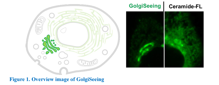
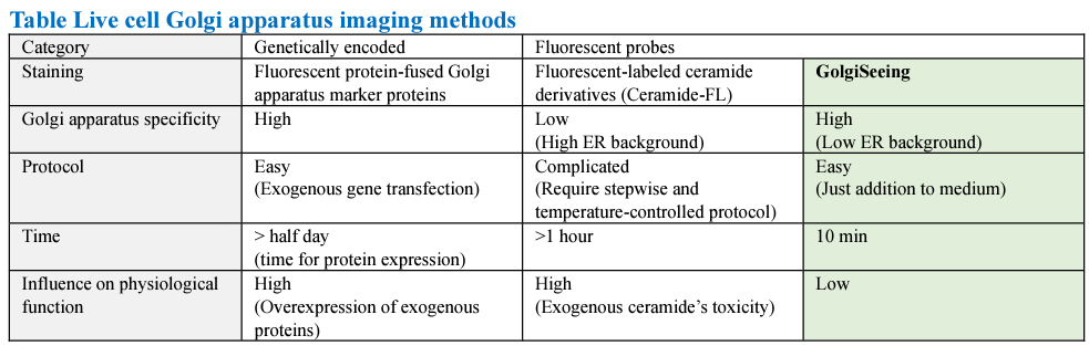
Two major methods have been used to stain the Golgi apparatus in living cells: The first is fluorescent staining using fluorescently labeled ceramide derivatives (hereafter ceramide-FL). Ceramide lipids accumulate in the Golgi apparatus during the metabolic pathway, and ceramide-FLs have been used to visualize the Golgi apparatus. However, 1) The Golgi selectivity of ceramide derivatives is low, 2) Ceramide derivatives also localize to the ER, etc., and 3) Ceramide derivatives have high cytotoxicity and are quickly metabolized intracellularly. The second staining method is to overexpress fluorescent proteins by fusing them to Golgi-specific expressed proteins (such as Giantin, Nacetylgalactosaminyltransferase, etc.). While this method allows visualization of the Golgi apparatus with a high degree of specificity, it requires prior plasmid transfection, making it impossible to immediately observe the Golgi apparatus when necessary. Furthermore, there are concerns overexpression of specific marker genes may affect physiological functions of the Golgi apparatus. GolgiSeeing is a novel small molecule fluorescent reagent that utilizes a Golgi apparatus selective localization motif discovered by Dr. Shinya Tsukiji's group at Nagoya Institute of Technology. Unlike conventional ceramide-FLs, it can stain the Golgi apparatus with a simple protocol that requires only 10 minutes of addition to the culture medium. It also shows higher Golgi specificity than ceramide-FLs, allowing Golgi apparatus-focused analysis (Figure 1). The GolgiSeeing can visualize the Golgi apparatus of target cells at any desired timing without genetic manipulation and without bias to physiological functions caused by overexpression, allowing the dynamic behavior of the Golgi apparatus to be observed under more physiological conditions. Under the non-wash protocol, GolgiSeeing visualizes not only the Golgi apparatus but also the plasma membrane. This property allows simultaneous imaging of the Golgi apparatus and cell morphology (judged by plasma membrane).
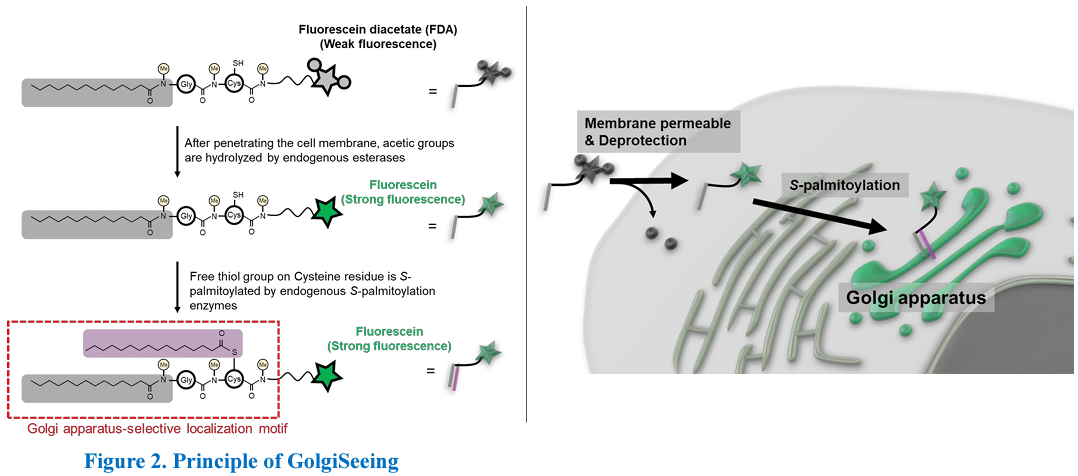
GolgiSeeing (original compound name; mgc3MeFDA in Ref.1) has a unique structure consisting of fluorescein diacetate (FDA) and N-myristoylated Gly-Cys dipeptide (mgc) linked by a linker, and three amide bonds in the mgc peptide chain are all methylated (Figure 2 left, upper). GolgiSeeing functions as a Golgi apparatus-selective fluorescent probe through several processes in the cell. FDA is a modified fluorescein quenched to improve cell membrane permeability, and GolgiSeeing emits slight fluorescence before use. After GolgiSeeing penetrates the cell membrane and enters the cell, acetyl groups are removed by endogenous esterases, and the green fluorescence of fluorescein is restored (Figure 2 left, middle). The deacetylated form of GolgiSeeing transiently localizes mainly to the ER and Golgi apparatus by the effect of N-myristoyl groups and is S-palmitoylation by endogenous Spalmitoylation enzymes. The Golgi apparatus selective localization motif is formed by adding palmitic acid to the Cys side chain by enzymatic action (Figure 2 left, lower). Although the intrinsic palmitoylation modification is known to localize to the Golgi apparatus and the plasma membrane, the effect of the three methyl groups biases the equilibrium toward the Golgi, which allows GolgiSeeing to selectively visualize the Golgi apparatus (Figure 2 right).
Description
Size: 0.1 mg
Molecular weight: 1258.5 g/mol
Solubility: Soluble in DMSO
Fluorescent characteristics:
Ex. 440-500 nm (maximum ~480 nm) / Em 500-560 nm (maximum ~520 nm)
Compatible with conventional FITC filter set
Compatible with conventional FITC filter set
*NOTE: GolgiSeeing has fluorescein diacetate (FDA), which is quenched by two acetates and emits a slight fluorescence. After hydrolysis of two acetic groups in cells by physiological esterases, fluorescein is exposed and restores strong green fluorescence.
Reconstitution and Storage
Reconstitution: Stock solution recommended concentration 1 mM in 100% DMSO. Add 79 μL of DMSO/vial to prepare 1 mM stock solution.
Storage (powder): Store powder at less than -20oC
Storage (solution): After reconstitution in DMSO, aliquot and store at less than -20°C. Avoid repeated freeze-thaw cycles and recommend single use of each aliquot.
Storage (powder): Store powder at less than -20oC
Storage (solution): After reconstitution in DMSO, aliquot and store at less than -20°C. Avoid repeated freeze-thaw cycles and recommend single use of each aliquot.
Important notice for stability
GolgiSeeing has a free-thiol group (See Figure 2 left), which is S-palmitoylated in cells and essential for Golgi apparatus localization. This thiol group is easily oxidized to form a homodimer in physiological pH buffers, and GolgiSeeing-dimer loses its Golgi apparatus localization function and may cause a background signal. GolgiSeeing working solution (such as GolgiSeeing-containing medium or buffers) should be prepared just before use. Importantly, GolgiSeeing may be gradually oxidized even in DMSO. After reconstitution of GolgiSeeing in DMSO, use within one month to maintain good staining results.
How to use
General procedure for Golgi apparatus-selective staining in live cells
*This procedure is an example of Golgi apparatus-selective staining
1. Prepare 10 μM GolgiSeeing in serum-free medium such as DMEM just before use NOTE: Empirically optimize and determine the concentration of GolgiSeeing for your experiments.
2. Remove the culture medium and wash cells with medium several times
3. Add GolgiSeeing-containing medium to cells
NOTE: Working solution of GolgiSeeing prepared in step-1 should be quickly used, as GolgiSeeing may be oxidazed to form inactive dimer in medium.
4. Incubate cells at RT for 10 min NOTE: Empirically optimize incubation time and temperature for your experiments. 5. Wash cells with 3 mg/ml BSA-containing medium over two times and add fresh serum-free and phenol red free medium
NOTE: GolgiSeeing is a highly hydrophobic compound and non-specifically absorbed to plastic or glass surface of imaging chambers. This non-specific absorption may cause the background signal of fluorescent images. To reduce non-specific signals, we highly recommend using a wash buffer containing BSA because BSA efficiently binds to excess GolgiSeeing and removes it from the surface of imaging chambers. 3 mg/ml BSA-containing basal medium is the preferred choice of GolgiSeeingwash buffer.
6. Observe cells under live cell condition
General procedure for Golgi apparatus and plasma membrane staining in live cells
*This procedure is an example of Golgi apparatus and plasma membrane staining
1. Prepare 2 μM GolgiSeeing in serum-free and phenol red-free medium such as DMEM just before use
NOTE: Empirically optimize and determine the concentration of GolgiSeeing for your experiments.
2. Remove the culture medium and wash cells with medium several times 3. Add GolgiSeeing-containing medium to cells
NOTE: Working solution of GolgiSeeing prepared in step-1 should be quickly used, as GolgiSeeing may be oxidazed to form inactive dimer in medium.
4. Incubate cells at RT for 10 min
NOTE: Empirically optimize incubation time temperature for your experiments.
6. Observe cells without wash-out step under live cell condition Important Noticeof Use
1) GolgiSeeing will selectively localize to the Golgi apparatus through the S-palmitoylation on the free-thiol group by endogenous S-palmitoylation enzymes, as mentioned in Figure 2. Note that any drugs inhibiting Spalmitoylation may influence the Golgi apparatus-selective staining property of GolgiSeeing. Especially, alkylation reagents of the free thiol (such as maleimide, iodoacetate, etc.) will critically inhibit S-palmitoylation and are incompatible with GolgiSeeing.
2) GolgiSeeing has no fixable functional groups in the molecule and is not fixed by either paraformaldehyde (PFA) or methanol. GolgiSeeing is a specialized reagent for live cell imaging applications, not compatible with fixed cell imaging and immunocytochemistry.
3) Long incubation time of GolgiSeeing may increase ER-derived fluorescent signal and reduce Golgi/ER ratio. Conduct empirical optimization ranges for your experiments' incubation time and observation time course.
Reference data
Comparison between GolgiSeeing and a ceramide-based reagent
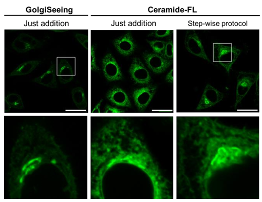
| HeLa cells were treated with 10 μM GolgiSeeing or 5 μM ceramide-FL (as BSA complex). In the case of GolgiSeeing, the protocol is a simple addition of GolgiSeeing into media final 10 μM and incubated for 10 min. After washing cells with 3 mg/ml BSA containing media, fluorescent images were captured by confocal laser microscopy (Ex 488 nm/Em 500-600 nm). On the other hand, ceramide-FL (BSA complex) -staining was performed by two protocols, simple addition or stepwise temperaturecontrolled protocol. The later protocol cells were incubated with ceramide-FL (BSA complex) for 30 min at 4oC in HBSS, washed with ice-cold HBSS, and incubated in a fresh culture medium for an additional 30 min at 37oC. Finally, cells were washed with a fresh medium again and observed by confocal laser microscopy. This stepwise protocol requires over 1 hour. The ceramide-FL probe stains not only the Golgi apparatus but also ER structure with high intensity nonspecifically. GolgiSeeing was able to visualize Golgi apparatus selectively and suppressed non-specific ER staining. |
 |
Organelle specificity
| HeLa cells were co-stained with GolgiSeeing and organelle markers. In the case of the Golgi apparatus marker, overexpression of mCherry-Giantin fusion protein was used. In the cases of ER, mitochondria, and lysosome, each organelle was stained by organelle-specific chemical probes. The fluorescent signal from GolgiSeeing is well corresponded with Golgi-marker and weakly overlaps with ER. However, the signals are not matched with mitochondria and lysosomes. | 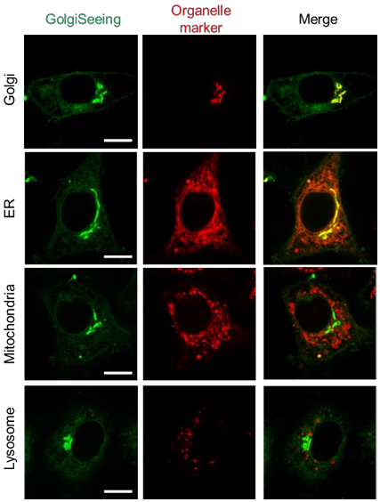 |
Effect of wash-out step on plasma membrane staining
| HeLa cells were treated with 10 μM GolgiSeeing for 10 min and observed by confocal laser microscopy (Ex 488 nm/Em 500-600 nm) with or without the wash step. For wash-out, 3 mg/ml BSA-containing medium was used as wash buffer. In the case of non-wash observation, the fluorescent signals of GolgiSeeing were observed in not only the Golgi apparatus but also the plasma membrane. As the Golgi apparatus is clearly distinguished from the plasma membrane, non-wash staining is useful for simultaneous observation of the Golgi apparatus and cell morphology (the shape of the plasma membrane). The wash-out step dramatically reduced the fluorescent signal from the plasma membrane. | 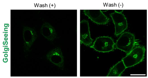 |
Various cell staining
| Four types of cultured cells (HeLa, COS-7, HEK293, and Jurkat) were treated with 10 μM GolgiSeeing for 10 min. After cell wash-out, cells were observed by confocal laser microscopy (Ex 488 nm/ Em 500-600 nm). For all cells tested here, GolgiSeeing highly selectively stained the Golgi apparatus. | 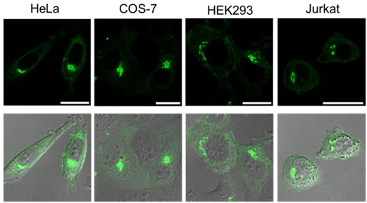 |
Cellular toxicity
| HeLa cells were seeded into a 12-well plate at 0.5 x 10⁵ cells/well and cultured for 48 hours with/without 10 μM GolgiSeeing. After 2, 24, and 48 hours, cell numbers were assessed by a cell counter. GolgiSeeing shows little effect on cell proliferation. | 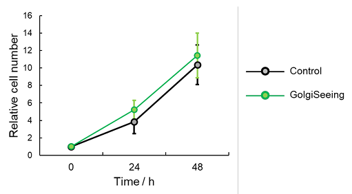 |
Application data
Live cell time-lapse imaging on Brefeldin A-induced collapse of Golgi apparatus
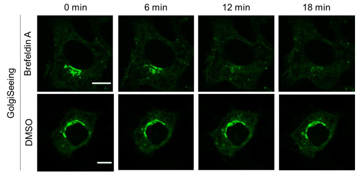
HeLa cells were seeded on glass bottom dishes and treated with 10 μM GolgiSeeing for 10 min. After washing cells with 3 mg/ml BSA-containing media, the cells were cultured in brefeldin A (final 1 μM in 0.1% DMSO)-containing medium or 0.1 % DMSO-containing medium as a negative control and observed under live cell condition by confocal microscopy (Ex 488 nm/Em 500-600 nm). In the brefeldin A-treated cells, the fluorescent signal from the Golgi apparatus gradually disappeared.
Live cell time-lapse imaging during cell division
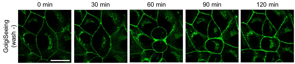
MDCK cells were seeded on a glass bottom dish, incubated with 2.5 μM GolgiSeeing, and observed for 2 hours by confocal laser microscopy (Ex 488 nm/ Em 500-600 nm) without washing step. As a non-wash procedure, the fluorescent signal of GolgiSeeing were observed not only in Golgi apparatus but also in plasma membrane. By imaging of plasma mebrane structures, cell morphology was easily observed. According to the progression of cell division, the Golgi apparatus disappeared (60 min) and was reconstituted in two daughter cells (90-120 min).
-----------------------------------------------------------------------------------------------------
Reference
1. Sawada et al.,ACS Chem. Biol., 18, 1047-1053 (2023) Palmitoylation-Dependent Small-Molecule Fluorescent Probes for Live-Cell Golgi Imaging.
------------------------------------------------------------------------------------------------------
Related products
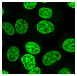
NucleoSeeing <Live Nucleus Green>
NucleoSeeing is DNA-responsive green dye for monitoring cell nucleus in live cells. As it shows low cytotoxicity and phototoxicity, it is very suitable for long-term live imaging of cell nucleus.
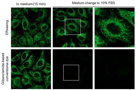
ERseeing <Endoplasmic reticulum Green>
| Catalog No. FNK-FDV-0029 Size 0.1 mg Features - Easy and quick procedure - Compatible with 10% FBS - Validated for both adherent cells and floating cells - Little influence on cellular functions - Ex/Em: 488 nm/520 nm (commercial FITC filters are available) Buy now =>  |
 |
| Catalog No. FNK-FDV-0038 Size 10 nmol Features - Recommended Ex/Em: 509 nm/524 nm - Lless pharmacological effect on ER proteins - Suitable for long-term live cell imaging Buy now => |
 |
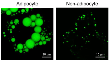
| Catalog No. FNK-FDV-0027 Size 0.1 mg Features - Recommended Ex/Em:400-500 nm / 490-550 nm - Enable to detect <1 μm lipid droplets - Suitable for long-term live cell imaging - Extremely photostable compared with conventional dyes - Compatible with both live and fixed cells Buy now => |
 |
<more info.>
Organelle Imaging Dye in Living Cells
 |
 |
 |
|
 |
|
 |
|
 |
Full PDF Document Download, Click here "Organelle Imaging Dye in Living Cells"
