Ponceau S staining solution

Product#: FNK-BCL-PSS-01
$63.59
Availability:
Ships in 1-2 Weeks

Pack size

500 mL
FNK-BCL-PSS-01
Staining Reagents

Ponceau-S Staining Solution

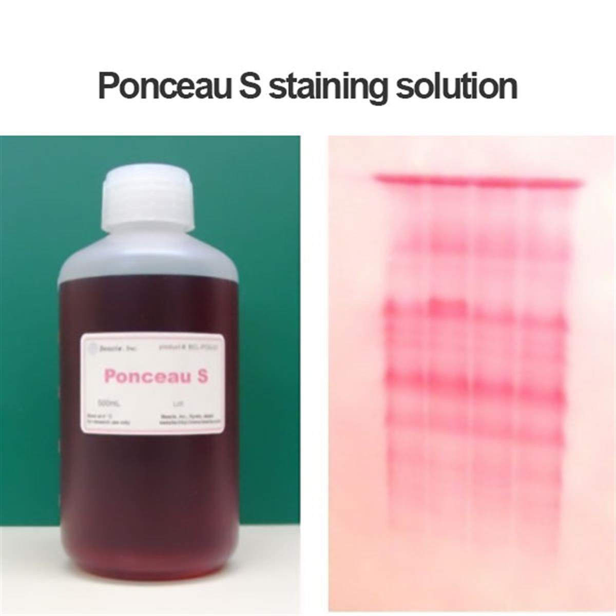
Ponceau-S Staining Solution (FNK-BCL-PSS-01) is a ready-to-use staining solution for proteins transferred to a membrane in Western blotting. Because the dye stains proteins quickly and is just as quickly removed, it lets you confirm transfer efficiency and the position of target proteins on the membrane — and because removal does not affect the antibody reaction, the check costs the blot nothing.

  • Ready-to-use formulation
  • Rapid staining — simply immerse the membrane for a few minutes
  • Reversible staining allows for immediate destaining
  • Safe to use, with no effect on subsequent antibody reactions after removal
  • Confirms transfer efficiency and the position of target proteins on the membrane
  • Store at room temperature
  • Supplied in 500 mL
SKU: FNK-BCL-PSS-01 UNSPSC: 12161500 Staining Solution
Ponceau-S Staining Solution — Ready-to-Use, 500 mL
  • Product numberBCL-PSS-01
  • FormatReady-to-use solution
  • Content500 mL
  • ApplicationProtein-transferred membrane staining, western blot
  • Staining modeReversible
  • Staining time2 to 5 min
  • Background decolorizationNormally 2 min (1% acetic acid)
  • Total decolorizationNormally 10 to 20 sec (0.1 M NaOH)
  • Washing2 min (DW)
  • StorageRoom temperature
RUO
Principle

Why the stain comes back off

Ponceau-S is a negatively charged chemical dye with a molecular weight of approximately 760.5. It stains proteins by interacting reversibly with the positively charged amino groups of the amino acids. That interaction is what makes the stain both fast to develop and fast to remove — and it is why the membrane can go straight on to blocking once the dye is washed out.

science

Reversible interaction

The dye binds the positively charged amino groups of amino acids rather than modifying the protein, so binding can be reversed.

bolt

Fast in both directions

Membranes stain by soaking for a few minutes, and normally 10 to 20 seconds in 0.1 M NaOH removes all staining.

verified

Antibody reaction unaffected

After decolorization the antibody reaction is not affected, so the membrane proceeds normally to blocking and detection.

visibility

Two things you learn

Staining reveals how efficiently protein transferred and where the target proteins sit on the membrane.

How to use

Staining and destaining

The full sequence, from blotting apparatus to the start of the antibody-reaction workflow.

  1. 1

    Removal of membrane

    Remove the protein-transferred membrane from the blotting apparatus.

  2. 2

    Staining — 2 to 5 min

    Put the membrane into a plastic box and pour in Ponceau-S staining solution so that the membrane is fully soaked. Let it stay for 2 to 5 min while shaking.

  3. 3

    Decolorization of background — normally 2 min

    Discard the staining solution and add 1% acetic acid solution. While gently shaking, remove excess staining. Normally 2 min is enough, but it is better to examine the state of staining by eye. To take pictures at this stage, move the membrane to wet filter paper — be careful not to dry the membrane.

  4. 4

    Total decolorization — normally 10 to 20 sec

    Discard the solution, then pour in 0.1 M NaOH solution (dissolve 4 g of NaOH in 1 L of DW) so that the membrane is fully soaked. While gently shaking, remove staining. Normally 10 to 20 sec is enough to remove all staining.

  5. 5

    Washing — 2 min

    Discard the 0.1 M NaOH solution and wash with DW by shaking for 2 min. The membrane is now ready to move to the antibody-reaction step, starting from membrane blocking.

Do not let the membrane dry when moving it to wet filter paper for photography.
Applications

Where it fits in the blot

Ponceau-S staining sits between transfer and blocking, as a check on the step that is otherwise invisible until detection.

Western Blot

Transfer Efficiency Check

Confirms how efficiently protein transferred from the gel to the membrane, before the blot is committed to antibody incubation.

Western blotTransfer check
Western Blot

Target Protein Positioning

Reveals where target proteins sit on the membrane, which helps with cutting, aligning, and interpreting the blot.

Band positionMembrane mapping
Documentation

Blot Photography

The stained membrane can be moved to wet filter paper and photographed before total decolorization.

Wet filter paperImaging
Workflow

Direct Hand-Off to Blocking

After the DW wash the membrane is ready for the antibody-reaction step, starting from membrane blocking.

BlockingImmunodetection
Product Information

Specifications

As documented in the manufacturer instruction sheet for BCL-PSS-01.

Product
Parameter Specification
Product name Ponceau-S staining solution
Product number BCL-PSS-01
Diagnocine SKU FNK-BCL-PSS-01
Content 500 mL
Format Ready-to-use solution
Dye & Mechanism
Parameter Specification
Dye Ponceau-S
Molecular weight Approximately 760.5
Dye charge Negatively charged
Binding Reversible interaction with positively charged amino groups of amino acids
Staining mode Reversible
Protocol Timings
Step Time
Staining 2 to 5 min
Decolorization of background (1% acetic acid) Normally 2 min
Total decolorization (0.1 M NaOH) Normally 10 to 20 sec
Washing (DW) 2 min
Storage & Regulatory
Parameter Specification
Storage Room temperature
Manufacturer Beacle, Inc., Kyoto, Japan
UNSPSC 12161500
Regulatory status Research use only; do not use for medical purpose
Documentation

Instructions and safety data

Safety Data Sheet

The SDS for this product can be downloaded from the manufacturer at beacle.com/download_en.

Instruction Sheet

Manufacturer instruction sheet Ver. 2.0E covers introduction, product information, storage, and the staining procedure.

Manufactured By

Beacle, Inc., 14-1 Yoshida Kawaracho, Sakyo-ku, Kyoto, 606-8305 Japan.

Technical Service

Ordering and product questions in the US: support@diagnocine.com or 551-245-1033.

FAQ

Frequently asked questions

Common questions about Ponceau-S Staining Solution BCL-PSS-01.

It stains proteins transferred to a membrane in Western blotting. Because the dye stains quickly and is easily and quickly removed, it is used to confirm the transfer efficiency and the position of target proteins on the membrane.
Ponceau-S is a negatively charged dye with a molecular weight of approximately 760.5. It stains proteins by interacting reversibly with the positively charged amino groups of the amino acids, which is why the staining can be removed again quickly.
No. After decolorization the antibody reaction is not affected. Following the DW wash, the membrane is ready to move to the antibody-reaction step starting from membrane blocking.
Staining takes 2 to 5 min while shaking. Background decolorization with 1% acetic acid normally takes 2 min, though it is better to examine the state of staining by eye. Total decolorization with 0.1 M NaOH normally takes 10 to 20 sec, followed by a 2 min DW wash.
No. This is a ready-to-use Ponceau-S solution. Pour it into the box so the membrane is fully soaked.
Yes. Taking pictures is recommended after background decolorization if needed. Move the membrane to wet filter paper to do so, and be careful not to dry the membrane.
Store at room temperature. The SDS for this product can be downloaded from the manufacturer at beacle.com/download_en.

Satisfaction
Quality Rating
Value Rating
Style Rating
X