Flamma® 675 Thiol
Size of product (mg)
-
1 mg

-
5 mg

-
25 mg

Flamma® 675 Thiol
Cat. No. List below
Description
Flamma® Fluors 675 Thiol is a sophisticated near-infrared (NIR) fluorescent dye designed for advanced bioimaging applications. This inactive form of the dye is derived from a benzindocyanine structure and offers exceptional stability in fluorescence signaling, making it an invaluable tool for researchers in various fields of life sciences.
The dye exhibits maximal excitation at 674 nm and emission at 691 nm, placing it in the same spectral range as other popular NIR dyes such as Alexa 680, Cy5.5, IRDye 680LT, and DyLight 680. This spectral similarity allows for seamless integration into existing imaging protocols and systems. Flamma 675 can be efficiently excited using a 633 nm laser line, with its emission occurring in the NIR region, which is particularly advantageous for biological tissue penetration.
The thiol group is attached to the Flamma 675 fluorophore through a spacer, providing a reactive site for further modifications. This thiol functionality allows the dye to form disulfide bonds with cysteine residues on biomolecules, offering a unique labeling strategy. Additionally, Flamma® Fluors 675 Thiol can serve as a reference standard for dye-conjugates in various experimental setups.
Flamma 675 Thiol excels in various biomedical research applications such as bioimaging (generates stable fluorescence signals for in vitro and in vivo imaging studies), protein labeling (can form disulfide bonds with cysteine residues in proteins), reference standard (useful as a control in experiments involving dye-conjugates), and nanoparticle functionalization (potential for attaching to gold nanoparticles or other thiol-reactive surfaces).
Flamma 675 Thiol offers comparable performance to other popular NIR dyes like Alexa 680, DyLight 680, Cy5.5, IRDye 680LT, and CF680, making it a suitable alternative or complement in multi-color imaging experiments.
While not explicitly stated in the provided specifications, it's worth noting that the thiol functionality of this dye opens up unique possibilities for bioconjugation. The ability to form disulfide bonds with cysteine residues allows for site-specific labeling of proteins, which can be particularly useful in studying protein structure and function.The NIR properties of Flamma 675 Thiol make it especially valuable for in vivo imaging applications. The biological tissue-permeable NIR region allows for deeper penetration and reduced scattering, enabling clearer imaging in complex biological systems such as small animal models. Researchers should be aware that working with thiol-containing compounds requires careful handling to prevent oxidation. It's advisable to use the dye in an oxygen-free environment when possible and to consider using reducing agents to maintain the thiol group's reactivity.
Flamma® Fluors 675 Thiol combines high sensitivity, stability, and versatility, making it an excellent choice for researchers in fields such as molecular biology, cell biology, and biomedical imaging. Its unique thiol reactivity and NIR properties enable advanced imaging studies, contributing to our understanding of complex biological processes at the molecular level.
- Fluorophore: Flamma® Fluors 675
- Functional group: Thiol
- Excitation/Emission Max.(nm): 674/691
- Spectrally similar dyes: Alexa680, DyLight680, Cy5.5, IRDye680LT, CF680
- Extinction coefficient: ≥ 200,000 cm-1M-1
- Appearance: Blue Solid
- Molecular Weight: 1004.24 g/mol
- Solubility: DMF, DMSO
- Storage conditions: -20 ℃, protect from light
| Quick link (Cat.#) | FSD series | EXmax (nm) | EMmax (nm) | Spectrally similar dyes |
| CWT1001 | Flamma® 496 Thiol | 494 | 520 | Alexa488, FITC, Cy2 |
| CWT1058 | Flamma® 552 Thiol | 550 | 564 | Alexa555, DyLight549, Cy3, ATTO550 |
| KWT1415 | Flamma® 581 Thiol | 578 | 593 | Alexa594, DyLight594 |
| KWT1042 | Flamma® 648 Thiol | 648 | 663 | Alexa647, DyLight650, Cy5 |
| PWT1415 | Flamma® 675 Thiol | 674 | 691 | Alexa680, DyLight680, Cy5.5, IRDye680LT, CF680 |
| PWT1215 | Flamma® 749 Thiol | 749 | 774 | Alexa750, DyLight755, Cy7.5, IRDye750 |
| PWT1515 | Flamma® 774 Thiol | 774 | 800 | Cy7.5, DyLight800, IRDye800 |
Background
Flamma® Fluors
BioActs offers a broad range of Flamma® Fluors dyes equipped with variety of reactive and functional groups, which can cover the full spectral range from UV to NIR with their excellent fluorescence performance. Characteristic features of these superior dyes are strong absorption, high fluorescence quantum yield and high photostability. Flamma® dyes maintain good fluorescence activity and stability after conjugation to biomolecules and allow the detection of low-abundance biological structures with great sensitivity. The dyes are compatible with optical conditions of most of fluorescent equipment and are ideal for any applications in biological studies.
- Covering the full spectral range from UV to NIR
- Equipped with a variety of reactive groups: NHS and Sulfo-NHS ester, Vinylsulfone, Maleimide, Click chemistry, isothiocyanate, hydrazide and hydrophobic substances.
- High quantum yields and photostability
- High purity and compatible with most of biomolecules
What are the advantages?
- Stability: Provides consistent and reliable fluorescence signals in bioimaging applications.
- Versatility: Compatible with thiol-specific conjugation methods and various biomolecules.
- NIR Emission: Allows for deep tissue penetration in biological samples, reducing background autofluorescence.
- Spectral Compatibility: Similar spectral properties to widely used NIR dyes, facilitating integration into existing protocols.
Flamma® Fluors Thiol
Flamma® Fluors thiol dyes have an attached primary thiol, which connected through a spacer. These thiol dyes can be labeled to biomolecules through disulfide bond formation with thiol of cysteine residue.

Figure 1. Absorption (upper) and emission (bottom) spectra overlap of Flamma® Fluors

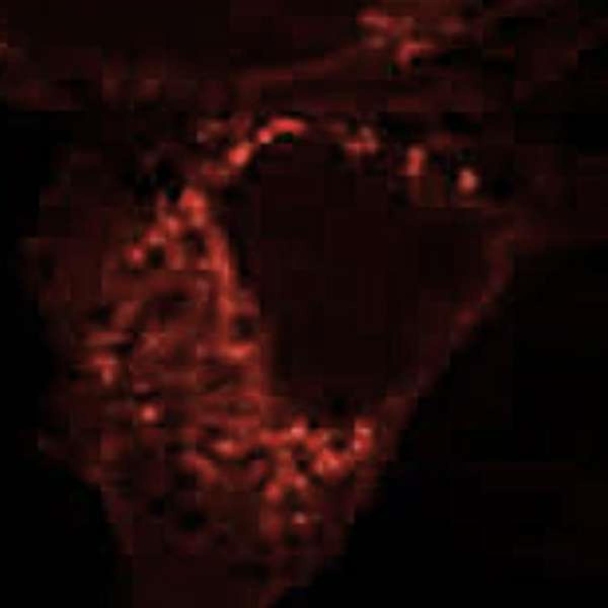
Figure 2. Immunofluorescence imaging and in situ hybridization imaging

Figure 3. Fluorescence images of Flamma® 749 (upper) and Flamma® 774 (bottom) carboxylic acid injected mouse model
Citation & Reference
1. Xu, Peisheng. Zwitterionic chitosan derivatives for pH-sensitive stealth coating. Biomacromolecules 11.9 (2010): 2352-2358.
2. Ibrahim, Basma M. A strategy to deliver genes to cystic fibrosis lungs: a battle with environment. Journal of controlled release 155.2 (2011): 289-295.
3. Oh, Keun Sang. Accurate sequential detection of primary tumor and metastatic lymphatics using a temperature-induced phase transition nanoparticulate system. International journal of nanomedicine 9 (2014): 2955.
4. Yhee, Ji Young. Tumor-targeting transferrin nanoparticles for systemic polymerized siRNA delivery in tumor-bearing mice. Bioconjugate chemistry 24.11 (2013): 1850-1860.
5. Yoon, Hong Yeol. Glycol chitosan nanoparticles as specialized cancer therapeutic vehicles: Sequential delivery of doxorubicin and Bcl-2 siRNA. Scientific reports 4 (2014).
6. Ryu, Ju Hee. Early diagnosis of arthritis in mice with collagen?induced arthritis, using a fluorogenic matrix metalloproteinase 3–specific polymeric probe. Arthritis & Rheumatism 63.12 (2011): 3824-3832.
7. Hollis, Christin P. In vivo investigation of hybrid paclitaxel nanocrystals with dual fluorescent probes for cancer theranostics. Pharmaceutical research 31.6 (2014): 1450-1459.
8. Koo, Heebeom. The movement of self-assembled amphiphilic polymeric nanoparticles in the vitreous and retina after intravitreal injection. Biomaterials 33.12 (2012): 3485-3493.
9. Zhu, Lei. Real-time monitoring of caspase cascade activation in living cells. Journal of controlled release 163.1 (2012): 55-62.
10. Yoon, Hong Yeol. Bioreducible hyaluronic acid conjugates as siRNA carrier for tumor targeting. Journal of Controlled Release 172.3 (2013): 653-661.
11. Yhee, Ji Young. Cancer-targeted MDR-1 siRNA delivery using self-cross-linked glycol chitosan nanoparticles to overcome drug resistance. Journal of Controlled Release 198 (2015): 1-9.
12. Park, Jin Woo. Wide-Ranged Fluorescent Molecular Weight Size Markers for Electrophoresis. Bulletin of the Korean Chemical Society 34.1 (2013): 29-30.
13. Huang, Xinglu. Multiplex Imaging of an Intracellular Proteolytic Cascade by using a Broad?Spectrum Nanoquencher. Angewandte Chemie International Edition 51.7 (2012): 1625-1630.