

ProteoCarry (Protein Transfection Reagent)
ProteoCarry (Protein Transfection Reagent)
Cat.No. FNK-FDV-0015
Size 1 Set
Store at -20°C
Description
ProteoCarry is a new type peptide base transfection reagent.
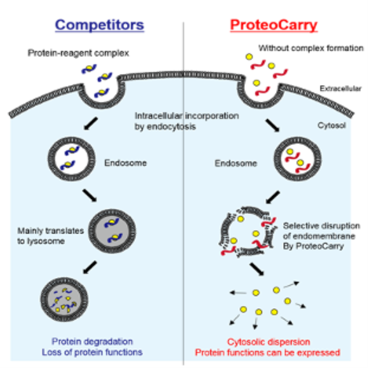
When using a conventional protein transfection reagent, transfected proteins are accumulated in endosome or lysosome, and most of such proteins are dissociated in lysosome proteolysis system. Contrary, ProteoCarry can deliver transfected proteins to cytoplasm effectively. Moreover, pre-incubation with proteins are NOT needed. Therefore effect to the protein functions can be minimized.
Product Background
ProteoCarry is a novel peptide-based transfection reagent for exogenous proteins such as functional proteins and antibodies with highly efficiency of cytosolic delivery. Protein transfection, intracellular delivery of proteins, is a powerful method to analyze cellular response to the protein of interest. While plasmid-based gene expression generally needs 12-24 hours to express proteins well, protein transfection reagents can immediately import functional proteins into living cells within several hours.
Although many protein transfection reagents including peptide-, cationic lipid- and polymer-based compounds have been developed to date, these reagents struggle to deliver proteins into “cytosol”. Commonly these reagents interact with
protein to form complexes and subsequently protein-reagent complexes are entered into cells via endocytosis pathway. But protein-reagent complexes are able to escape endosomes with little efficiency and consequently transported to lysosomes to be degraded. Highly effective endosomal escape of proteins is the most important subject of protein transfection reagents.
ProteoCarry can overcome this problem based on a novel and potent pHdependent endosomal membrane-lytic activity. Proteins and ProteoCarry can be delivered into endosomes by endocytosis pathway and also escape to cytosol by its highly membrane-lytic activity with low cytotoxicity.
Features
- Highly soluble in water
- High efficiency of delivering proteins to cytoplasm
- Fast - 1 hour incubation
- No pre-incubation with proteins needed
- Effect to the function of proteins cab be minimized
- Low cytotoxicity
- Polysaccharide can be also transfected.
Kit components
- ProteoCarry, 4mg in vial (vial A)
- FITC-dextran as positive control, 2 mg in vial (vial B)
Storage
- Shipping : Room temperature
- Store : -20℃
- HeLa, SW280, COS7, NIH3T3, HUVEC<>li/

Many reagents have been developed so far, such as peptide-base, cationic lipid-base, and polymer base. Proteins transfected by such reagents generally form complex with proteins and incorporated to cells by endocytosis. After that, the complex breaks endosome membrane and transported to cytoplasm.

Left: Problems of conventional competitors Right : Principle of ProteoCarry
However, current these reagents have weak activity for disrupting membranes.
Therefore, such arrested proteins cannot escape from endosome. Arrested proteins are considered to move to lysosome proteolysis system.
Therefore, transfection reagents should have strong activity for disrupting membranes so that transfected proteins could be functioned and transported in cytoplasm effectively.
ProteoCarry, a new peptide-base transfection reagent has a strong and selective membrane-disrupting activity. ProteoCarry does not require a forming of protein-reagent complex. Therefore, not only proteins, but also biopolymers can also be transfected to cells.
Preparation of 50x ProteoCarry stock solution
- Add 581 µl of sterile ultrapure water to vial A and mix gently well to be 50x concentrated solution.
- Stock solution should be stored at -20oC for up to 6 months in small aliquots to avoid repeated freeze and thaw cycles.
- Add 1 mL of sterile PBS to vial B and mix gently well to prepare 10x stock solution (2 mg/ml).
- Stock solution should be stored at -20oC.
- Prepare 10x concentrated protein solution with sterile PBS. 10x solution should be prepared at time of use.
- If it is challenging to prepare 10x concentrated solution, please optimize Table 2 below
|
|
Final conc. |
10x conc. |
|
Antibody |
50-250 µg/ml |
500-2500 µg/ml |
|
Proteins |
|
|
|
1-10 µg/ml |
10-100 µg/ml |
|
10-100 µg/ml |
100-1000 µg/ml |
|
Positive control |
|
|
|
200 µg/ml |
2 µg/ml |
fluorescent signal from endosome may be higher than the signal from cytosol.
Transfection protocol
Transfection efficiency is highly affected by protein’s profile such as molecular weight and net charge, celltype, and confluency of cell. To obtain best results, please optimize condition for your experiments.
number for each experiment.
|
Scale |
50x ProteoCarry(µL) |
10x Protein soln (µL) |
Base medium volume (µL) |
Total volume (µL) |
|
96 well |
2 |
10 |
88 |
100 |
|
24 well |
10 |
50 |
440 |
500 |
|
12 well |
20 |
100 |
880 |
1000 |
|
6 well |
40 |
200 |
1760 |
2000 |
transfection. Delivery efficiency through ProteoCarry is not affected by serum (<10% FBS). In some
cases, PBS give the best results. Please select a basal medium according to your cells of tested.
Memo
Mechanism of intracellular delivery of proteins by ProteoCarry is based on endocytosis and
macropinocytosis pathways. Please avoid to use inhibitors for endocytosis and/or macropinocytosis
pathway during transfection.
Reference
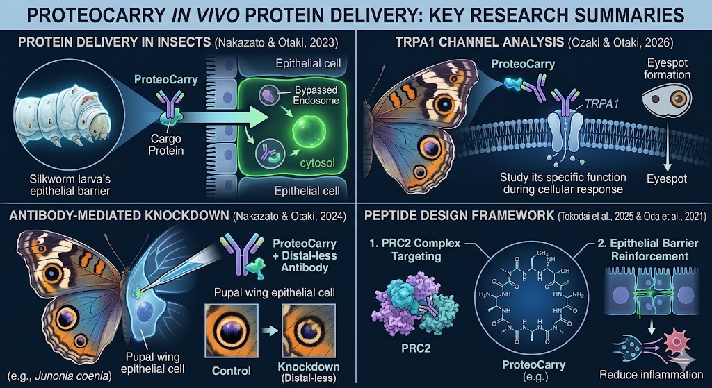
- Nakazato, Y., & Otaki, J. M. (2023). Protein delivery to insect epithelial cells in vivo. BioTech, 12(2), 28. https://doi.org/10.3390/biotech12020028
- Nakazato, Y., & Otaki, J. M. (2024). Antibody-mediated protein knockdown reveals Distal-less functions for eyespots and parafocal elements in butterfly wing color pattern development. Cells, 13(17), 1476. https://doi.org/10.3390/cells13171476
- Ozaki, M., & Otaki, J. M. (2026). TRPA1 for butterfly eyespot formation. International Journal of Molecular Sciences, 27(3), 1420. https://doi.org/10.3390/ijms27031420
- Tokodai, Yasuaki, et al. "Structure-Guided Design and Development of Cyclic Peptide Allosteric Activators of Polycomb Repressive Complex 2." bioRxiv, 2025. https://pmc.ncbi.nlm.nih.gov/articles/PMC12244496/.
- Oda, Yukako, et al. "Discovery of Anti-Inflammatory Physiological Peptides That Promote Tissue Repair by Reinforcing Epithelial Barrier Formation." Science Advances, vol. 7, no. 47, 2021, p. eabj6895. PMC8597994
Key Publication Summaries
-
Protein Delivery to Insect Epithelial Cells in Vivo (Nakazato & Otaki, 2023)
-
Summary: Validates the real-world stability and effectiveness of ProteoCarry by successfully delivering functional proteins directly into living insect epithelial cells in vivo, bypassing standard mammalian cell restrictions.
-
-
Antibody-Mediated Protein Knockdown in Butterfly Wings (Nakazato & Otaki, 2024)
-
Summary: Demonstrates ProteoCarry's high-efficiency cytosolic transfection of active antibodies into butterfly cells. This allowed researchers to successfully achieve targeted protein knockdown to study specific wing color pattern development (Distal-less genes).
-
-
TRPA1 for Butterfly Eyespot Formation (Ozaki & Otaki, 2026)
-
Summary: Extends the in vivo application of ProteoCarry to investigate cellular responses and specific channel functions (TRPA1) in living butterfly wing tissue models.
-
-
Structure-Guided Design of Cyclic Peptide Activators (Tokodai et al., 2025) * Summary: Focuses on the structural design and optimization of advanced cyclic peptides, establishing the broader scientific framework for targeting complex systems like the Polycomb Repressive Complex 2 (PRC2).
-
Discovery of Anti-Inflammatory Physiological Peptides (Oda et al., 2021)
-
Summary: Explores the biological foundations of cell-penetrating and therapeutic peptides that reinforce epithelial barriers to prevent inflammatory degradation and accelerate tissue repair.
-
Application data
|
1. FITC-dextran (positive control) Cell: HeLa (80% confluency) Cargo : FITC-dextran (200 mg/ml) ProteoCarry : 1x concentration Transfection time : 1 hour |
2. Fluorofore-labeled IgG Cell: HeLa (80% confluency) Cargo : Fluorofore-labeled IgG (250 ?g/ml) ProteoCarry : 1x concentration Transfection time : 1 hour |
|
|
|


