

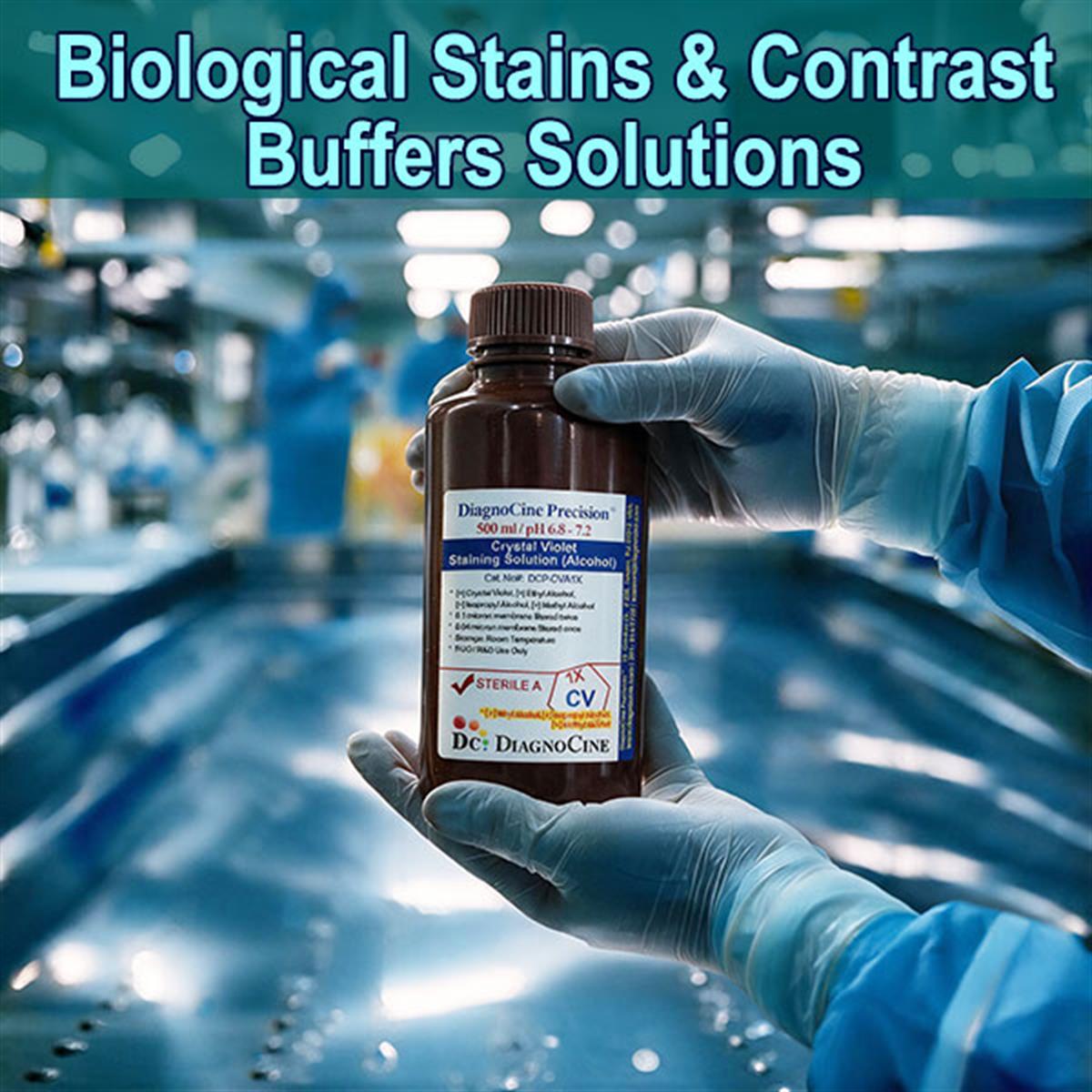
Biological Stains & Contrast Solutions - Category Selection Guide
Seven stains, one page. Pick the solution your protocol calls for and go straight to its product page — the staining chemistry, the history, and the application guidance follow below.
| Stain / Solution | Cat. No. | Primary Applications | Product Page |
|---|---|---|---|
| Histopathological Tissue Staining | HistopathologicalTissueStaining | Optimizes nucleus-vs-cytoplasm contrast in biopsies and FFPE sections | Viewarrow_forward |
| Crystal Violet Staining Solution (Water-Based) | DCP-CVW1X | Cell viability & biofilms; monolayers, biofilm visualization, colony / focus quantification | Viewarrow_forward |
| Crystal Violet Staining Solution (Alcohol-Based) | DCP-CVA1X | Classic Gram-stain primary stain; bacteriology | Viewarrow_forward |
| Acridine Orange Solution (10 mg/mL) | DCP-AO10X | Differential DNA / RNA fluorescence; cell-cycle, viability, and apoptosis studies | Viewarrow_forward |
| Protein Detection Stains Variations | ProteinDetectionStains | Electrophoresis band visualization in polyacrylamide gels | Viewarrow_forward |
| Methylene Blue Staining Solution | DCP-MB1X | Versatile counterstain; tissue morphology, blood smears, yeast viability | Viewarrow_forward |
| Ponceau S staining solution | FNK-BCL-PSS-01 | Not described in the category source text — see the product page for its specification. | Viewarrow_forward |
Not sure which stain? See what each stain is asked to do · See applications by workflow · Read the FAQ
Why a transparent cell needs a dye
Even perfectly fixed and sectioned tissue is nearly invisible under a standard light microscope. Cells are mostly water, so their internal components share almost identical refractive indices and produce little optical contrast. Staining solutions and buffers bind selectively to specific structures — DNA, proteins, lipid membranes — imparting color or fluorescence that lets researchers map cellular architecture and diagnose disease.
The chemistry that decides which structure a dye lands on — and the numbers a protocol actually turns on.
- Seven solutions stocked: Histopathological Tissue Staining, Crystal Violet Staining Solution (Water-Based), Crystal Violet Staining Solution (Alcohol-Based), Acridine Orange Solution (10 mg/mL), Protein Detection Stains Variations, Methylene Blue Staining Solution, and Ponceau S staining solution.
- Visual contrast comes two ways: dyes absorb specific wavelengths (colorimetric) or emit light on excitation (fluorometric), sharply defining otherwise invisible boundaries.
- Chemical selectivity is charge engineering: basic (cationic) dyes bind acidic components such as DNA and RNA; acidic (anionic) dyes bind basic cytoplasmic proteins. That affinity is what makes staining structure-specific.
- Pathological identification: some stains react selectively with abnormal structures — bacterial walls, amyloid, atypical nuclei — making them essential for diagnosis.
- The carrier is part of the method: the same cationic dye behaves differently in water and in solvent, which is why crystal violet is stocked as both a water-based and an alcohol-based solution.
- Metachromasia gives one dye two readouts: acridine orange fluoresces green on double-stranded DNA and shifts to orange-red when stacked on single-stranded nucleic acid.
- Gel-compatible chemistry is its own class: protein detection stains bind and reveal protein bands directly in polyacrylamide gels after a run.
- Products in this category7
- Acridine Orange solution strength10 mg/mL
- Acridine orange excitation (double-stranded)~500 nm
- Acridine orange emission on dsDNA~525 nm (green)
- Acridine orange emission on ssRNA~650 nm (orange-red)
- Gram-stain sequencecrystal violet → iodine → alcohol → counterstain
- Gram stain introduced1884
- First differential nuclear staining shown1858 (carmine)
- Basic (cationic) dyes bindacidic components — DNA / RNA
- Acidic (anionic) dyes bindbasic cytoplasmic proteins
Microscopic structures are too small and transparent to generate contrast on their own
Each of the mechanisms below solves a different half of that problem — one makes a structure visible, the next decides which structure becomes visible.
The refractive-index problem
Cells are mostly water, so their internal components share almost identical refractive indices and produce little optical contrast. Even perfectly fixed and sectioned tissue is nearly invisible under a standard light microscope.
Visual contrast
Dyes absorb specific wavelengths (colorimetric) or emit light on excitation (fluorometric), sharply defining otherwise invisible boundaries.
Chemical selectivity
Basic (cationic) dyes bind acidic components like DNA and RNA; acidic (anionic) dyes bind basic cytoplasmic proteins. This charge / affinity engineering is what makes staining structure-specific.
Pathological identification
Some stains react selectively with abnormal structures — bacterial walls, amyloid, atypical nuclei — making them essential for diagnosis.
The carrier changes the job
Crystal violet is a cationic (basic) dye in both of its stocked forms. The aqueous solution gently stains monolayers without stripping fragile structures; in the alcohol-based solution the carrier aids penetration and fixation in bacteriology.
Metachromasia
Acridine orange is metachromatic: bound to double-stranded nucleic acid it excites around 500 nm and emits green (~525 nm); stacked on single-stranded nucleic acid the emission shifts toward red (~650 nm). One dye, two readouts.
Read the mechanism, not the color
Two solutions of the same dye are not interchangeable, and one dye can report two different things. Crystal violet in water and crystal violet in alcohol are both cationic, but the first is the biofilm and monolayer reagent and the second is the classic Gram-stain primary stain. Acridine orange changes color with the strandedness of what it binds, so its published peak positions vary with dye:base ratio, pH, and ionic strength and should be treated as nominal.
How staining became a reproducible science
Three developments turned dye chemistry from an accident of the anatomy bench into the classification tool still used at the microscope today.
-
1
Mid-19th century Natural dyes and the first selective stain
Anatomists experimented with natural dyes like carmine and hematoxylin. In 1858, Joseph von Gerlach showed that carmine selectively colored cell nuclei — one of the first demonstrations of differential biological staining, with dye binding structure-selectively rather than uniformly.
-
2
1884 The Gram stain
Hans Christian Gram, working in Carl Friedländer’s Berlin lab on lung tissue from pneumonia patients, developed the crystal-violet / iodine / alcohol / counterstain procedure that bears his name. It created the Gram stain — still the foundational method for classifying bacteria as Gram-positive or Gram-negative and guiding antibiotic choice.
-
3
Late 19th century Synthetic dye chemistry
The synthetic coal-tar dye industry gave microbiologists — Paul Ehrlich, Robert Koch, and others — new tools like methylene blue and crystal violet to classify bacteria and visualize pathology. This marked histology’s transformation from a craft using natural pigments into a rigorous, reproducible science built on synthetic dye chemistry.
What a staining solution is actually asked to do
Three requirements, three different pieces of dye chemistry. Use this table to translate a protocol requirement into the class of stain that satisfies it.
| Requirement | How the dye delivers it | Result on the slide |
|---|---|---|
| Visual contrast | Dyes absorb specific wavelengths (colorimetric) or emit light on excitation (fluorometric) | Sharply defines otherwise invisible boundaries |
| Chemical selectivity | Basic (cationic) dyes bind acidic components like DNA / RNA; acidic (anionic) dyes bind basic cytoplasmic proteins | This charge / affinity engineering is what makes staining structure-specific |
| Pathological identification | Some stains react selectively with abnormal structures (bacterial walls, amyloid, atypical nuclei) | Makes them essential for diagnosis |
Which stain for which bench workflow
Select a workflow to see the applications the source assigns to it, and the stocked solutions that serve them.
- Differentiating Gram-positive vs. Gram-negative bacteria to guide treatment
- Crystal Violet (Alcohol-Based) — classic Gram-stain primary stain; the alcohol carrier aids penetration and fixation in bacteriology
- Grading cancer cells and identifying tissue damage in biopsy sections
- Histopathology Tissue Stain — clinical evaluation; optimizes nucleus-vs-cytoplasm contrast in biopsies and FFPE sections
- Methylene Blue — versatile counterstain for tissue morphology and blood smears
- Tracking viability, membrane integrity, and nucleic-acid content in real time
- Acridine Orange (10 mg/mL) — differential DNA / RNA fluorescence: bound to dsDNA it fluoresces green (~525 nm emission); bound to ssRNA it shifts to orange-red (~650 nm emission)
- Used for cell-cycle, viability, and apoptosis studies
- Visualizing and quantifying protein bands directly in polyacrylamide gels
- Protein Detection Stains — gel-compatible protein-binding chemistry (e.g., Coomassie-based); binds and reveals protein bands in polyacrylamide gels after a run
- Distinguishing live from dead cells by dye exclusion or metachromasia
- Crystal Violet (Water-Based) — cell viability and biofilms; gently stains monolayers without stripping fragile structures; common for biofilm visualization and colony / focus quantification
- Methylene Blue — yeast viability: dead cells stain blue, live cells exclude the dye
Staining mechanism and best use, side by side
The source’s product comparison, reproduced row-for-row. Product labels are the source’s own.
| Product | Staining Mechanism | Best Use |
|---|---|---|
| Crystal Violet (Water-Based) | Cationic (basic) dye, aqueous | Cell viability & biofilms — gently stains monolayers without stripping fragile structures; common for biofilm visualization and colony / focus quantification |
| Crystal Violet (Alcohol-Based) | Cationic dye, solvent carrier | Classic Gram-stain primary stain — the alcohol carrier aids penetration and fixation in bacteriology |
| Acridine Orange (10 mg/mL) | Nucleic-acid-selective metachromatic fluorophore | Differential DNA / RNA fluorescence — bound to dsDNA it fluoresces green (~525 nm emission); bound to ssRNA it shifts to orange-red (~650 nm emission). Used for cell-cycle, viability, and apoptosis studies |
| Methylene Blue | Thiazine cationic dye | Versatile counterstain — tissue morphology, blood smears, and yeast viability (dead cells stain blue, live cells exclude the dye) |
| Histopathology Tissue Stain | Multi-component morphology system | Clinical evaluation — optimizes nucleus-vs-cytoplasm contrast in biopsies and FFPE sections |
| Protein Detection Stains | Gel-compatible protein-binding (e.g., Coomassie-based) | Electrophoresis band visualization — binds and reveals protein bands in polyacrylamide gels after a run |
Frequently asked questions
The questions that come up most often when a staining protocol meets a purchasing spec.
The primary literature behind these methods
The source’s reference list, reproduced and re-verified against the primary literature. DOIs are shown only where a real one exists.
- Gram, H. C. (1884). Über die isolierte Färbung der Schizomyceten in Schnitt- und Trockenpräparaten. Fortschritte der Medizin, 2, 185–189. — The original paper introducing crystal-violet differential staining of bacteria (the Gram stain).
- Darzynkiewicz, Z., Traganos, F., Sharpless, T., & Melamed, M. R. (1975). Thermal denaturation of DNA in situ as studied by acridine orange staining and automated cytofluorometry. Experimental Cell Research, 90(2), 411–428. — A foundational study of acridine orange’s differential (metachromatic) interaction with double- vs. single-stranded nucleic acids, the basis for AO cell-cycle and chromatin-structure assays. doi:10.1016/0014-4827(75)90331-6
- Kiernan, J. A. (2015). Histological and Histochemical Methods: Theory and Practice (5th ed.). Scion Publishing. — The standard modern reference on the chemistry and practice of tissue staining and contrast methods.