Anti-Laminin γ2 N-terminal fragment, Human, Mouse-Mono (Clone P2H)
Anti-Laminin γ2 N-terminal fragment, Human, Mouse-Mono (Clone P2H)
General information
| Cat. No. | :FNK-FDV-0025 |
| Size | :100 ul |
| Format | :Mouse ascites |
| Formulation | :Ascites without any additives |
| Host Species and Clonality | :Mouse monoclonal |
| Isotype and Subclass | :IgG2a |
| Purification | :No purification |
| Lot Number | :see vial label |
| Specificity | :Human, other species not tested yet |
| Antigen | :recombinant human γ2pf containing domain IV + V |
| Epitope | :γ2 domain V, NE2 |
| Application |
|
| Storage | :For short-term storage, -20oC. For long-term storage, -80oC storage is preferable. Avoid repeated freeze-thaw cycles and avoid storage at 4oC |
Description
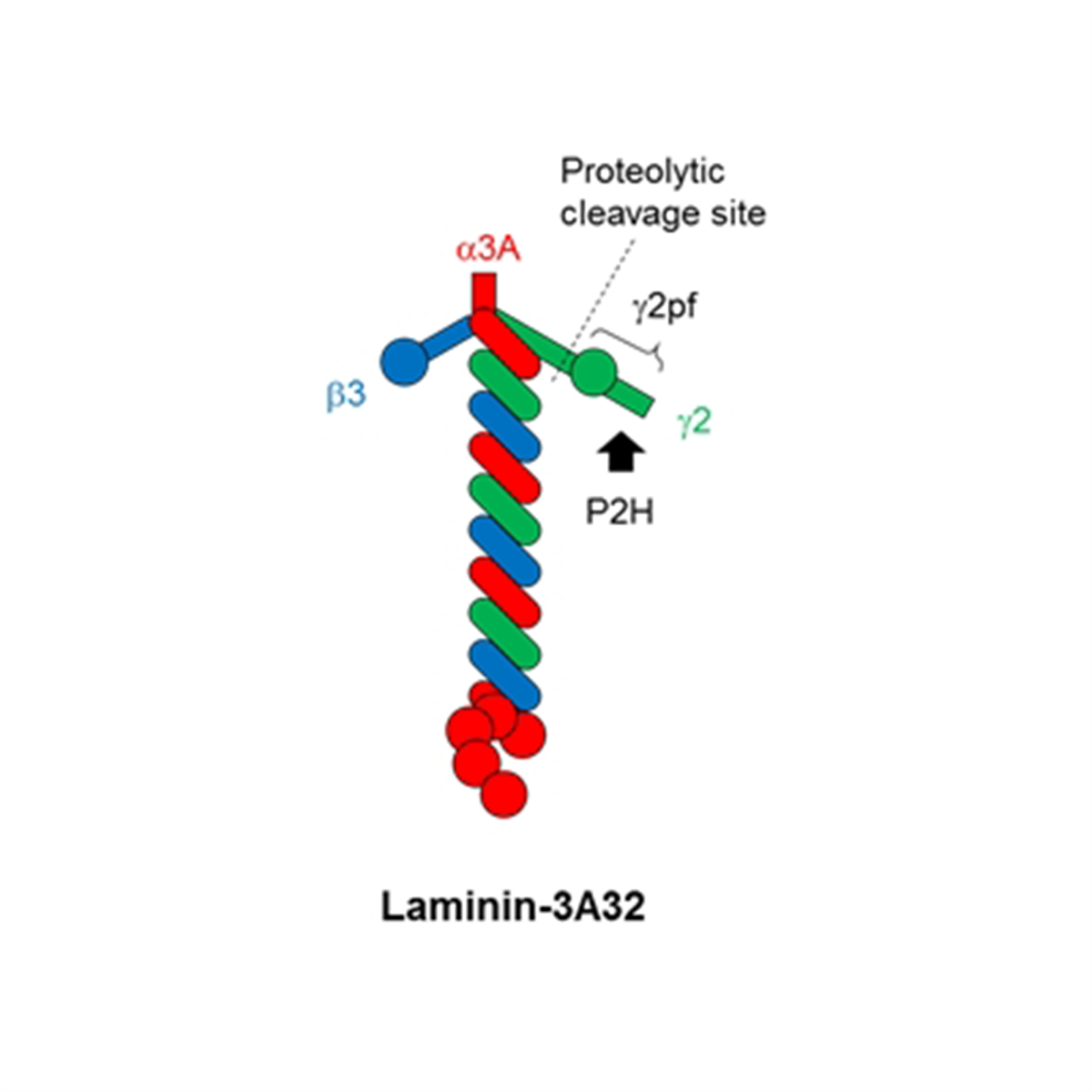
Laminins, which consist of three subunits called α, β and γ chains, are major cell-adhesive components of extracellular matrix, especially basement membranes (BMs). The laminin family is constituted of over 15 isoforms, expressed in a tissue-specific manner and plays differential roles in each tissue. Laminin γ2 chain is a subunit of laminin-332 isoform, formerly laminin-5, but it is frequently overexpressed as a monomer form or the β3-γ2 heterodimer in invasive cancers. Laminin γ2 is a protein of approximately 150 kDa and cleaved at its short arm mainly by bone morphogenic protein-1 (BMP1) in cancer cells, releasing an N-terminal proteolytic fragment (γ2pf) of 45 kDa. The short arm and γ2pf are also cleaved by MMPs and serine proteinases, releasing further small N-terminal fragments. Recently, Dr. Kaoru Miyazaki and co-workers generated a novel mouse monoclonal antibody against γ2pf, clone P2H, which can efficiently detect invasive cancer cells. Clone P2H recognizes laminin γ2 chain of laminin-332 on normal epithelial basement membranes, while in malignant cancer tissues it strongly immunostains the cytoplasm of cancer cells at invasion fronts. Thus, P2H is an important antibody capable of highly detecting the tumor invasion marker laminin γ2 chain. P2H is an only commercially available antibody to detect the N-terminal proteolytic fragments of laminin γchain in cancer tissues. It is useful for pathological assessment of invasiveness and malignancy in various types of human cancers. P2H is applicable to not only immunohistochemistry, but also immunoblotting, ELISA and immunoprecipitation.
 |
|
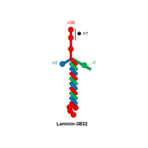
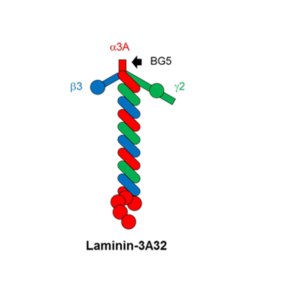
| Figure 1. Protein structure of laminin-3A32s and cleavage site/N-terminal fragment of γ2 chain |
Application examples
| Western blot | |
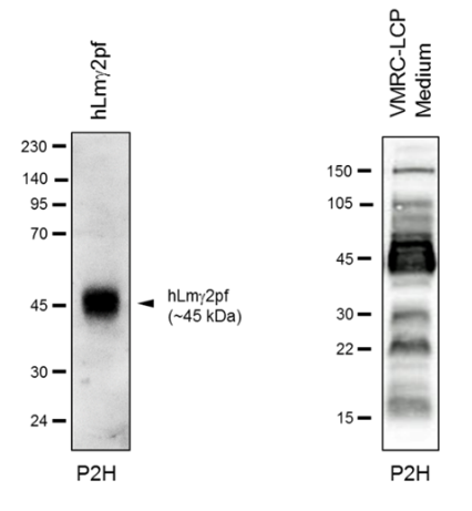
| Samples : (left) recombinant human γ2pf under reducing conditions (right) conditioned medium of VMRC-LCP lung cancer cells. Small γ2 N-terminal fragments are seen below the γ2pf band (45 kDa), and 15-kDa bands corresponds to NE1-2 and NE2-3. Gel conc. : 10% (left), gradient gel (right) 1 st Antibody : clone P2H, 1/1,000 dilution 2 nd Antibody : anti-mouse IgG (H+L)-HRP conjugate |
 |
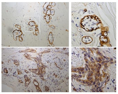
| Immunohistochemistry : Normal mammary gland and breast cancer | |
| Upper panel : Normal mammary gland Lower panel : Breast cancer P2H stains basement membranes surrounding mammary glands in normal tissue, whereas in a breast cancer tissue cytoplasmic accumulation of γ2pf or its fragments is detected with P2H. |
 |
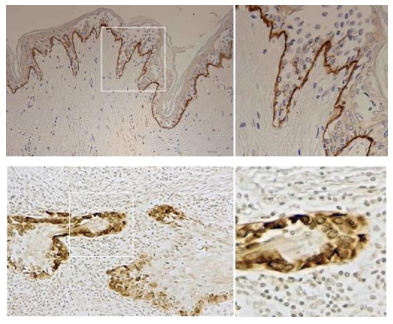
| Immunohistochemistry : Normal skin and skin cancer | |
| Upper panel : Normal skin tissue Lower panel : Skin cancer tissue Although P2H stains epithelial basement membranes in normal skin, in a skin cancer tissue it strongly stains invading cancer cells or their cytoplasm. |
 |
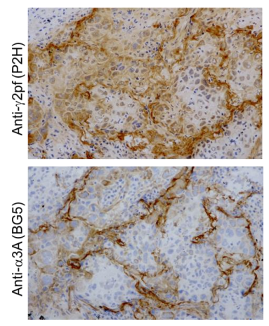
| Immunohistochemistry : Lung cancers - SCC??????? | |
| Right upper panel : Stained by P2H (anti-γ2pf) Right lower panel : Stained by BG5 (anti-α3A, cat. No. FDV-0024) Although BG5 detected basement membrane (BM)-like structure surrounding tumor cells, P2H detected not only BMs but also the cytoplasm of cancer cells. |
 |
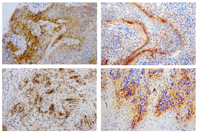
| Immunohistochemistry : Lung SCC and ADC | |
| Upper two panels : Invasive lung SCC Lower two panels : Invasive lung ADC P2H strongly detected cytoplasm of cancer cells at invasion fronts. |
 |
Reference
- Miyazaki et al., Cancer Sci., 107, 1909-1918 (2016) Highly sensitive detection of invasive lung cancer cells by novel antibody against amino-terminal domain of laminin gamma2 chain.
Related products
|
Clone |
BG5 |
F7 |
P2H |
12D |
|
Target |
Laminin-α3A chain |
Laminin-α3B chain |
Laminin-γ2 chain N-terminal region |
Laminin-511 trimer |
|
Normal Tissue |
|
|
|
|
|
Epitope |
|
|
|
|
|
Cancer Tissue |
|
|
|
|


Outline of four clones
|
Description |
Anti-Laminin alpha 3A |
Anti-Laminin alpha 3B |
Anti-Laminin gamma 2 N-terminal Fragment |
Anti-Laminin-511 |
|
Clone |
BG5 |
F7 |
P2H |
12D |
|
Specificity |
Laminin alpha 3A*1 |
Laminin alpha 3B |
Laminin gamma 2 Domain V-NE2 |
Laminin-511 |
|
Host and Type |
Mouse Monoclonal |
|||
|
Subclass |
IgG2a |
IgG1 |
IgG2a |
IgG2b |
|
Reactivity |
Human |
Human |
Human and Mouse |
Human |
|
Form |
Mouse Ascites (Unpurifed) |
|||
|
Application |
ELISA, IHC, IP, WB |
IHC (frozen & paraffin sections), WB |
IHC (frozen), IP, WB (Non-reducing) *2 |
|
|
Code |
FDV-0024 |
FDV-0023 |
FDV-0025 |
FDV-0026 |
|
Size |
100 uL |
|||
|
Storage |
-20oC *3 |
|||
*1 Clone BG5 only detects alpha 3A in IHC. However, this clone may react with alpha 3B in ELISA, IP and Western Blot.
*2 Clone 12D can be only used for IHC (Frozen) and under non-reducing condition for Western Blot.
*3 Non-preservative added. So these 4 clones can be degraded at 4oC. We recommend -20oC storage.
Aliases for LAMC2 Gene
- Laminin Subunit Gamma 2 2 3 5
- Large Adhesive Scatter Factor 140 KDa Subunit 3 4
- Cell-Scattering Factor 140 KDa Subunit 3 4
- Laminin Subunit Gamma-2 3 4
- Epiligrin Subunit Gamma 3 4
- Ladsin 140 KDa Subunit 3 4
- Kalinin Subunit Gamma 3 4
- Nicein Subunit Gamma 3 4
- CSF 140 KDa Subunit 3 4
- Laminin B2t Chain 3 4
- Laminin, Gamma 2 2 3
- BM600-100kDa 2 3
- LAMB2T 3 4
- LAMNB2 3 4
- Laminin, Gamma 2 (Nicein (100kD), Kalinin (105kD), BM600 (100kD), Herlitz Junctional Epidermolysis Bullosa)) 2
- Kalinin/Nicein/Epiligrin 100 KDa Subunit 4
- Laminin-5 Subunit Gamma 4
- Kalinin-105kDa 2
- Nicein-100kDa 2
- BM600 3
- EBR2A 3
- LAMC2 5
- EBR2 3
- B2T 3
- CSF 3