Anti-Laminin 511, Human, Mouse-Mono (12D)
Product#: FNK-FDV-0026
$478.40
Availability:
Ships in 1-2 Weeks
Anti-Laminin 511, Human, Mouse-Mono (12D)
General information
| Cat. No. | :FNK-FDV-0026 |
| Size | :100 ul |
| Format | :Mouse ascites |
| Formulation | :Ascites without any additives |
| Host Species and Clonality | :Mouse monoclonal |
| Isotype and Subclass | :IgG2a |
| Purification | :No purification |
| Lot Number | :see vial label |
| Specificity | :Human, other species not tested yet |
| Application |
|
| Storage | :For short-term storage, -20oC. For long-term storage, -80oC storage is preferable. Avoid repeated freeze-thaw cycles and avoid storage at 4oC |
Description
Useful monoclonal antibodies for the research of cancer pathology 4 unique monoclonal antibodies against cancer-related laminin chains
Laminins are major extracellular matrix proteins containing over 15 members. Laminins are large hetero-trimers consisting of 5 alpha chains, 3 beta chains, and 3 gamma chains. Due to its structural varieties, it has been challenging to detect each laminin isoform by conventional antibodies.
Funakoshi commercializes 4 unique antibodies, originally developed by Dr. Kaoru Miyazaki, Professor Emeritus of Yokohama City University in Japan.
Our antibodies can separately recognize specific isoforms or structures of laminin.

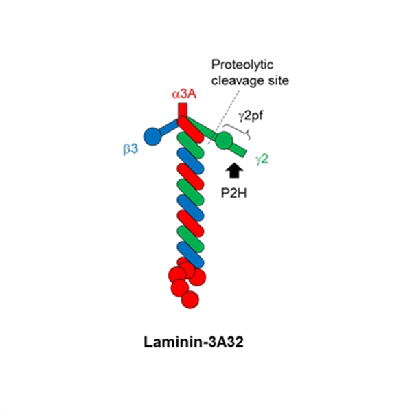
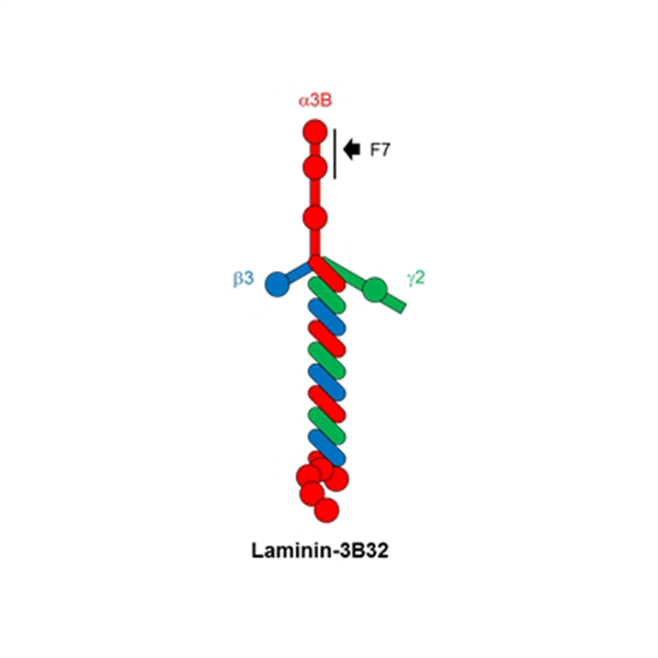
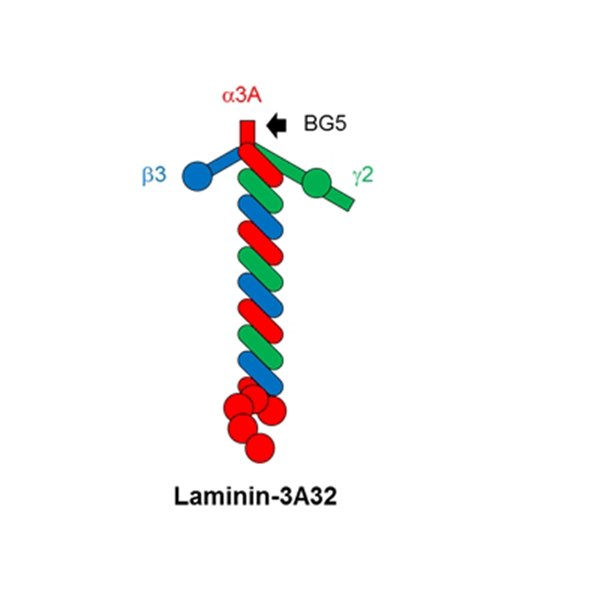
Fig.1 Structure of Laminin 3A32, 3B32 and γ2pf fragment with epitopes of 3 clones (F7, BG5 and P2H).
Features
- BG5/F7 : For laminin alpha-3 chains
- Laminin alpha 3 chain, a major component of Laminin-332, has two splicing variants, alpha 3A and alpha 3B
- Clone BG5 is specific antibody for alpha 3A
- Clone F7 is the world first commercially available antibody for alpha 3B
- P2H : For laminin gamma-2 chain
- Laminin gamma2, an only component of Laminin-332, is a marker gene of invasive cancer tissue
- Laminin gamma-2 chain is activated via proteolytical cleavage at N-terminal
- N-terminal proteolytic fragment of gamma-2, called γ2pf, is highly accumulated into cytosol under malignant cancer
- Clone P2H is the world first antibody for γ2pf
- 12D : For laminin-511
- There are two similar isoforms, laminin-511 and laminin-521 and it is difficult for conventional antibodies such as anti-alpha 5 chain to separate them.
- Clone 12D specifically react with trimeric structure of laminin-511, not react with each components alpha 5, beta 1, gamma 1 and similar isoform laminin-511.
Laminin 511 is used as a culture substrate for ES and iPS cells. (See our iMatrix-511 / Easy iMatrix-511).

Fig.2 Structure of Laminin-511
|
Clone |
BG5 |
F7 |
P2H |
12D |
|
Target |
Laminin-α3A chain |
Laminin-α3B chain |
Laminin-γ2 chain N-terminal region |
Laminin-511 trimer |
|
Normal Tissue |
|
|
|
|
|
Epitope |
|
|
|
|
|
Cancer Tissue |
|
|
|
|


Outline of four clones
|
Description |
Anti-Laminin alpha 3A |
Anti-Laminin alpha 3B |
Anti-Laminin gamma 2 N-terminal Fragment |
Anti-Laminin-511 |
|
Clone |
BG5 |
F7 |
P2H |
12D |
|
Specificity |
Laminin alpha 3A*1 |
Laminin alpha 3B |
Laminin gamma 2 Domain V-NE2 |
Laminin-511 |
|
Host and Type |
Mouse Monoclonal |
|||
|
Subclass |
IgG2a |
IgG1 |
IgG2a |
IgG2b |
|
Reactivity |
Human |
Human |
Human and Mouse |
Human |
|
Form |
Mouse Ascites (Unpurifed) |
|||
|
Application |
ELISA, IHC, IP, WB |
IHC (frozen & paraffin sections), WB |
IHC (frozen), IP, WB (Non-reducing) *2 |
|
|
Code |
FDV-0024 |
FDV-0023 |
FDV-0025 |
FDV-0026 |
|
Size |
100 uL |
|||
|
Storage |
-20 ℃ *3 |
|||
*1 Clone BG5 only detects alpha 3A in IHC. However, this clone may react with alpha 3B in ELISA, IP and Western Blot.
*2 Clone 12D can be only used for IHC (Frozen) and under non-reducing condition for Western Blot.
*3 Non-preservative added. So these 4 clones can be degraded at 4C. We recommend -20C storage.
Data Examples

Fig.3 IHC image of skin cancer tissue in clone BG5 and F7
Left : Clone BG5, Right : Clone F7
Both clones detected basal membrane like structure around the tumor but F7 clone also detected vascular basement membrane (arrow heads)

Fig.4 IHC image of tissues in clone P2H
Upper images : Normal mammary gland
Lower images : Breast cancer
P2H stains basement membranes surrouding mammary glands in normal tisue, whereas in a breast cancer tissue cytoplasmic accumulation of γ2pf orits fragments is detected.

Fig.5 IHC image of breast cancer tissue in clone 12D
A : Vascular basement membrane
B : Mammary gland basement membrane
C : Basement membrane-like structures surrounding tumor cells

Fig.6 Specificity of clone 12D against laminin isoforms
Seven recombinant human laminins were coated on a multi-well plate and detected by clone 12D. The data shows clone 12D specifically reacts with Laminin 511.
Reference
- Kariya et al., J. Biol. Chem., 279, 24774-24784 (2004) Characterization of laminin-5B (a3Bß3r2) and NH2- terminal proteolytic fragment of its a3B chain: Promotion of cellular adhesion, migration and proliferation.
- Kariya et al., J. Mol. Histol., 39, 435-446 (2008) Localization of laminin alpha3B chain in vascular and epithelial basement membranes of normal human tissues and its down-regulation in skin cancers.
- Mori et al., J. Biol. Chem., 285, 35068-35078 (2010) Laminin-3B11, a novel vascular type laminin capable of inducing prominent lamellipodial protrusions in microvascular endothelial cells.
- Mori et al, Camcer Sci., 102, 1095-1100 (2011) Downregulation of a newly identified laminin, laminin-3B11, in vascular basement membranes of invasive human breast cancers.
- Miyazaki et al., Cancer Sci., 107, 1909-1918 (2016) Highly sensitive detection of invasive lung cancer cells by novel antibody against amino-terminal domain of laminin gamma2 chain.

Related Products
Anti-Laminin α3B, Human, Mouse-Mono (Clone F7)
$478.40
$478.40
Anti-Laminin α3A, Human, Mouse-Mono (Clone BG5)
$478.40
$478.40