Anti-Cofilin Rabbit Polyclonal Antibody
Product#: SB-GB112496
$560.00
Availability:
Ships in 1-2 Weeks
Anti-Cofilin Rabbit Polyclonal Antibody
General information
| Cat. No. | :SB-GB112496 |
| Size | :100 uL |
| Protein full name | :Cofilin-1 |
| Synonym | :Cofilin, non-muscle isoform, Cfl1, HEL-S-15, epididymis secretory protein Li 15, p18 |
| Immunogen | :Recombinant protein corresponding to Mouse Cofilin/KLH conjugated Synthetic peptide corresponding to Mouse Cofilin |
| Isotype | :IgG |
| Purity | :Affinity purification |
| Predicted MW. | :19 kDa |
| Observed MW. | :19 kDa |
| Uniprot ID | :P23528, P18760, P45592 |
| Storage | :Store at -20 ℃ for one year. Avoid repeated freeze/thaw cycles. |
| Shippping Condition | :Dry Ice |
| Storage Buffer | :PBS with 0.02% sodium azide,100 μg/ml BSA and 50% glycerol. |
Application
| Applications | Species | Dilution | Positive Tissue |
| WB | Human, Mouse, Rat | 1: 500-1: 1000 |
liver |
Description
Controls reversibly actin polymerization and depolymerization in a pH-sensitive manner. It has the ability to bind G- and F-actin in a 1:1 ratio of cofilin to actin. It is the major component of intranuclear and cytoplasmic actin rods.
 |
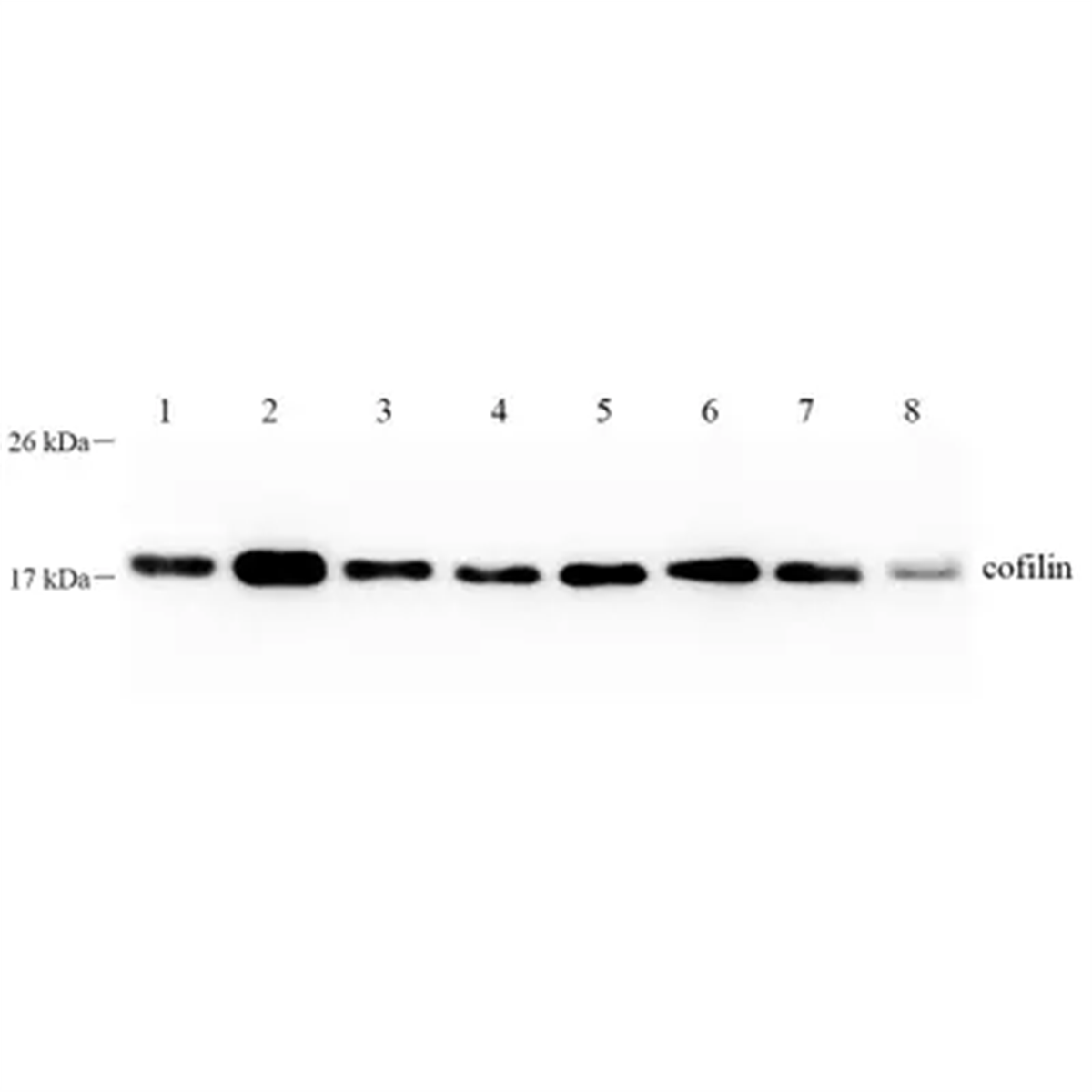
Western blot analysis of Cofilin (GB112496) at dilution of 1: 1000 Lane 1: HeLa cell lysate Lane 2: MCF7 cell lysate Lane 3: 293T cell lysate Lane 4: 293 cell lysate Lane 5: A431 cell lysate Lane 6: NIH3T3 cell lysate Lane 7: SH-SY5Y cell lysate Lane 8: Mouse liver tissue lysate |
 |
Western blot analysis of Cofilin (GB112496) at dilution of 1: 1000 Lane 1: HeLa cell lysate Lane 2: HepG2 cell lysate Lane 3: MCF7 cell lysate Lane 4: 293T cell lysate Lane 5: SH-SY5Y cell lysate Lane 6: A431cell lysate Lane 7: Caco2 cell lysate Lane 8: NIH3T3 cell lysate Lane 9: C6 cell lysate |
Human Diseases
- Cancer: Cofilin-1 is implicated in tumor progression and metastasis due to its role in actin dynamics.
- Muscular Dystrophies: Increased expression in LMNA mutant cells suggests a protective role against degradation.
- Alzheimer's Disease: Altered cofilin-1 levels associated with tau pathology.
Cellular Signaling Pathways
- Regulates actin filament turnover by severing F-actin, essential for cell motility and morphology.
- Involved in the ERK1/2 signaling pathway, which phosphorylates cofilin-1, affecting its stability and function.
- Modulates responses to extracellular signals through actin cytoskeleton dynamics.
Aliases for Cofilin Gene
- GeneCards Symbol: CFL1 2
- Cofilin 1 2 3 5
- CFL 3 4 5
- Cofilin 1 (Non-Muscle) 2 3
- 18 KDa Phosphoprotein 3 4
- Cofilin-1 3 4
- P18 3 4
- Epididymis Secretory Protein Li 15 3
- Cofilin, Non-Muscle Isoform 4
- HEL-S-15 3
- Cofilin 3
References
- Ullo, Maria F, and Jeremy S Logue. “ADF and Cofilin-1 Collaborate to Promote Cortical Actin Flow and the Leader Bleb-Based Migration of Confined Cells.” eLife, eLife Sciences Publications, Ltd, 25 June 2021, elifesciences.org/articles/67856.
- Vignier, Nicolas, et al. “The Non-Muscle ADF/Cofilin-1 Controls Sarcomeric Actin Filament Integrity and Force Production in Striated Muscle Laminopathies.” Cell Reports, U.S. National Library of Medicine, 24 Aug. 2021, www.ncbi.nlm.nih.gov/pmc/articles/PMC8411111/.
- Cofilin 1 Protein: Human Recombinant.” Cytoskeleton, Inc., www.cytoskeleton.com/cf01.
- Author links open overlay panelAnnie Cristhine Moraes Sousa-Squiavinato, et al. “Cofilin-1 Signaling Mediates Epithelial-Mesenchymal Transition by Promoting Actin Cytoskeleton Reorganization and Cell-Cell Adhesion Regulation in Colorectal Cancer Cells.” Biochimica et Biophysica Acta (BBA) - Molecular Cell Research, Elsevier, 5 Oct. 2018, www.sciencedirect.com/science/article/pii/S0167488918304294.
Recently Viewed
m-PEG2-NHS ester
$400.00

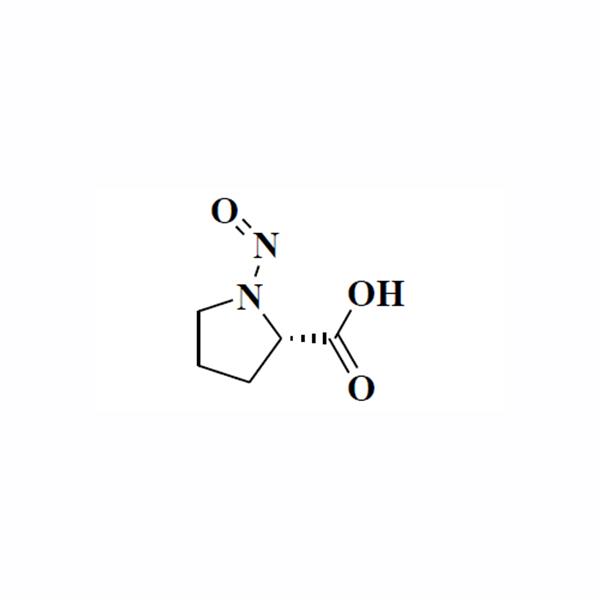
N-Nitroso-L-Proline
$1700.00
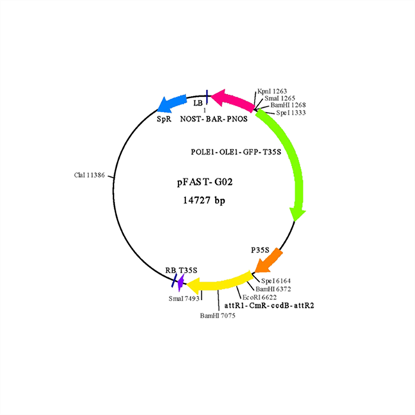
pFAST-G02 Vector(for Academic)
$2080.62

pBIDAVL-GWR1 Vector
$693.56
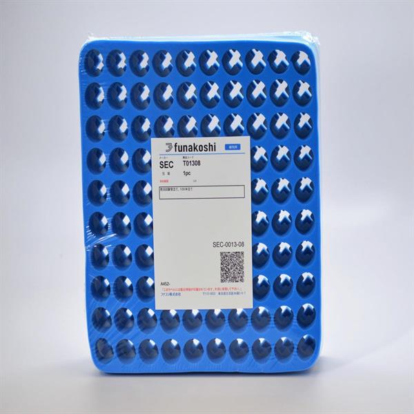
Foam Rack (100 tubes)
$52.67
Foam Rack (50 tubes)
$30.08

Foam Rack (150 tubes)
$61.64