iPGell MOQ/Special Order
iPGell MOQ/Special Order
Shortcut link
- Product ID/SKU : MOQ_Special_5 MOQ FNK-PG20-1
- Product ID/SKU : MOQ_Special_5 MOQ FNK-SG-01
MOQ : iPGell
Cat. No. MOQ_Special_FNK-PG20-1
| Size : MOQ // if order only 1 Kit, Please, Click Go button => | .png) |
https://diagnocine.com/Product/iPGell/52457 |
Storage -20 oC
Description
Making a section of floating cells or 3D cultured cells (such as embryoid body, spheroid, etc) is burdensome work for researchers.
Conventional methods are cytospin, smear slide, or agarose gel but these method gives damages to your cell sample as the cells are dried or heated during sample preparaiton.
Moreover, for cytospin or smear slide method, you need to start again from sample preparation.
iPGell will greatly help you to achieve this just by using two components.
Cells can be rapidly jellified with unheated condition. Any toxic components affect to cells are not contained.
Jellified sample can be soaked into fixation solution and then you can make a sample block (frozen / paraffin) like the way to make tissue block.
Features
-
Rapid and Easy procedure
- Just mixing solution A and B, ~2 minutes protocol. -
Lower damage to Cells (RNA and Protein)
- as sample is not heated. - Various analysis can be done from same sample
- frozen/paraffin blocks enable you to make a section when you need.

- Primary cultured cell
- Embryoid body
- Spheroid
- iPS cells / ES cells
- Specific cell population sorted by cell sorter
Experiment Examples
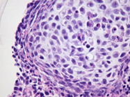
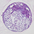
- Morphorogy of mouse derived embryoid body by HE staining
|
|
|
|
|
|
Embryoid body A (x100) |
Embryoid body A (x400) |
Embryoid body B (x100) |
Embryoid body C (x100) |


-
Expression analysis of 3D cultured (spheroid culture) cell

IHC (Fixed frozen section, x 1000)
Antibody D (Brown) / Counter Stain (Blue) -
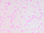
Expression analysis of cultured cell by in situ hybridization
Gene (Blue) / Counter Stain (Pink)
Gene (Blue) / Counter Stain (Pink)
Gene (Blue) / Counter Stain (Pink)

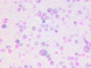
Gene E (x400)
Gene F (x400)
Negative Control (x400)
For more information please see the video :
References
- Preparation of novel RGD-conjugated thermosensitive mPEG-PCL composite hydrogels and in vitro investigation of their impacts on adhesion-dependent cellular behavior. Journal of Industrial and Engineering Chemistry, Volume 84, 25 April 2020, Pages 226-235.
- Injectable hydrogels based on MPEG-PCL-RGD and BMSCs for bone tissue engineering,Biomaterials Science, Biomaterials Science, 2020 Aug 7;8(15):4334-4345
- Malignant subclone drives metastasis of genetically and phenotypically heterogenous cell clusters through fibrotic niche generation. Nature Communications volume 12, Article number: 863. Published: 08 February 2021 (2021)
==========================================================
 |
This products with ICE or DRY ICE shipping requirements, DO NOT select Ground Shipping. |
MOQ Smear Gell
Cat. No. MOQ_Special_5 MOQ-FNK-SG-01
| Size : MOQ 5 kits. // if order only 1 Kit, Please, Click Go button => | |
https://diagnocine.com/Product/Smear-Gell/52446 |
Storage -20°C
Description
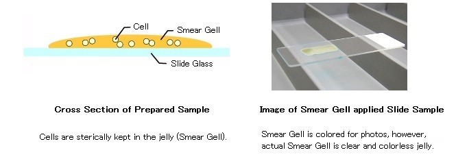
It is easy to make slide preparations from extremely small amounts of cells and non-adherent cells. Smear Gell is a reagent kit for attaching cells and making a slide preparation with easy handling with no equipment necessary such as a cytospin centrifuge. After mixing the cell suspension and solution on a slide glass, the mixture is made into a form of gel immediately and attaches on the slide glass without changing its morphology. This kit is suitable for morphological analysis or immunostaining of cells in culturing, before/after processing cell sorting.
Next generation of smear preparation reagentSmear Gell
Smear Gell is a reagent for making a slide sample of low number cells in suspension by jellifying.
Just mix the cell suspension with Smear Gell and spread it on the slide to make a slide sample very quickly.
Features of Smear Gell
- No hazardous components for cells are contained.
- Cells can be attached to the slide as they do not use cytospin. Special technique for smear preparation is not required.
- No need to prepare paraffin or frozen tissue block.
- Easy to handle for staining after fixation.
Examples of samples
- Sorted Cells
- Non-adherent Cells
- Clump of Cells (< 50 um, such as cultured Spheroids)
- Giemsa stain
- HE Stain
- Immunocytochemistry (Chromogenic / Fluorescence)

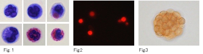
Fig.1 : Wright Giemsa Staining of rat peritonial infiltrative cells.
Fig.2 : Texas Red Staining of rat peritonial infiltrative cells.
Fig.3 : DAB Staining of cultured spheroid