

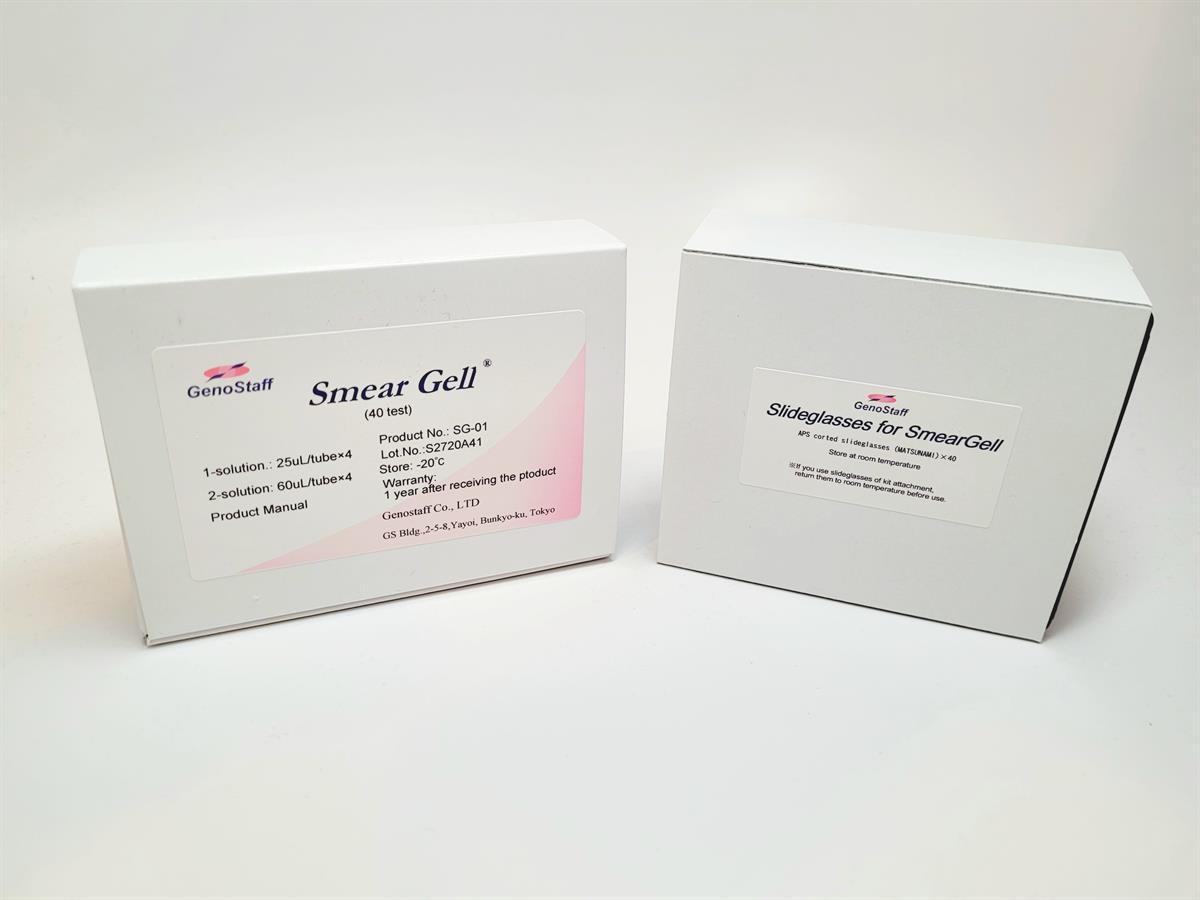
Smear Gell
 |
This products with ICE or DRY ICE shipping requirements, DO NOT select Ground Shipping. |
Smear Gell
Cat. No. FNK-SG-01
Size 1 kit
Storage -20°C
Description
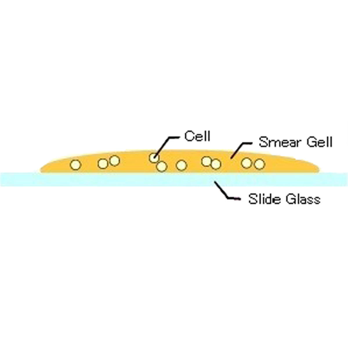
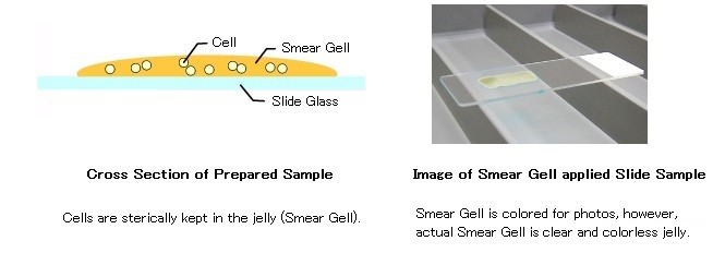
It is easy to make slide preparations from extremely small amounts of cells and non-adherent cells. Smear Gell is a reagent kit for attaching cells and making a slide preparation with easy handling with no equipment necessary such as a cytospin centrifuge. After mixing the cell suspension and solution on a slide glass, the mixture is made into a form of gel immediately and attaches on the slide glass without changing its morphology. This kit is suitable for morphological analysis or immunostaining of cells in culturing, before/after processing cell sorting.
Next generation of smear preparation reagentSmear Gell
Smear Gell is a reagent for making a slide sample of low number cells in suspension by jellifying.
Just mix the cell suspension with Smear Gell and spread it on the slide to make a slide sample very quickly.
Features of Smear Gell
- No hazardous components for cells are contained.
- Cells can be attached to the slide as they do not use cytospin. Special technique for smear preparation is not required.
- No need to prepare paraffin or frozen tissue block.
- Easy to handle for staining after fixation.
Examples of samples
- Sorted Cells
- Non-adherent Cells
- Clump of Cells (< 50 um, such as cultured Spheroids)
- Giemsa stain
- HE Stain
- Immunocytochemistry (Chromogenic / Fluorescence)

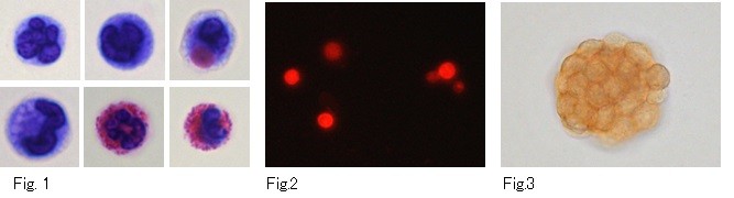
Fig.1 : Wright Giemsa Staining of rat peritonial infiltrative cells.
Fig.2 : Texas Red Staining of rat peritonial infiltrative cells.
Fig.3 : DAB Staining of cultured spheroid
Reference
- Wang, K., Guzman, A. K., Yan, Z., Zhang, S., Hu, M. Y., Hamaneh, M. B., ... & Bouhassira, E. E. (2019). Ultra-high-frequency reprogramming of individual long-term hematopoietic stem cells yields low somatic variant induced pluripotent stem cells. Cell Reports, 26(10), 2580–2592.e7.
- Ikami, K., Shoffner-Beck, S., Weh, M. T., Schnell, S., Yoshida, S., Miranda, E. A. D., ... & Lei, L. (2023). Branched germline cysts and female-specific cyst fragmentation facilitate oocyte determination in mice. Proceedings of the National Academy of Sciences (PNAS), 120(20), e2219683120.
- Wang, K., Schriver, B. J., Aschar-Sobbi, R., Yi, A. Y., Feric, N. T., & Graziano, M. P. (2023). Human engineered cardiac tissue model of hypertrophic cardiomyopathy recapitulates key hallmarks of the disease and the effect of chronic mavacamten treatment. Frontiers in Bioengineering and Biotechnology, 11, 1227184.
- Lei, L., Ikami, K., Miranda, E. A. D., Ko, S., Wilson, F., Abbott, H., ... & Jin, S. (2024). The mouse Balbiani body regulates primary oocyte quiescence via RNA storage. Communications Biology, 7(1), 1247.
