

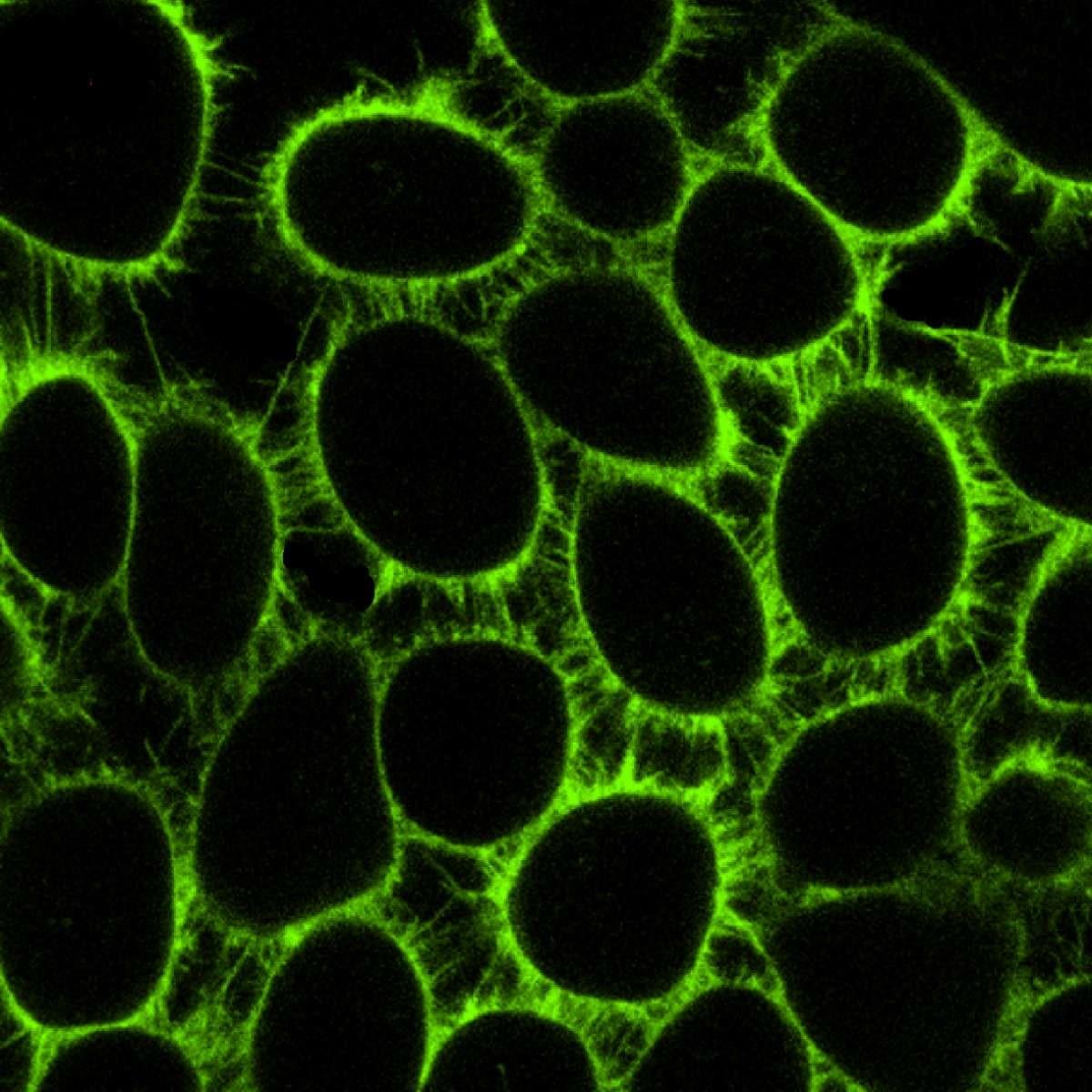
No More Sales-CytoFlamma® 552 Cell-membrane (1 mM)
Product#: inactivated-RCS1322
$99,999.00
Availability:
Ships in 1-2 Weeks
CytoFlamma® 552 Cell-membrane (1 mM)
Cat. No. List below
Description
CytoFlamma® Fluor Cell-labeling solution is a lipophilic fluorescence dye for labeling hydrophobic structures in membranes and other cells. CytoFlamma® 552 Cell-labeling solution, with its low toxicity, can label cells by being applied at water-soluble culture medium, and it can also be applied for tracking of cell-cell fusion, cell adhesion and metastasis. CytoFlamma® Fluor Cell-labeling solution of BioActs that is prepared in a variety of fluorescent colors provides a great convenience for experimental design of multicolor imaging. Its water soluble nature of the dye in culture medium suggests straightforward staining protocol without the need for complex preparation steps. Its high photostability allows it to be resistant to photobleaching, promoting a long-term imaging and its strong absorption and high quantum yield provides bright fluorescence signals
Specifications
- Fluorophore: Flamma® 552
- Application: Cell membrane imaging
- Excitation/Emission Max.(nm): 549/564 nm, suitable for green-imaging
- Product form: 1 mM solution in DMSO facilitates its solubility and cellular uptake
- Storage conditions: -20 ℃, protect from light
- Concentration: 1 mM solution allows for flexible dilution to achieve optimal staining concentration for different types of cells or experimental conditions
Table 1. List of CytoFlamma® cell membrane
| Quick link (Cat.#) | Series | λEx (nm) | λEm (nm) | Packing unit |
| RCS1322 | CytoFlamma® 552 Cell-membrane (1 mM) | 549 | 564 | 1 mL |
| RCS1323 | CytoFlamma® 552 Cell-membrane (5 mM) | 549 | 564 | 1 mL |
| RCS1422 | CytoFlamma® 648 Cell-membrane (1 mM) | 648 | 672 | 1 mL |
| RCS1423 | CytoFlamma® 648 Cell-membrane (5 mM) | 648 | 672 | 1 mL |
| RCS1622 | CytoFlamma® 749 Cell-membrane (1 mM) | 756 | 785 | 1 mL |
| RCS1623 | CytoFlamma® 749 Cell-membrane (5 mM) | 756 | 785 | 1 mL |
Background
Lipophilic organic dyes bear some structural resemblance to natural lipids and can be incorporated into cell wall and liposome. BioActs developed CytoFlamma® cell membrane series as versatile membrane selective fluorescent probes. CytoFlamma® dyes exhibit strong absorption, high fluorescence quantum yield, and high photostability. These properties are maintained even after conjugation to biomolecules, making them excellent for imaging applications. The probes consist of Flamma dye attached with hydrophobic lipid analogs. They can be used in studies for biophysical analysis of membranes, for tracing lipid transport and metabolism in live cells, and for lipid-mediated signal transduction processes. Flamma® dyes display low toxicity, strong absorption, high fluorescence quantum yield and high photostability, and they maintain good fluorescence activity and stability after conjugation to biomolecules. The probes are stable under photooxidation effects induced by ultraviolet excitation and are resistant to spontaneous oxidation. We offer CytoFlamma® cell membrane series as selective fluorescent probes for analyzing cell structure by incorporating into plasma membrane. Advantages to CytoFlamma® are: straightforward staining protocol and no complex preparation steps required, fllexible dilution allowing optimzarion for different cell types and experimental conditions, long term cell tracing because of its stable retention in membrances allows for extended tracking and finally good endocytosis and exocytosis markers which are used to study membrane dynamics in real time. In comparison to other labeling methods, CytoFlamma® does not reuqire genetic modication of cells, provides faster labeling compared to transfection-based methods and it offers potentially less interference with cellular functions compared to antibody-based labeling.
Lipophilic organic dyes bear some structural resemblance to natural lipids and can be incorporated into cell wall and liposome. BioActs developed CytoFlamma® cell membrane series as versatile membrane selective fluorescent probes. CytoFlamma® dyes exhibit strong absorption, high fluorescence quantum yield, and high photostability. These properties are maintained even after conjugation to biomolecules, making them excellent for imaging applications. The probes consist of Flamma dye attached with hydrophobic lipid analogs. They can be used in studies for biophysical analysis of membranes, for tracing lipid transport and metabolism in live cells, and for lipid-mediated signal transduction processes. Flamma® dyes display low toxicity, strong absorption, high fluorescence quantum yield and high photostability, and they maintain good fluorescence activity and stability after conjugation to biomolecules. The probes are stable under photooxidation effects induced by ultraviolet excitation and are resistant to spontaneous oxidation. We offer CytoFlamma® cell membrane series as selective fluorescent probes for analyzing cell structure by incorporating into plasma membrane. Advantages to CytoFlamma® are: straightforward staining protocol and no complex preparation steps required, fllexible dilution allowing optimzarion for different cell types and experimental conditions, long term cell tracing because of its stable retention in membrances allows for extended tracking and finally good endocytosis and exocytosis markers which are used to study membrane dynamics in real time. In comparison to other labeling methods, CytoFlamma® does not reuqire genetic modication of cells, provides faster labeling compared to transfection-based methods and it offers potentially less interference with cellular functions compared to antibody-based labeling.
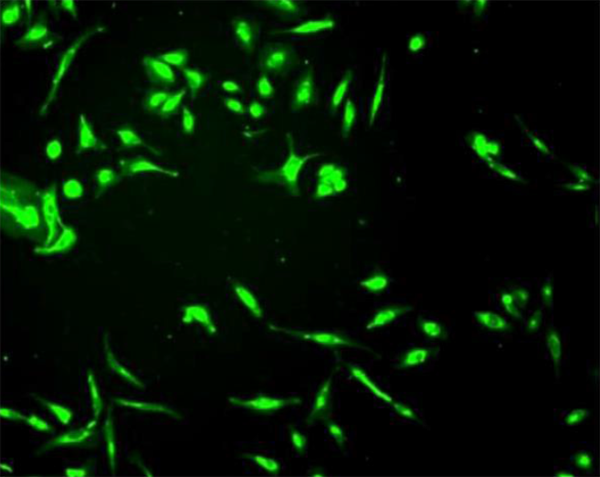
Figure 1. Imaging of CytoFlamma®496 cell membrane staining
Detaching HeLa cell from plate and treating CytoFlamma®496 cell membrane and staining for 5 min, then divided to plate again and cultivated for 24 h, image obtained with FITC filter
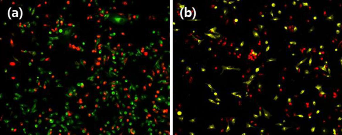
Figure 2. Imaging of Cultured cell after CytoFlamma® cell membrane staining
Image obtained from cell cultivation after staining HeLa cell treated with CytoFlamma® series. (a) CytoFlamma® 496 (green) and CytoFlamma® 648 (red). (b) CytoFlamma® 552(yellow) and CytoFlamma® 648 (red)
※ CytoFlamma® 496 cell membrane is no longer be available because it is discontinued products.
Experimental Protocol
Suspension cell label
- Immerse cells in serum-free medium, HBSS or PBS at a density of 1 × 10⁶/mL.
- Add 5 μL of CytoFlamma® cell membrane solution (1mM) per mL and mix well by pipetting.
- Cover the mixture for about 20 minutes at 37 °C. The optimal labeling time varies depending on the cell type, and usually uniformly labeled in about 20 minutes.
- Centrifuge the labeled cells for 5 minutes at 1,500 rpm, 37 °C.
- Remove the supernatant and resuspend in suitable volume using pre-warmed medium.
- Repeat steps 4 and 5 twice.
Adherent cell label
- Culture cells on a Chamber slide (Nunc ™ Lab-Tek ™ Chamber Slide ™ System).
- Prepare a staining medium by adding 5 μL of CytoFlamma® cell membrane solution (1mM) per mL to medium.
- Remove the culture medium from chamber, and add the staining medium into the chamber.
- Cover the mixture for about 20 minutes at 37 °C. The optimal labeling time varies depending on the cell type, and usually uniformly labeled in about 20 minutes.
- Remove staining medium and add pre-warmed medium and incubate at 37 ℃ for 10 minutes.
- Repeat step 5 twice.
Citation & Reference
1. Wheat Germ Agglutinin Staining as a Suitable Method for Detection and Quantification of Fibrosis in Cardiac Tissue after Myocardial Infarction. B. Emde. Eur J Histochem.2014 Oct 22; 58(4): 2448.
2. Labeling membrane glycoproteins or glycolipids with fluorescent wheat germ agglutinin. Chazotte B. Cold Spring Harb Protoc.2011 May1;2011(5)
