

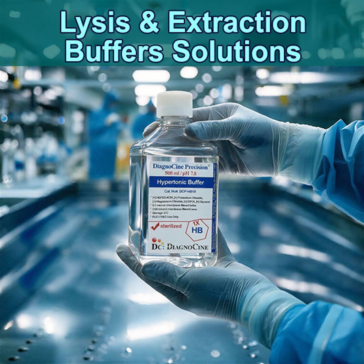
Lysis & Extraction Buffers Solutions
Select Buffers-Group
- see below the table
Lysis & Extraction Buffers Solutions
Four buffer chemistries, one page. Pick the chemistry your target needs and go straight to its product page — the mechanism, history, applications, and product-by-product comparison follow below.
| Product | Base Chemistry & Filtration | Primary Use | Product Page |
|---|---|---|---|
|
filter_altFluxMPS™ · Quadruple-Stage Filtration · 3 products
Published process for the three products in this band: filtered 0.1 µm twice, then 0.04 µm twice, in a sterile environment. See the filtration architecture.
|
|||
| Hypertonic BufferProduct#: DCP-HB1X · FluxMPS™filter_altQuadruple-Stage | High-salt (KCl) · HEPES-KOH pH 7.8 · MgCl2 · EDTA0.1 µm ×2 + 0.04 µm ×2 | Cell extraction, particularly effective for nuclear protein extraction | Viewarrow_forward |
| Tris Based Lysis/Extraction BuffersProduct#: TrisBasedLysis/ExtractionBuffers · FluxMPS™filter_altQuadruple-Stage | Tris-based · clear, colorless liquid0.1 µm ×2 + 0.04 µm ×2 | Versatile protein extraction and lysis; maintains physiological pH and accepts a wide range of additives | Viewarrow_forward |
| Phosphate Based Lysis/Extraction BuffersProduct#: PhosphateBasedLysis/ExtractionBuffers · FluxMPS™filter_altQuadruple-Stage | Phosphate-based · pH 7.4 · store at 4 °C0.1 µm ×2 + 0.04 µm ×2 | Protein isolation where physiological conditions must be maintained, or when working with phosphate-binding proteins | Viewarrow_forward |
|
inventory_2Ready-to-Use 1X Protein Extraction Buffer · 1 product
Listed as its own band, outside the FluxMPS™ quadruple-stage group above. The source states no 0.1 µm or 0.04 µm membrane process for this product, so no filtration specification is given for it here.
|
|||
| RIPA and UltraRIPA Lysis Buffers - Ready-to-Use 1x Protein Extraction BuffersProduct#: Buffers-RIPA-UltraRIPA-LysisBuffersinventory_2Ready-to-Use 1X | Ionic / non-ionic detergent blend · ready-to-use 1XFiltration: not specified in the source | Difficult extractions — lipid-rich tissue and membrane-bound proteins — with low background for quantitative immunodetection | Viewarrow_forward |
Not sure which chemistry? Compare the four products side by side · Read the filtration architecture · See applications by workflow · Read the FAQ
What a lysis buffer has to do, and what these four are specified at
To study intracellular proteins, nucleic acids, or organelles, you first have to break the cell open. Lysis and extraction buffers are formulations engineered to disrupt cell membranes and nuclear envelopes, releasing intracellular contents into solution while preserving the target molecules’ stability and integrity for downstream analysis.
- Four products in this group: Hypertonic Buffer, Tris Based Lysis/Extraction Buffers, Phosphate Based Lysis/Extraction Buffers, and RIPA and UltraRIPA Lysis Buffers — Ready-to-Use 1x Protein Extraction Buffers.
- Chemical, not mechanical. Detergents insert into the lipid bilayer, break up its structure, and form soluble micelles around membrane lipids and proteins — without the heat and shear that mortar-and-pestle, blender, French press, or sonication methods generate.
- Harshness is a choice. Mild non-ionic buffers leave proteins in native, active conformations and preserve interactions; harsh ionic buffers fully denature and extract even tightly membrane-bound targets.
- Self-digestion is the failure mode. Lysis releases endogenous proteases and phosphatases; controlled pH and ionic strength, chelators such as EDTA, and added protease / phosphatase inhibitors keep the sample from degrading.
- Buffer base sets the pH platform. Tris-HCl holds a stable pH near 7.5–8.0; the phosphate-based buffer is specified at pH 7.4 to mimic intracellular ionic conditions.
- Nuclear work is a separate chemistry. The Hypertonic Buffer uses potassium chloride to create the high-salt environment needed to extract proteins from nuclei, buffered by HEPES-KOH at pH 7.8.
- Sterile-filtered, ready to use. The three FluxMPS™ buffers are filtered through 0.1 µm membranes twice and 0.04 µm membranes twice in a sterile environment.
- Products in this group4
- Hypertonic Buffer catalog no.DCP-HB1X
- Hypertonic Buffer buffering agentHEPES-KOH, pH 7.8
- Hypertonic Buffer saltpotassium chloride (KCl)
- Hypertonic Buffer additivesMgCl2, EDTA
- Phosphate-based buffer pH7.4
- Phosphate-based buffer storage4 °C
- Appearance (Tris- and phosphate-based)clear, colorless liquid
- Filtration (FluxMPS™ buffers)0.1 µm ×2 + 0.04 µm ×2
- Fill environment (FluxMPS™ buffers)sterile environment
Cells are built to keep their contents in
Cells are enclosed by hydrophobic lipid bilayers (and sometimes rigid walls) that resist leakage. Each card below is one barrier the buffer has to overcome — or one way the extraction quietly fails.
Membrane disruption
Detergents insert into the lipid bilayer, break up its structure, and form soluble micelles around membrane lipids and proteins. This is what replaces physical force as the way into the cell.
Target solubilization
Buffers are tuned to keep the molecule of interest stable. Mild (non-ionic) buffers leave proteins in native, active conformations and preserve interactions; harsh (ionic) buffers fully denature and extract even tightly membrane-bound targets.
Protection from self-digestion
Lysis releases endogenous proteases and phosphatases. Controlled pH and ionic strength, chelators such as EDTA — which sequester the divalent cations many nucleases and metalloproteases require — and added protease / phosphatase inhibitors keep the sample from degrading.
What mechanical lysis costs you
Mortar and pestle, blenders, French press, and sonication are effective but crude: they generate heat and shear that denature proteins and shear genomic DNA, limiting recovery of intact targets.
Detergent class and CMC
Detergent class, concentration, and critical micelle concentration (CMC) govern membrane solubilization. That physical chemistry — placed on a rigorous footing in 1975 — is the theoretical basis for every modern detergent-based lysis buffer.
The buffer base is a decision, not a detail
A Tris-HCl-stabilized system holds a stable pH around 7.5–8.0 and preserves protein integrity for colorimetric assays and gel electrophoresis. A phosphate-stabilized system mimics intracellular ionic conditions and suits general tissue extraction where preserving enzyme activity matters.
Match the buffer harshness to the target, not to habit
The field shifted from destructive mechanical lysis to tunable chemical lysis precisely so that researchers could match buffer harshness to the target and the assay. A mild non-ionic buffer that preserves a co-immunoprecipitation complex is the wrong tool for a tightly membrane-bound protein, and an ionic RIPA-type buffer that solubilizes that protein will not leave the complex intact.
From brute force to tunable chemistry
Each step below explains a choice you still make at the bench today — why the buffer is chemical rather than mechanical, why the detergent class is specified, and why nuclear and cytoplasmic fractions are prepared separately.
-
1
Mid-20th century Physical disruption
Early biochemists relied on physical disruption — mortar and pestle, blenders, French press, or sonication. Effective but crude: these methods generated heat and shear that denatured proteins and sheared genomic DNA, limiting recovery of intact targets.
-
2
1970s The physical chemistry of detergents
The physical chemistry of detergent–membrane interactions was placed on a rigorous footing, most notably by Helenius & Simons’ 1975 landmark review in BBA. It explained how detergent class, concentration, and critical micelle concentration (CMC) govern membrane solubilization — the theoretical basis for all modern detergent-based lysis.[1]
-
3
1970s–1980s Ionic, non-ionic, and RIPA
Ionic detergents (SDS, sodium deoxycholate) and non-ionic detergents (Triton X-100, NP-40) were adopted to selectively solubilize lipid bilayers. Multi-detergent formulations — notably RIPA (radioimmunoprecipitation assay) buffer — became standard, dissolving tough membranes while keeping immunoprecipitation backgrounds low. This shifted the field from destructive mechanical lysis to tunable chemical lysis, letting researchers match buffer harshness to the target and assay.[3]
-
4
1983 The reference fractionation protocol
Dignam, Lebovitz & Roeder published their method for preparing transcriptionally active nuclear and cytoplasmic extracts from mammalian cells. It established the reference protocol for subcellular fractionation and remains a cornerstone of nuclear-extract preparation today.[2]
Quadruple-stage filtration on the three FluxMPS™ buffers
The published sterility statement for Hypertonic Buffer (DCP-HB1X), Tris Based Lysis/Extraction Buffers, and Phosphate Based Lysis/Extraction Buffers is the same: filtered through a 0.1-micron membrane twice and a 0.04-micron membrane, in a sterile environment.
-
1
0.1 µm Pre-filtration I
First pass through a 0.1-micron membrane.
-
2
0.04 µm Pre-filtration II
First pass through a 0.04-micron membrane.
-
3
0.1 µm Sterile-filtration I
Second pass through a 0.1-micron membrane, performed in a sterile environment.
-
4
0.04 µm Sterile-filtration II — Final Polish
Final 0.04-micron polishing pass in a sterile environment. The buffer is delivered as a clear, colorless liquid.
Why a sub-0.1 µm buffer matters for a lysate
A lysis buffer is added directly to the sample and travels with it through every downstream step, so any particulate it carries becomes part of the lysate. An ultra-filtered, ultra-low-particulate buffer keeps that background out of colorimetric assays, gel electrophoresis, and quantitative immunodetection, and keeps it out of microfluidic and organ-on-a-chip (OoC) sample handling.

Which workflow you are lysing for
Select a workflow to see what the lysis step has to deliver for it. Every application listed is reproduced from the source.
- Western blotting & ELISA — extracting total cellular protein from cultured cells or tissue biopsies
- Immunoprecipitation (IP / Co-IP) — gentle, non-denaturing extraction to pull down intact multi-protein complexes
- Reporter gene assays — lysing cells to measure intracellular enzyme activity (luciferase, β-galactosidase)
- Subcellular fractionation — separating nuclear, cytoplasmic, mitochondrial, or membrane fractions from a single lysate
- Kinase / phosphoprotein analysis — phosphatase-inhibited lysis to preserve phosphorylation states
- DNA/RNA extraction — releasing and stabilizing nucleic acids for downstream molecular workflows
How the four buffers compare
Core attributes and best use, product by product, reproduced from the source comparison table.
| Product | Core Attributes | Best Use |
|---|---|---|
| Hypertonic Buffer | High-salt (KCl) matrix, buffered by HEPES-KOH at pH 7.8, with MgCl2 and EDTA | Nuclear protein extraction — the high potassium chloride concentration creates the high-salt environment needed to extract proteins from nuclei, the extraction step of a fractionation workflow such as the Dignam method. Magnesium ions act as cofactors for many enzymes and help stabilize nuclear structures; EDTA binds metal ions, inhibiting metal-dependent enzymes that could degrade proteins or nucleic acids. |
| Tris Based Lysis/Extraction Buffers | Tris-HCl-stabilized surfactant system | Versatile protein extraction — customizable, reliable for standard mammalian cell lysis; stable pH (~7.5–8.0) preserves protein integrity for colorimetric assays and gel electrophoresis. |
| Phosphate Based Lysis/Extraction Buffers | Phosphate-stabilized surfactant system, pH 7.4 | Physiological optimization — mimics intracellular ionic conditions; well suited to general tissue extraction where preserving enzyme activity matters, and to work with phosphate-binding proteins. |
| UltraRIPA Buffer | Advanced ionic/non-ionic detergent blend | Premium standard for difficult extractions — an optimized RIPA that thoroughly solubilizes challenging samples (lipid-rich tissue, membrane-bound proteins) while minimizing background for high-yield, quantitative immunodetection. |
Frequently asked questions
The questions that come up most often when a target protein meets a buffer choice.
The literature behind the chemistry
The three primary sources the source description cites, verified against the published record.
- Helenius, A., & Simons, K. (1975). Solubilization of membranes by detergents. Biochimica et Biophysica Acta (BBA) – Reviews on Biomembranes, 415(1), 29–79. — The definitive review on how detergent classes interact with and dismantle lipid bilayers; foundational to all detergent-based lysis.doi:10.1016/0304-4157(75)90016-7
- Dignam, J. D., Lebovitz, R. M., & Roeder, R. G. (1983). Accurate transcription initiation by RNA polymerase II in a soluble extract from isolated mammalian nuclei. Nucleic Acids Research, 11(5), 1475–1489. — The reference protocol for preparing functional nuclear and cytoplasmic extracts; a cornerstone of subcellular fractionation.doi:10.1093/nar/11.5.1475
- Kessler, S. W. (1975). Rapid isolation of antigens from cells with a staphylococcal protein A–antibody adsorbent: parameters of the interaction of antibody-antigen complexes with protein A. Journal of Immunology, 115(6), 1617–1624. — Established the protein A–based immunoprecipitation method that underpins the radioimmunoprecipitation (RIPA) workflow the buffer is named for.doi:10.4049/jimmunol.115.6.1617