


LiveReceptor mGluR1, Endogenous mGluR1 Labeling Reagent
Product#: FNK-FDV-0018C
$717.60
Availability:
Ships in 1-2 Weeks
LiveReceptor mGluR1, Endogenous mGluR1 Labeling Reagent
Cat No.: FNK-FDV-0018C
Size: 10 µg
Storage: -20°C
Formulation: C51H46FN9O11S
Molecular Weight: 1012.04 g/mol
Visibility: Orange lyophilized powder
Solubility: Soluble in DMSO
*This compound has water-solubility but it can be easily degraded in water and culture medium. Please avoid store in water.
Background
Neurotransmitter receptors including glutamate receptors and GABA receptors etc. located on post-synapse in neuronal cells play various roles in brain functions. To understand physiological roles of neurotransmitter receptors, live cell imaging is one of the powerful approaches. Conventional imaging methods on live cellsrelay on a genetically engineered proteins fused with fluorescent proteins such as GFP. However, one serious problem is that the functions and movement of over-expressed neurotransmitter receptors with non-physiological tags are not precisely correlated with endogenous native receptors. The labeling methods for endogenous receptors are desirable to observe physiological functions of receptors.
LiveReceptor isthe world first reagent series for target-specific receptor labeling. The principle of LiveReceptor is based on ligand-directed acyl imidazole (LDAI) chemistry (ref.1,2). LDAI-based chemical labelling is driven by selective ligand-protein recognition, which facilitates an acyl substitution reaction of labeling reagents on nucleophilic amino acid residues including Lys, Ser and Tyr located near ligand-binding domain. After wash out, the labelled receptors which have free ligand-binding pockets are observed on live cells. Furthermore, based on pHdependent fluorescent property of fluorescein, fluorescent signal of labeled receptors in endocytosis pathway are highly quenched and only cell surface receptors can be observed. LiveReceptors are powerful tools to monitor reduction of cell surface receptors by endocytosis upon extracellular stimulation.
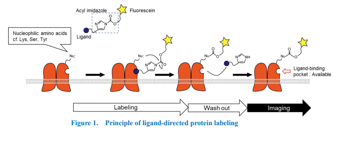
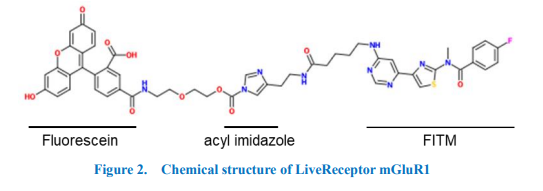
“LiveReceptor mGluR1” is a specific labeling reagent for cell-surface metabotropic glutamate receptor 1 (mGluR1) which is the G-protein Gq family coupled receptor, is strongly expressed in Purkinje cells in the cerebellum, and plays important roles in cerebellar functions. LiveReceptor mGluR1 has three domains including FITM as an affinity ligand for mGluR1, fluorescein and acyl imidazole (a related compound was published in ref.3). Only when FITM binds to mGluR1, nucleophilic amino acid residues (Lys, Ser or Tyr) located near ligand-binding domain on mGluR1 are attacked acyl imidazole and fluorescein is transferred into mGluR1. After removing excess reagents and resultant ligand moiety, labeled mGluR1 can be observed in both live and fixed cells. The protocol is very simple, no genetic manipulation and additional treatment are required. Because LiveReceptor mGluR1 shows no cell membrane permeability, only cell surface mGluR1 are labelled.
Spectrum
Excitation/Emission: 495/515 nm
*Compatible with FITC filter
Application
- Live vell imaging
- Immunocytochemistry with specific antibodies
- Immunoprecipitation with anti-fluorescein antibody
- Immunoblotting with anti-fluorescein antibody
Reconstitution :
Reconstitute at 0.1 mM - 1 mM in 100% DMSO. Please optimize the final concentration of DMSO
depended on your experiments. Before reconstitution, please spin down to collect the orange lyophilized powderon the bottom of a tube. Carefully add DMSO into the tube and vigorously mix to completely dissolve the
powder.
Storage:
(powder) Store at -20oC. Protected from light.
(solution) DMSO stock solution is stable at least for 1 year at -80oC. Please make aliquots and avoid freeze and throw. Protected from light.
(solution) DMSO stock solution is stable at least for 1 year at -80oC. Please make aliquots and avoid freeze and throw. Protected from light.
How to use
General procedure for mGluR1 labeling
- Prepare 10-100 nM of LiveReceptor mGluR1 in the appropriate medium.*
- Replace media of cultured cells to LiveReceptor mGluR1 containing medium.
- Culture cells with LiveReceptor mGluR1 for 1-4 hours at 17-37oC.
- After labeling, wash cells several times or perfused continuously to remove excess reagents.
- Labelled mGluR1 can be observed.
Application data
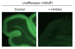
Live cell imaging of labelled mGluR1 in acute mouse cerebellum brain slice
Acute mouse cerebellum brain slices from 3 week-old mouse treated with 10 nM LiveReceptor mGluR1 in artificial cerebrospinal fluid (ACSF) for 4 hours at RT. After then, slices were washed three times by ACSF and fluorescent signal was observed by epi-fluorescent microscopy. Strong fluorescent signal was observed in molecular layer and Purkinje cells. When the slice was pretreated with FITM, an allosteric mGluR1 inhibitor, fluorescent signal was clearly suppressed.
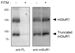
Validation of mGluR1 labeling in acute mouse cerebellum brain slice by western blotting
Acute mouse cerebellum brain slices from 3 week-old mouse treated with 10 nM LiveReceptor mGluR1 in artificial cerebrospinal fluid (ACSF) for 4 hours at RT in the absence or presence of 500 nM FITM. After then, slices were washed three times by ACSF and tissue lysate was prepared. The lysate was analyzed western blotting using antifluorescein (FL) or anti-mGluR1 antibodies. Two bands were observed by anti-FL antibody and these bands were well corresponded with antimGluR1. FITM-treatment clearly reduced the signal of anti-FL antibody.
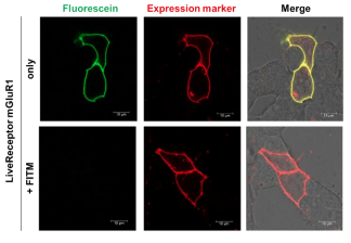
Live cell imaging of labelled mGluR1 in mGluR1-expressed HEK293
mGluR1-expressed HEK293 cells were treated with 100 nM of LiveReceptor mGluR1 in the absence or presence of 2 ?M FITM for 4 hour at 17oC and washed out two times with the basal medium. FITM-treatment clearly reduced the green fluorescent signal. (scale bars, 10 ?m).