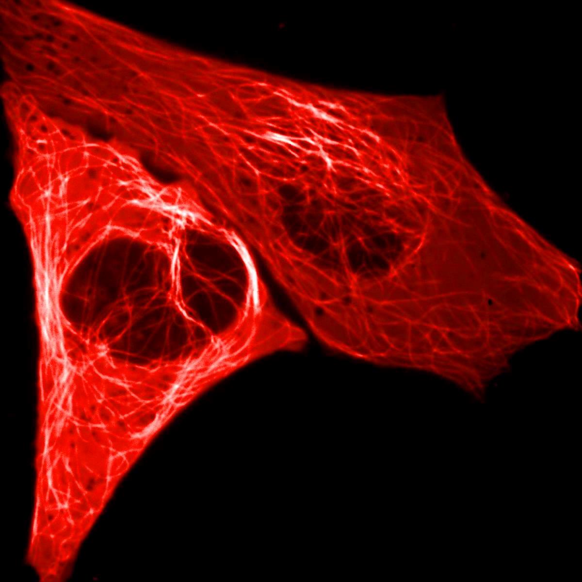
Description
Lectin-FSD 647 is an advanced fluorescence-labeled carbohydrate binding protein designed for the precise detection and visualization of cellular carbohydrates. This innovative probe combines a lectin protein covalently attached to the FSD Fluor™ 647 fluorophore, creating a powerful tool for researchers studying glycobiology and cell surface interactions.
Key Features:
1. Lectin protein with high affinity for specific sugar residues
2. FSD Fluor™ 647 fluorophore with excitation/emission maxima at 653/671 nm
3. Molecular weight of approximately ≥71 kDa
4. Blue liquid appearance
5. Concentration of 1 mg/mL in an appropriate buffer
6. Storage at 4°C, protected from light
Applications:
1. Microscopy imaging of cellular carbohydrates
2. Histochemistry studies of tissue glycosylation patterns
3. Flow cytometry analysis of cell surface glycans
4. Detection of subtle alterations in glycosylation profiles
5. Identification and localization of specific sugar residues in cells and tissues
6. Studying changes in glycosylation during cellular processes or disease states
Advantages:
1. High specificity: Lectins display high, specific affinity to sugar residues, enabling the distinction of subtle alterations in glycosylation that may be otherwise indistinguishable.
2. Bright fluorescence: FSD Fluor™ 647 is a bright red dye developed using BioActs' cutting-edge technology, offering excellent optical properties compared to spectrally similar dyes.
3. Versatility: Fluorescence-conjugated lectins serve as versatile carbohydrate detecting probes across various analytical techniques.
4. Sensitivity: The bright fluorescence of FSD Fluor™ 647 allows for the detection of low-abundance glycan structures.
5. Compatibility: Suitable for use with common fluorescence imaging systems and flow cytometers equipped with far-red filter sets.
6. Stability: The covalent attachment of the FSD fluorophore to the lectin ensures a stable, reliable probe for extended experimental use.
7. Non-invasive detection: Allows for the study of carbohydrate structures on cell surfaces without the need for cell permeabilization.
8. Multiplexing capability: The far-red emission profile of FSD Fluor™ 647 enables multi-color imaging when combined with other fluorophores in different spectral ranges.
Lectin-FSD 647 serves as a valuable tool for researchers studying glycobiology, cell surface interactions, and disease-related changes in glycosylation patterns. Its ability to selectively bind and visualize specific carbohydrate structures, combined with the bright and stable fluorescence of FSD Fluor™ 647, makes it an invaluable asset in the fields of cell biology, immunology, and cancer research.
Specifications
Functionality: Fluorescent protein
Fluorophore: FSD Fluor™ 647
Reacting with: Carbohydrate
Excitation/Emission Max.(nm): 653/671
Molecular weight: ~≥ 71 KDa
Appearance: Blue Liquid
Concentration: 1 mg/mL in proper buffer
Storage conditions: 4 ℃, protect from light
Citation & Reference
1. Furlan M. Staining of glycoproteins in polyacrylamide and agarose gels with fluorescent lectins. Anal Biochem 96.1 (1979): 208-14.
2. Xiao-Peng He. Fluorescent glycoprobes: a sweet addition for improved sensing. Chem. Commun 53 (2017): 82-90.
3. Xiuli Dan. Development and Applications of Lectins as Biological Tools in Biomedical Research. Med Res Rev 36.2 (2016): 221-47.
4. Gijs Hendrik KleinJan. Fluorescent Lectins for Local in Vivo Visualization of Peripheral Nerves. Molecules 19.7 (2014): 9876-92.
5. Bird-Lieberman EL. Molecular imaging using fluorescent lectins permits rapid endoscopic identification of dysplasia in Barrett's esophagus. Nat Med 18.2 (2012): 315-21.