

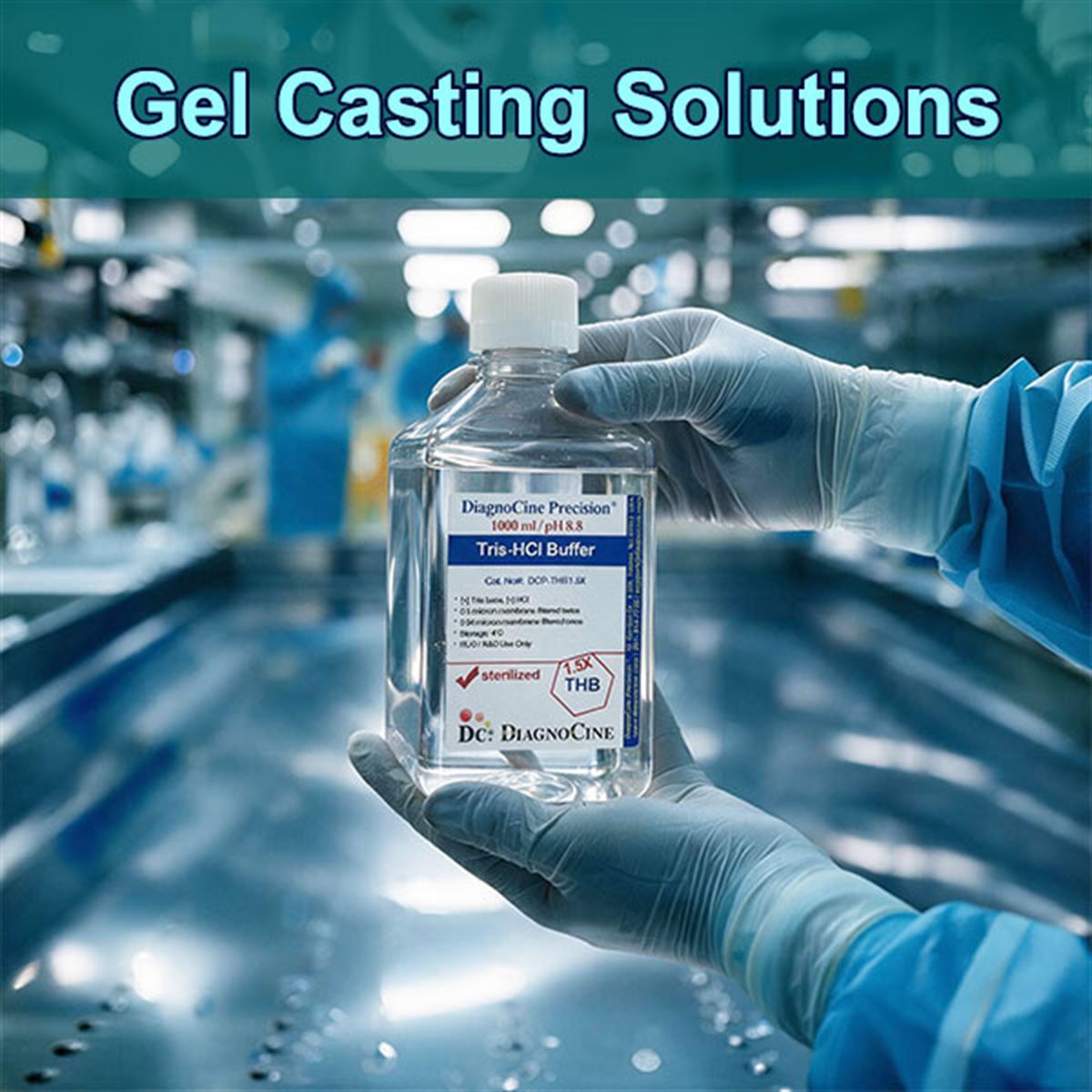
Gel Casting Buffers — A Selection Guide for the Research Laboratory
Three Tris-HCl formulations for pouring the gel itself. Pick the one your layer needs and go straight to its product page — the discontinuous-system reference, the history, and the application guidance follow below.
| Product | Composition | Best Use | Product Page |
|---|---|---|---|
| Tris-HCl Buffer, Sterile [0.5X] | Low-concentration sterile Tris-HCl | Custom dilution workflows; low-ionic-strength casting adjustments; native-PAGE layers where minimized background salt is preferred | Viewarrow_forward |
| Tris-HCl Buffer, Sterile [1.5X] | Concentrated sterile Tris-HCl, pH ~8.8 | The classic resolving-gel buffer — used for casting the main separating portion of SDS-PAGE or native-PAGE gels | Viewarrow_forward |
| Tris-HCl with SDS Buffer | Tris-HCl pre-blended with sodium dodecyl sulfate | All-in-one denaturing casting buffer; combines the Tris system with the correct SDS proportion to keep proteins linearized during polymerization, streamlining standard SDS-PAGE prep | Viewarrow_forward |
Casting a two-layer gel? See the discontinuous system reference · Browse applications · What a casting buffer must deliver · Read the FAQ
Sharp bands or a smeared mess — the gel decides before the run starts
Gel electrophoresis is the workhorse technique for separating DNA, RNA, and proteins by size and charge. Gel casting buffers are the concentrated formulations used to pour agarose or polyacrylamide gel matrices — they set the pH, ionic strength, and denaturing conditions that determine whether molecules resolve into sharp, distinct bands or a smeared mess.
- Three formulations stocked: Tris-HCl sterile 0.5X, Tris-HCl sterile 1.5X (pH ~8.8), and Tris-HCl pre-blended with SDS.
- A bare gel matrix cannot separate molecules cleanly on its own — the casting buffer is what makes the matrix work.
- Controlled polymerization: in polyacrylamide gels, crosslinking (typically catalyzed by APS and TEMED) requires a precise pH environment, so pore size stays consistent throughout the gel.
- Discontinuous band sharpening: shifting pH between the stacking and resolving gel changes the ionization state of trailing ions (e.g., glycine), stacking sample proteins into a tight starting zone.[2,3]
- Denaturation maintenance: buffers that incorporate SDS coat proteins in a uniform negative charge and keep them fully unfolded, so separation depends on molecular mass alone rather than native 3D shape.[4]
- The Laemmli system — a stacking gel at pH ~6.8 and a resolving gel at pH ~8.8 — became, and remains, the default for essentially all modern SDS-PAGE protein separation.[4]
- Both matrix types are served: polyacrylamide for vertical protein work and agarose for horizontal nucleic acid separation.
- Products in this group3
- Resolving gel pH (Laemmli)~8.8
- Stacking gel pH (Laemmli)~6.8
- Resolving-gel buffer offeredTris-HCl sterile 1.5X
- Low-ionic-strength optionTris-HCl sterile 0.5X
- Denaturing all-in-oneTris-HCl with SDS
- Polymerization catalysts (typical)APS and TEMED
- Trailing ion, disc systemglycine
- Matrix types servedpolyacrylamide, agarose
- Applications listed6
Six things the casting buffer — not the gel — controls
A bare gel matrix cannot separate molecules cleanly on its own. Everything below is a property the buffer supplies, and each one is a way the run can fail if the buffer is wrong.
pH, ionic strength, denaturing conditions
Gel casting buffers are the concentrated formulations used to pour agarose or polyacrylamide gel matrices — they set the pH, ionic strength, and denaturing conditions that determine whether molecules resolve into sharp, distinct bands or a smeared mess.
Controlled polymerization
In polyacrylamide gels, crosslinking (typically catalyzed by APS and TEMED) requires a precise pH environment; casting buffer keeps this reaction uniform so pore size stays consistent throughout the gel.
Discontinuous band sharpening
Shifting pH between the stacking and resolving gel changes the ionization state of trailing ions (e.g., glycine), stacking sample proteins into a tight starting zone before they enter the separating matrix — this is the core insight of the Laemmli/Ornstein-Davis system.[2,3,4]
Denaturation maintenance
Buffers that incorporate SDS coat proteins in a uniform negative charge and keep them fully unfolded, so separation depends on molecular mass alone rather than native 3D shape.[4]
Pore size is a buffer problem
The arrival of polyacrylamide as the dominant matrix created the need for precisely buffered casting solutions to control pore size and polymerization — a requirement the earlier starch and paper matrices never imposed.[1]
Ion mobility concentrates the sample
Shifting pH and ion mobility between stacking and resolving zones concentrates samples into ultra-thin starting bands — the effect Ornstein explained with rigorous electrochemical theory and Davis put into practice.[2,3]
Two layers, two pH values, one reason
The discontinuous system exists because the sample has to be compressed before it is separated. A stacking gel at pH ~6.8 concentrates proteins into an ultra-thin starting zone; a resolving gel at pH ~8.8 then separates them. That two-pH arrangement, described in Laemmli's 1970 methods section and grounded in Ornstein's and Davis's 1964 theory, became — and remains — the default buffer system for essentially all modern SDS-PAGE protein separation.[2,3,4]
From starch to the Laemmli system in twenty years
Every buffer on this page traces to one of these four moments. Reproduced from the source's era table, one step per era.
-
1
1950s Starch and paper matrices
Early protein/nucleic acid electrophoresis relied on starch or paper matrices, which yielded poor, diffuse resolution. This set the stage for a synthetic, reproducible matrix.
-
2
1959 Polyacrylamide arrives
Raymond & Weintraub published a one-page Science note describing acrylamide gel as a stable, transparent, reproducible supporting medium for zone electrophoresis. Independently, Ornstein and Davis were developing a related polyacrylamide approach the same year. This established polyacrylamide as the dominant matrix for protein electrophoresis, replacing starch gel, and created the need for precisely buffered casting solutions to control pore size and polymerization.[1]
-
3
1964 Disc electrophoresis theory
Ornstein published the theoretical foundation for “disc electrophoresis” (discontinuous buffer systems), and Davis published the companion methods paper applying it to human serum proteins. Together they explained, with rigorous electrochemical theory, how shifting pH and ion mobility between stacking and resolving zones concentrates samples into ultra-thin starting bands.[2,3]
-
4
1970 Laemmli's SDS-PAGE system
Ulrich Laemmli published his landmark Nature paper — technically about bacteriophage T4 assembly, but the methods section became the most-cited buffer system in protein biochemistry — describing the discontinuous SDS-PAGE system: a stacking gel at pH ~6.8 and a resolving gel at pH ~8.8. It became, and remains, the default buffer system for essentially all modern SDS-PAGE protein separation.[4]
Which layer, which pH, which buffer
The framework the whole category is built on, with what this catalog currently covers for each part of it.
| Layer / matrix | pH and role as the source states it | Covered by this catalog |
|---|---|---|
| Stacking gel (polyacrylamide) | pH ~6.8. Shifting pH changes the ionization state of trailing ions (e.g., glycine), stacking sample proteins into a tight starting zone before they enter the separating matrix[2,3,4] | No dedicated pH ~6.8 stacking buffer is listed — see the note below |
| Resolving / separating gel (polyacrylamide) | pH ~8.8. The main separating portion, where molecules are actually resolved by size[4] | Tris-HCl Buffer, Sterile [1.5X] — the classic resolving-gel buffer, for SDS-PAGE or native-PAGE |
| Denaturing (SDS-containing) layers | SDS coats proteins in a uniform negative charge and keeps them fully unfolded, so separation depends on molecular mass alone rather than native 3D shape[4] | Tris-HCl with SDS Buffer — the all-in-one denaturing casting buffer |
| Low-ionic-strength / native layers | Non-denaturing casting where minimized background salt is preferred | Tris-HCl Buffer, Sterile [0.5X] — custom dilution workflows and low-ionic-strength adjustments |
| Agarose (horizontal, nucleic acid) | Dissolving agarose for horizontal nucleic acid (DNA/RNA) separation | Listed as an application of this category; no agarose-specific casting buffer is in this group |
Six workflows these buffers are cast for
The source's full application list, grouped by the kind of gel being poured. Select a group to see what it covers.
- SDS-PAGE gel preparation — pouring stacking and resolving polyacrylamide layers for vertical, denaturing protein separation
- Western blot sample prep gels — casting the resolving gel that precedes transfer to membrane
- Native-PAGE gel casting — non-denaturing gels for studying intact protein complexes, oligomeric states, and enzymatic activity
- Isoelectric focusing / 2D gel first-dimension casting — where pH-controlled polyacrylamide is used ahead of a second denaturing dimension
- Agarose gel preparation — dissolving agarose for horizontal nucleic acid (DNA/RNA) separation
- Clinical/diagnostic serum protein electrophoresis — the original application described by Raymond, Weintraub, Ornstein, and Davis[1,2,3]
What a casting buffer has to deliver, and what happens if it doesn't
The source names three essentials. Each maps to a specific failure mode on the gel and to a specific product in this catalog.
| Function | Mechanism as the source states it | Where it shows up |
|---|---|---|
| Controlled polymerization | In polyacrylamide gels, crosslinking (typically catalyzed by APS and TEMED) requires a precise pH environment; casting buffer keeps this reaction uniform so pore size stays consistent throughout the gel | Uneven pore size across the gel — inconsistent migration between lanes |
| Discontinuous band sharpening | Shifting pH between the stacking and resolving gel changes the ionization state of trailing ions (e.g., glycine), stacking sample proteins into a tight starting zone before they enter the separating matrix — the core insight of the Laemmli/Ornstein-Davis system[2,3,4] | Diffuse, smeared starting zone instead of ultra-thin bands |
| Denaturation maintenance | Buffers that incorporate SDS coat proteins in a uniform negative charge and keep them fully unfolded, so separation depends on molecular mass alone rather than native 3D shape[4] | Migration reflecting native shape and charge, not molecular mass |
Frequently asked questions
The questions that come up most often when a gel is poured and the bands do not cooperate.
The four papers this category rests on
The primary record behind the statements above, reproduced from the source guide. Each entry was checked against the published article before this page was written.
- Raymond, S., & Weintraub, L. (1959). Acrylamide gel as a supporting medium for zone electrophoresis. Science, 130(3377), 711. — The original report establishing polyacrylamide as a practical electrophoresis matrix.doi:10.1126/science.130.3377.711.a
- Ornstein, L. (1964). Disc electrophoresis — I. Background and theory. Annals of the New York Academy of Sciences, 121(2), 321–349. — Rigorous electrochemical theory behind discontinuous buffer systems and pH-driven band sharpening.doi:10.1111/j.1749-6632.1964.tb14207.x
- Davis, B. J. (1964). Disc electrophoresis — II. Method and application to human serum proteins. Annals of the New York Academy of Sciences, 121(2), 404–427. — Companion methods paper putting Ornstein's theory into practice.doi:10.1111/j.1749-6632.1964.tb14213.x
- Laemmli, U. K. (1970). Cleavage of structural proteins during the assembly of the head of bacteriophage T4. Nature, 227(5259), 680–685. — One of the most-cited papers in biology; its methods section defined the modern discontinuous Tris-glycine-SDS buffer system used in virtually all SDS-PAGE today.doi:10.1038/227680a0