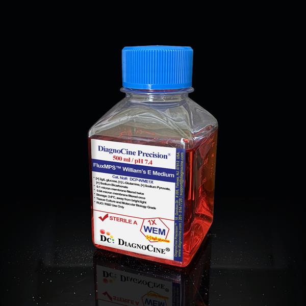
Tris-HCl Buffer, Sterile [0.5X]
FluxMPS™ Tris-HCl Buffer, Sterile [0.5X]
A 0.5 M Tris base buffer precisely titrated to pH 6.8 with HCl, purified through Diagnocine's quadruple-stage 0.1 µm and 0.04 µm membrane filtration for ultra-clean, sterile performance. Designed as a stacking gel buffer for SDS-PAGE, Native PAGE, and related electrophoretic workflows, it is manufactured under ISO 13485-certified, CE-approved facilities and tested free of detectable DNase and RNase activity.
- Quadruple-stage filtration: 0.1 µm membrane twice and 0.04 µm membrane twice
- Sterile, ultrapure formulation — world's cleanest buffer for cell and molecular biology work
- 0.5 M Tris base buffered to pH 6.8 with HCl for stacking gel discontinuity
- No detectable DNase or RNase activity after 18 hr incubation at room temperature
- Manufactured under ISO 13485-certified and CE-approved facilities
- Supplied sterile at 1000 mL, stored at 4°C with a 2-year shelf life
- Custom pH, concentration, and additive formulations available on request
- pH6.8
- Molarity0.5 M Tris base
- AppearanceClear, Colorless Liquid
- Filtration0.1 µm x2 + 0.04 µm x2
- SterilityFiltered, sterile environment
- DNase ActivityNot Detected
- RNase ActivityNot Detected
- Storage4°C
- Shelf Life2 Years
- Size1000 mL
Engineered where standard stacking gel buffers fail
Conventional 0.22 µm-filtered Tris-HCl buffer can carry subvisible particulate and residual nuclease activity into sensitive electrophoresis and downstream molecular biology work. FluxMPS™ buffers are purified further and quality-tested for nuclease activity so your gels and assays stay clean.
Microchannel-safe purity
Quadruple-stage 0.1 µm and 0.04 µm membrane filtration reduces particulate load beyond a single 0.22 µm pass, protecting fine gel-casting and microfluidic channels alike.
Precise, stable pH
Formulated as 0.5 M Tris base titrated with HCl to pH 6.8, creating the pH discontinuity needed for tight protein stacking at the resolving gel interface.
Ultrapure-grade water
Prepared with Ultrapure Type 1 water (18.2 MΩ·cm) as the base for a low-background, reagent-grade buffer.
Low background for sensitive assays
Absence of SDS and tested nuclease activity help preserve native protein structure, supporting activity assays and protein-protein interaction studies.
Defined, traceable composition
Simple, quality-controlled 0.5 M Tris base / HCl formulation with documented pH, appearance, DNase, and RNase test results.
Customization on demand
Alternative concentrations, pH values, and additions of chemicals, compounds, proteins, or supplements can be arranged — contact support@diagnocine.com.
Quadruple-stage filtration system
This buffer is filtered through a four-stage membrane sequence — 0.1 µm membrane twice and 0.04 µm membrane twice — in a sterile environment, then tested for DNase and RNase activity to guard against nuclease contamination in molecular biology work.
- 1
0.1 µm Pre-filtration I
Removes large particulate and aggregates, extending the life of downstream filters.
- 2
0.04 µm Pre-filtration II
Retains fine particulates and bioburden, including organisms near the size of the smallest known mycoplasma, approximately 0.2 micron.
- 3
0.1 µm Sterile-filtration I
A second 0.1 µm pass provides redundancy ahead of final polishing.
- 4
0.04 µm Sterile-filtration II — Final Polish
A second 0.04 µm pass performed in a sterile environment for the final polish prior to fill.
Performance vs. conventional buffer
Sequential 0.1 µm and 0.04 µm filtration removes finer particulates than a single 0.22 µm pass typical of conventional buffer.

© Diagnocine® — DCP-THB0.5X
Where this buffer performs
Tris-HCl buffer is widely used across molecular biology techniques including gel electrophoresis, nucleic acid work, and protein extraction and purification. As a stacking gel buffer, it creates a pH discontinuity between the stacking and resolving gels that concentrates proteins into sharp bands.
Automated Bioreactors & Robotics
For labs automating buffer dispensing, gel casting, or robotic liquid handling, an optional 0.01 µm (10 nm) ultra-filtered variant of this buffer can be arranged to further protect fine-bore valves, sensors, and automated dispensing hardware.
- Total Particulate Exclusion: finer membrane options minimize particulate carryover into automated systems
- Valve & Sensor Protection: reduced particulate load helps protect precision dispensing components
- Extended Perfusion Stability: cleaner buffer supports more stable long-run automated protocols
Inquiry Required: the 0.01 µm ultra-filtered grade is available on request; contact support@diagnocine.com.
SDS-PAGE Stacking Gel Buffer
A crucial component in SDS-PAGE, the most widely used form of protein electrophoresis, contributing to sharper band resolution.
Native PAGE & Complex Preservation
Used in native PAGE to separate proteins while preserving native conformation and enzymatic activity, without SDS.
Second-Dimension Separation
Used in the second dimension of two-dimensional electrophoresis techniques for high-resolution polypeptide separation.
Protein Sizing & MW Determination
The stacking effect contributes to more accurate protein sizing during SDS-PAGE-based molecular weight determination.
Protein Purification by Electrophoresis
The concentrating effect of the stacking gel is useful for purifying proteins during preparative electrophoresis.
Nucleic Acid & Protein Workflows
Broadly used in gel electrophoresis, nucleic acid work, and protein extraction and purification across molecular biology protocols.
Product specifications
Specifications reflect the current published quality-control results for this buffer.
| Parameter | Specification |
|---|---|
| Formulation / Composition | 0.5 M Tris base, HCl to adjust pH |
| Appearance | Clear, Colorless Liquid |
| pH (USP <791>) | 6.8 |
| Molarity / Concentration | 0.5 M Tris base |
| Parameter | Specification |
|---|---|
| Sterility USP | Filtered 0.1 micron twice and 0.04 micron twice in a sterile environment |
| DNase Activity | None detected (18 hr incubation with plasmid DNA at room temperature) |
| RNase Activity | None detected (18 hr incubation with ribosomal RNA at room temperature) |
| Manufacturing Standard ISO | ISO 13485-certified, CE-approved facilities |
| Parameter | Specification |
|---|---|
| Storage Temperature | 4°C |
| Shelf Life | 2 years |
| Parameter | Specification |
|---|---|
| Manufacturing QMS ISO | ISO 13485-certified facility |
| Regulatory Alignment | CE-approved facilities |
| Production Method | Final packaging, quality assurance, and testing performed at the Diagnocine R&D and Quality Testing Center; customization and assembly performed at Diagnocine Precision, Totowa, New Jersey, USA |
| Intended Use | Research Use Only (RUO) |
Full composition
A simple, two-component Tris-HCl buffering system, released per lot against the specifications above.
| Component | CAS Number | Concentration |
|---|---|---|
| Tris base | 77-86-1 | 0.5 M |
| Hydrochloric Acid (HCl) | 7647-01-0 | Added to adjust pH to 6.8 |
Manufacturing & compliance
Manufactured, packaged, and tested under a controlled quality system with full traceability.
ISO 13485:2016 QMS
Manufactured under ISO 13485-certified and CE-approved facilities, suppliers of Diagnocine Precision.
Ultrapure Type 1 Water
Prepared using Ultrapure Type 1 water (18.2 MΩ·cm) as the aqueous base.
Sterile Fill & Finish
Filtered through the quadruple-stage 0.1 µm / 0.04 µm membrane sequence in a sterile environment prior to fill.
Micro-Batch Precision
Final packaging, quality assurance, and testing are performed at the Diagnocine R&D and Quality Testing Center; customization is completed at Diagnocine Precision, Totowa, New Jersey, USA.
Endotoxin USP <85>
Endotoxin testing data is not part of the current published specification for this SKU; contact support@diagnocine.com for the latest lot-specific Certificate of Analysis.
Particulate USP <788>
Sub-visible particulate testing data is not part of the current published specification for this SKU; available upon request via CoA.
Osmolality USP <785>
Osmolality data is not part of the current published specification for this SKU.
Documentation / CoA
A Certificate of Analysis covering appearance, pH, sterility/filtration, DNase, and RNase testing is available upon request.
How DCP-THB0.5X compares
A side-by-side look at how this buffer's manufacturing differs from conventional stacking gel buffer.
| Parameter | DCP-THB0.5X (FluxMPS™) | Conventional Buffer | Standard Alternative |
|---|---|---|---|
| Final filtration pore size | 0.04 µm | 0.22 µm | 0.22 µm |
| Number of filtration stages | 4 | 1 | 1 |
| DNase / RNase tested | check_circle | cancel | cancel |
| Ultrapure Type 1 water | check_circle | cancel | cancel |
| Manufacturing QMS | ISO 13485-certified | Unspecified | Unspecified |
| pH 6.8 stacking-gel formulation | check_circle | check_circle | cancel |
| Custom formulation available | check_circle | cancel | cancel |
Frequently asked questions
Answers to common questions about this Tris-HCl stacking gel buffer.
Supporting literature
Curated references relevant to Tris-HCl stacking gel buffer chemistry and protein electrophoresis.
- Laemmli, U.K. Cleavage of structural proteins during the assembly of the head of bacteriophage T4. Nature. doi:10.1038/227680a0
- Ornstein, L. Disc electrophoresis. I. Background and theory. Ann N Y Acad Sci. doi:10.1111/j.1749-6632.1964.tb14126.x
- Davis, B.J. Disc electrophoresis. II. Method and application to human serum proteins. Ann N Y Acad Sci. doi:10.1111/j.1749-6632.1964.tb14213.x
- Schagger, H. & von Jagow, G. Blue native electrophoresis for isolation of membrane protein complexes. Anal Biochem. doi:10.1016/0003-2697(91)90094-A
- O'Farrell, P.H. High resolution two-dimensional electrophoresis of proteins. J Biol Chem. doi:10.1016/S0021-9258(19)41496-8
- Gallagher, S.R. One-dimensional SDS gel electrophoresis of proteins. Curr Protoc Mol Biol. doi:10.1002/0471142727.mb1004s96
- Good, N.E. et al. Hydrogen ion buffers for biological research. Biochemistry. doi:10.1021/bi00866a011
- Low, J.A. & Tagle, D.A. Organs-on-chips: into the next decade. Nat Rev Drug Discov. doi:10.1038/s41573-020-0079-3