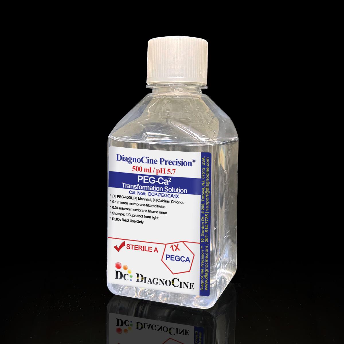
FluxMPS™, PEG‑Ca²⁺ Transformation Solution
FluxMPS™PEG-Ca2+ Transformation Solution
A ready-to-use reagent optimized for PEG-mediated DNA delivery into plant protoplasts. Combines 40% PEG-4000, mannitol osmotic support, and CaCl₂ for efficient, reproducible transient transformation.
- 4-stage filtration process (0.1 µm → 0.04 µm, followed by sterile 0.1 µm → 0.04 µm)
- PEG-4000: Drives membrane perturbation and DNA-membrane association, enabling DNA uptake while allowing recovery when exposure time is carefully controlled.
- Mannitol: Provides osmotic support during the PEG pulse so protoplasts do not swell or burst under the harsh conditions of transformation.
- CaClâ‚‚: Supplies calcium ions that promote DNA binding to the cell surface and assist in PEG-induced membrane fusion events, enhancing transformation efficiency at this pH.
- Ultrapure Type 1 water (18.2 MΩ·cm)
- ISO Class 5 laminar-flow fill & finish
- Customizable formulation available upon request
- pH5.7
- AppearanceClear Solution
- Filtration0.1µm×2 + 0.04µm×2
- Storage2-8 °C
- Shelf Life1 year
Engineered for maximum purity & performance
Standard laboratory reagents filtered at 0.22 µm leave sub-micron particulates, mycoplasma, and aggregates that interfere with sensitive downstream applications. FluxMPS™ PEG-Ca2+ Transformation Solution is manufactured under ISO 13485 quality systems with multi-stage ultra-filtration to eliminate these failure modes.
40% PEG-4000 DNA delivery
High-concentration PEG promotes close contact between plasmid DNA and the protoplast plasma membrane, enabling efficient DNA uptake.
Mannitol osmotic protection
0.2 M mannitol prevents protoplast swelling and lysis during the transient stress of PEG-mediated transformation.
CaCl₂ membrane fusion
0.1 M calcium chloride facilitates DNA binding to the cell surface and assists PEG-induced membrane fusion events.
Reproducible transformation
Designed for use with MMG and W5 buffers in standard protoplast workflows for promoter-reporter, localization, and signaling assays.
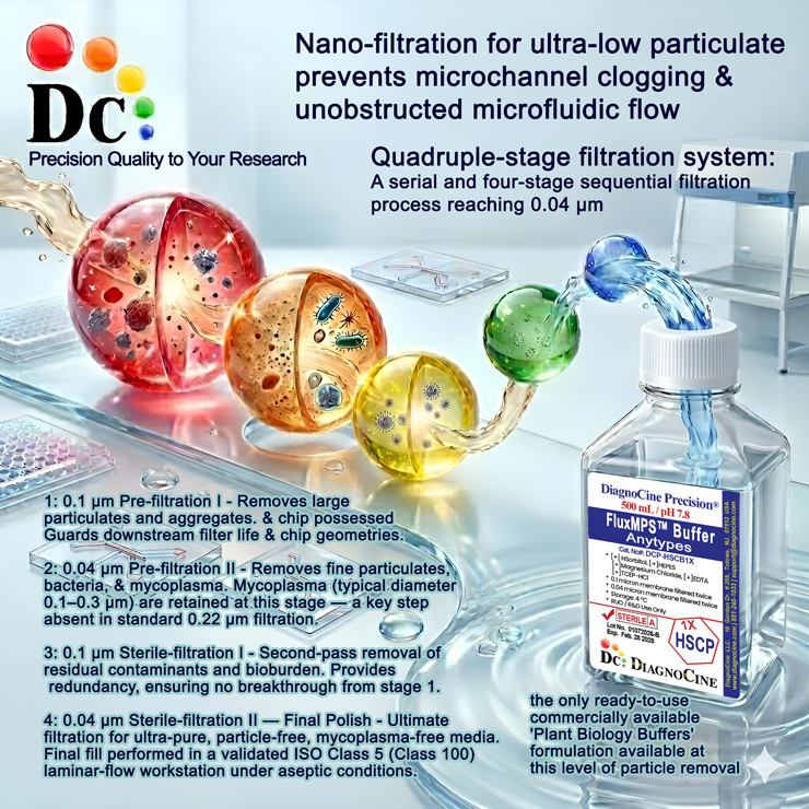
Quadruple-stage filtration system
A serial four-stage sequential filtration process reaching 0.04 µm—the only commercially available buffer at this level of particle removal.
-
1
0.1 µm Pre-filtration I
Removes large particulates and aggregates. Primary guard to extend downstream filter lifetime.
-
2
0.04 µm Pre-filtration II
Removes fine particulates, bacteria, and mycoplasma (0.1–0.3 µm)—a key step absent in standard 0.22 µm filtration.
-
3
0.1 µm Sterile-filtration I
Second-pass removal of residual contaminants and bioburden with full redundancy.
-
4
0.04 µm Sterile-filtration II — Final Polish
Ultimate filtration for particle-free, mycoplasma-free product. Final fill in validated ISO Class 5 laminar-flow workstation.
Performance vs. conventional reagents
Standard 0.22 µm single-pass filtration leaves particles in the 0.05–0.22 µm range. FluxMPS™ removes these contaminants for cleaner results and higher reproducibility.
Validated applications
- Promoter-reporter assays (e.g., luciferase, GUS, fluorescent reporters).
- Signaling and pathway activation studies.
- Subcellular localization and protein-protein interaction analyses.
- Rapid functional screening of gene constructs before stable transformation.
Composition & specifications
| Component |
|---|
| PEG-4000 |
| Mannitol |
| Calcium Chloride |
Protocol & usage instructions
- Isolate protoplasts using standard enzymatic digestion and wash in W5 or equivalent buffer.
- Resuspend protoplasts in MMG Protoplast Buffer at the desired density (e.g., 1-2 x 10⁵ cells per reaction volume, adjust per your protocol).
- In a suitable tube, mix protoplasts in MMG with plasmid DNA (typically 5-20 µg per reaction, depending on construct and assay).
- Add an appropriate volume of Transformation Solution (commonly 1:1 with the protoplast-DNA mixture or as specified in your protocol), and gently invert or flick to mix; avoid vigorous vortexing.
- Incubate for the defined PEG exposure time (often 5-20 minutes at room temperature, optimized per system).
- Gradually dilute the PEG mixture with W5 (or similar) buffer to reduce PEG concentration, then gently pellet and resuspend protoplasts in an appropriate incubation or culture medium.
- Incubate under suitable light and temperature conditions for expression (typically 12-24 hours for many reporters) before analysis.
Quality assurance & logistics
ISO 13485:2016 Certified
Manufactured under certified QMS with full traceability and lot documentation.
Made in USA
All manufacturing, QA testing, and final assembly at DiagnoCine Precision, Totowa, NJ.
Supporting literature
- Yoo SD, Cho YH, Sheen J. Arabidopsis mesophyll protoplasts: a versatile cell system for transient gene expression analysis. Nat Protoc. 2007;2(7):1565-1572.
- Sheen J. Signal transduction in maize and Arabidopsis mesophyll protoplasts. Plant Physiol. 2001;127(4):1466-1475.
- Abel S, Theologis A. Transient transformation of Arabidopsis leaf protoplasts: a versatile experimental system to study gene expression. Plant J. 1994;5(3):421-427.