


Fluorescent Cy2-labelled MAD Reagent
Product#: FNK-BCL-MC-001
$99,999,999.00
Availability:
Ships in 1-2 Weeks
Fluorescent Cy2-labelled MAD Reagent
Cat. No.: FNK-BCL-MC-001
Size: 50μg
Storage: -20℃
For inquiries regarding the price, please contact us at support@diagnocine.com
Introduction:
The BNC is a nano-sized (55 nm) particle consisted of about 100 proteins displaying antibody binding sites. The particle labelled with molecules for signal generation (such as HRP and Fluorescent dyes) is referred to as the MAD (Multi Antibody Detection) reagent. The representative of the MAD reagent is HRP-labelled at about 100 HRP molecules per particle. The MAD reagent binds to antibodies at high affinity, and can be used for various immunoassys as high sensitivity.
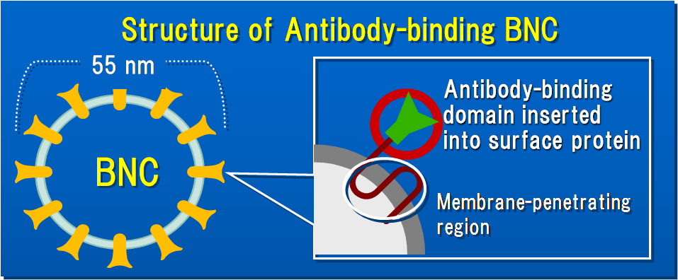
Principle:
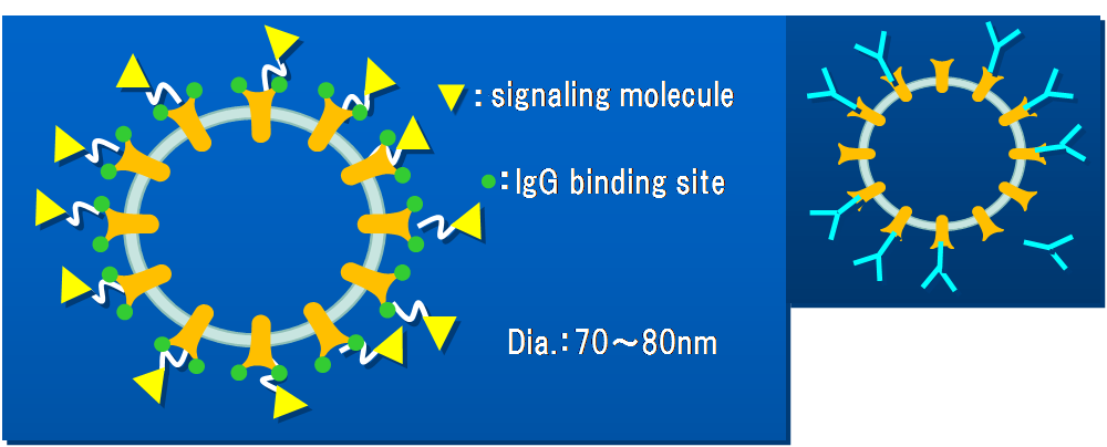
Antibody-binding BNC has 200 to 400 antibody-binding sites per particle depending on the type. The antibody binding site originates from the biding domain of antibody-binding protein, protein A and/or protein G. Because antibodies bind to these binding sites through their Fc region, the antigen binding ability of them is little affected by the binding. When BNC and IgG are mixed, they form a complex which displays many Fab domains outside as shown in the right figure. The MAD reagent is antibody-binding BNC labeled with molecules for signal generation, such as HRP and fluorescent dye. For example, HRP-labelled MAD reagent is composed of antibody-binding BNC which is labeled about 100 HRP molecules per particle, providing highly sensitive antibody detection.
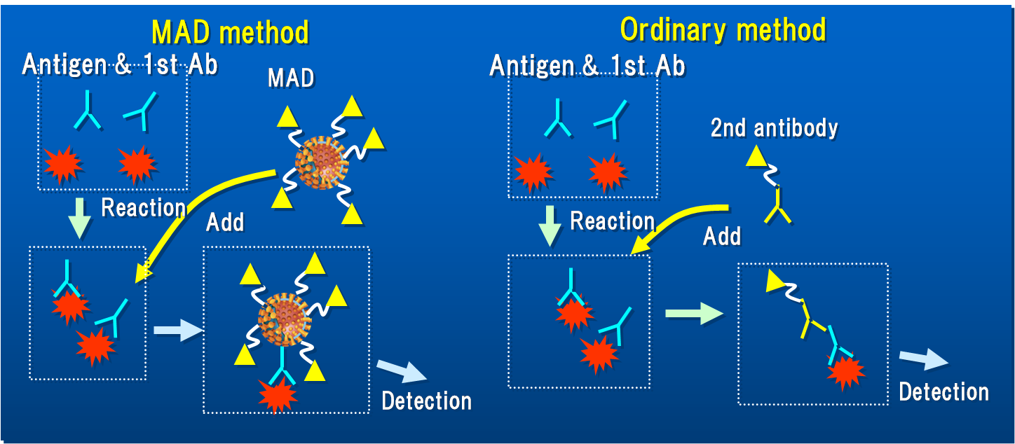
As seen in the below figures, in ordinary method, antibodies bound to antigen is detected by 2nd antibody, such as HRP-labelled antibody. In the MAD method, the antibodies are detected by the MAD reagent. Because the MAD reagent has much more antibody-binding sites (more than 100-fold) and is labelled with much more HRP (at least 50-fold more) than the 2nd antibody, the MAD reagent works as a more sensitive antibody sensor than 2nd antibodies. Furthermore, the MAD reagent provides a one-step antigen detector by using the MAD-antibody complex.
The product is BNC-ZZ labeled with 20 ALP (alkaline phosphatase) molecules per particle. It can be used as the substitute of 2nd antibody in Western blot, antibody-detecting ELISA , and antigen-detecting ELISA using direct method.
Uses
| Fluorescent MAD reagent | Antigen detecting ELISA by direct method | Sensitivity increase (further increased by simultaneous use with 2nd antibody) |
| Western blot | Simultaneous detection of several proteins with multicolor (IRODORI) | |
| Flow cytometry | Simultaneous immunolabeling of multiple antigens | |
| Immunohistochemistry | Simultaneous immunolabeling of multiple antigens |
Protocol for IRODORI (Multi-color Western blot system)
Example of IRODORI
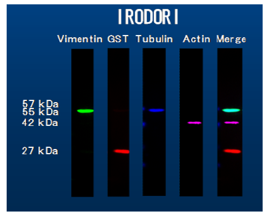
In the example 4 antigens (vimentin, GST, tubulin, actin) were detected using each antigen-specific antibodies which is attached to fluorescent MAD reagent labeled with 4 different Cy dyes.
Materials
-fMAD (fluorescent MAD reagent with Cy dye-labeled):
-4 different MAD types are available (Cy 2, Cy3, Cy4, Cy7).
-BNC-ZZ (non labeled)
-5 mM BS3 dissolved in PBS
-10 mM glycine
-Skim milk
-TBST (0.02M Tris-HCl, 0.14M NaCl, 0.05% Tween 20, pH 7.4)
-Protein A Sepharose
-PBS (pH 7.4)
-Antibodies
-4 different MAD types are available (Cy 2, Cy3, Cy4, Cy7).
-BNC-ZZ (non labeled)
-5 mM BS3 dissolved in PBS
-10 mM glycine
-Skim milk
-TBST (0.02M Tris-HCl, 0.14M NaCl, 0.05% Tween 20, pH 7.4)
-Protein A Sepharose
-PBS (pH 7.4)
-Antibodies
1. Preparation of antibody-fMAD complex (fMAD complex)
For multicolor western, prepare multiple complexes made of different antibody and labeled with Cy dyes of different fluorescence properties.
1) Dissolve 5µg of fMAD in adequate amount of PBS. To the fMAD solution, add 5 µL of 1mg/mL of Antibody solution*, and 5 µL of 5mM BS3 solution. Adjust the volume to be 50 µL. *: If the concentration of antibody solution less than 1mg/ml, increase the volume of antibody solution so that the amount of antibody should be 5 µg.
2) Incubate the solution for 30min at RT, and the at the end of incubation add 0.5µL of 10mM glycine to stop the reaction
3) Add all the solution to 100µL of Protein A Sepharose resin, incubate for15min at RT, and centrifuge at 10000 rpm for 2 min at 4℃.
4) Transfer 50µL of the supernatant add 5µg of BNC-ZZ , and incubation for 30 min at RT. 5) The antibody-fMAD complex is now ready.
2) Incubate the solution for 30min at RT, and the at the end of incubation add 0.5µL of 10mM glycine to stop the reaction
3) Add all the solution to 100µL of Protein A Sepharose resin, incubate for15min at RT, and centrifuge at 10000 rpm for 2 min at 4℃.
4) Transfer 50µL of the supernatant add 5µg of BNC-ZZ , and incubation for 30 min at RT. 5) The antibody-fMAD complex is now ready.
2. Multi-color western
1) Prepare protein-transferred membrane by standard method.
2) Block the membrane by incubating with Blocking buffer (0.05% Skim milk dissolved in TBST ) for 1hr.
3) When using multiple antibody-fMAD complexes, combine them. Then, dilute the antibody-fMAD complex solution to 500µL by Blocking buffer .
4) Incubate the membrane with the diluted antibody-fMAD complex solution for 1hr at RT.
5) Take the fluorescent (pseudo-color) image of the membrane using corresponding excitation and emission wave lengths of each antibody-fMAD complex.
6) Combine the captured pseudo-color image.
2) Block the membrane by incubating with Blocking buffer (0.05% Skim milk dissolved in TBST ) for 1hr.
3) When using multiple antibody-fMAD complexes, combine them. Then, dilute the antibody-fMAD complex solution to 500µL by Blocking buffer .
4) Incubate the membrane with the diluted antibody-fMAD complex solution for 1hr at RT.
5) Take the fluorescent (pseudo-color) image of the membrane using corresponding excitation and emission wave lengths of each antibody-fMAD complex.
6) Combine the captured pseudo-color image.
Reference
Fluorophore-labeled nanocapsules displaying IgG Fc-binding domains for the simultaneous detection of multiple antigens
M.Iijima, T. Matsuzaki, N. Yoshimoto, T. Niimi, K. Tanizawa, S.Kuroda (2011) Biomaterials 32, 9011-9020
Recently Viewed


iMatrix-332, 6x175 µg
$1803.20

iMatrix-111, 6x175 µg
$1803.20

Plant DNA Maxi Kit (10 Preps)
$120.00
*Centrifuge Tubes*
$0.00
iMatrix-332, 2x175 µg
$670.43
iMatrix-111, 2x175 µg
$670.43

No More Sales- HydroCell (Flask)
$99999.00