

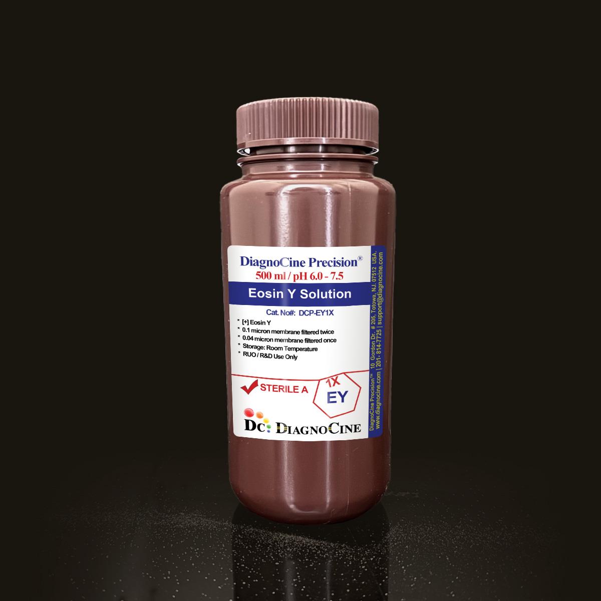
Eosin Y Solution
FluxMPS™ Eosin Y Solution
FluxMPS™ Eosin Y Solution is a pH-neutral, alcohol-free, acetic-acid-free 1% aqueous histological counterstain manufactured with quadruple-stage 0.1 µm and 0.04 µm membrane filtration for ultra-low particulate clarity. Formulated for laboratories that need consistent cytoplasmic staining and microchannel-safe purity across routine histology, immunohistochemistry, and organ-on-a-chip (OoC) tissue-section workflows.
- Quadruple-stage filtration: 0.1 µm membrane (twice) and 0.04 µm membrane (twice) for ultra-low particulate clarity
- Sterile, ultrapure formulation manufactured with Ultrapure Type 1 water (18.2 MΩ·cm)
- pH 6.0–7.5 (unmodified), alcohol-free and acetic-acid-free for stable, reproducible staining[1]
- Manufactured in an ISO Class 5 (Class 100) aseptic fill environment
- Compatible with microfluidic tissue-section processing and organ-on-a-chip (OoC) histology workflows[6]
- Tested free of DNase and RNase activity following 18-hour incubation at room temperature
- Custom concentrations, pH, and additive formulations available on request
- FormulationEosin Y, 1%
- AppearancePink to orange-pink
- pH6.0–7.5 (unmodified)
- Filtration0.1 µm membrane twice, 0.04 µm membrane twice
- SterilityFilter-sterilized
- DNase ActivityNone detected
- RNase ActivityNone detected
- StorageRoom temperature, away from light
- Shelf Life6 months unopened
- Size500 mL
Engineered where standard stains fall short
Conventional 0.22 µm-filtered eosin formulations can carry subvisible particulates, precipitation risk, and pH drift that introduce background and inconsistency into cytoplasmic staining. FluxMPS™ Eosin Y Solution is built to remove those failure modes at the source.
Microchannel-safe purity
Quadruple-stage 0.1 µm and 0.04 µm membrane filtration reduces particulate load beyond a single 0.22 µm pass, supporting clean slide backgrounds and microfluidic tissue-section handling.
pH-neutral stability
Unmodified pH of 6.0–7.5, with no glacial acetic acid, minimizes precipitation risk and extends usable shelf life without sacrificing staining clarity.
Ultrapure-grade formulation
Prepared with Ultrapure Type 1 water (18.2 MΩ·cm) and sterile filtration to support reproducible, low-background staining lot to lot.
Low background for imaging
Alcohol-free formulation minimizes tissue distortion in delicate samples such as renal biopsies and cytology smears, supporting clean chromogen-based IHC readouts.
Defined, traceable composition
Single-component 1% Eosin Y formulation, tested free of DNase and RNase activity, with quality control performed at the DiagnoCine R&D and Quality Testing Center.
Customization on demand
Alternative concentrations, pH, and additional chemicals, compounds, proteins, or supplements are available on inquiry.
Quadruple-stage filtration system
Every lot of FluxMPS™ Eosin Y Solution is filter-sterilized using a 0.1 µm membrane twice and a 0.04 µm membrane twice, removing progressively finer particulates than a single conventional filtration pass.
-
1
0.1 µm Pre-filtration I
Removes large particulates and aggregates, extending the working life of downstream filters.
-
2
0.04 µm Pre-filtration II
Retains fine particulates and bioburden ahead of final sterile filtration.
-
3
0.1 µm Sterile-filtration I
Provides second-pass filtration redundancy ahead of the final polish.
-
4
0.04 µm Sterile-filtration II — Final Polish
Final polishing filtration performed under ISO Class 5 (Class 100) aseptic fill conditions.
Performance vs. conventional staining solution
Sequential 0.1 µm and 0.04 µm filtration removes finer particulates than a single 0.22 µm pass used in conventional eosin formulations, supporting cleaner backgrounds in downstream imaging and IHC workflows.

© Diagnocine® — DCP-EY1X
Where FluxMPS™ Eosin Y Solution is used
A pH-neutral, alcohol-free counterstain suited to routine histology, specialized immunostaining, and microfluidic tissue-section workflows.
Automated Slide Stainers & Robotics
An optional 0.01 µm (10 nm) ultra-filtered variant is available for automated histology stainers and robotic slide-processing systems that are sensitive to fine particulates.
- Total Particulate Exclusion for high-throughput staining lines
- Valve & Sensor Protection in automated liquid-handling systems
- Extended Reagent Line Stability during continuous processing
Inquiry Required: Contact support@diagnocine.com to request the 0.01 µm ultra-filtered grade.
H&E Counterstaining
Counterstains nuclei following hematoxylin staining with minimal cytoplasmic overstaining.
Fiber & Matrix Visualization
Highlights collagen, muscle, and elastic fibers in pink-red hues for connective tissue evaluation.
Chromogen-Based IHC
Compatible with chromogen-based immunohistochemistry protocols, including DAB detection systems.
Renal & Cytology Sample Processing
Alcohol-free formulation minimizes tissue distortion in delicate samples such as renal biopsies and cytology smears.
Teaching Laboratories
Extended usability and stable performance make this formulation well suited to educational and training histology labs.
Organ-on-a-Chip Tissue Sections
Microchannel-safe filtration supports counterstaining of tissue sections derived from organ-on-a-chip (OoC) and microphysiological system (MPS) models.
Specification summary
Key physical, sterility, storage, and traceability parameters for DCP-EY1X.
| Parameter | Specification |
|---|---|
| Formulation / Composition | Eosin Y, 1% |
| Appearance | Pink to orange-pink |
| pH (USP <791>) | 6.0–7.5 (unmodified) USP <791> |
| Parameter | Specification |
|---|---|
| Sterility | Filtered 0.1 µm membrane twice and 0.04 µm membrane twice USP <71> |
| Water Quality | Ultrapure Type 1 water (18.2 MΩ·cm) USP <85> |
| Manufacturing Standard | ISO 13485-certified, CE-approved facilities ISO 13485 |
| DNase Activity | None detected after 18-hour incubation of plasmid DNA at room temperature |
| RNase Activity | None detected after 18-hour incubation of ribosomal RNA at room temperature |
| Parameter | Specification |
|---|---|
| Storage Temperature | Room temperature, away from bright light |
| Shelf Life | 6 months unopened |
| Expiry | Use before expiry date printed on product label |
| Parameter | Specification |
|---|---|
| Manufacturing QMS | ISO 13485-certified facilities (Suppliers of DiagnoCine Precision) |
| Regulatory Alignment | CE-approved |
| Production Method | Filter-sterilized: 0.1 µm membrane twice, 0.04 µm membrane twice |
| Traceability | Final packaging, quality assurance, and testing performed at the DiagnoCine R&D and Quality Testing Center; customization and assembly performed at DiagnoCine Precision, Totowa, New Jersey, USA |
| Intended Use | Research Use Only (RUO); histological counterstaining, connective tissue studies, and chromogen-based IHC workflows |
Full composition
Single-component aqueous Eosin Y formulation, manufactured under per-lot quality control.
| Component | CAS Number | Concentration |
|---|---|---|
| Eosin Y | 17372-87-1 | 1% |
Manufacturing & compliance
Manufactured, packaged, and tested under a documented quality system with full traceability.
ISO 13485:2016 QMS
Manufactured under ISO 13485-certified and CE-approved facilities (Suppliers of DiagnoCine Precision).
Ultrapure Type 1 Water
Formulated with Ultrapure Type 1 water (18.2 MΩ·cm) per USP <85>.
ISO Class 5 Fill & Finish
Final sterile filtration and fill performed in an ISO Class 5 (Class 100) aseptic environment.
Micro-Batch Precision
All final packaging, quality assurance, and testing are completed at the DiagnoCine R&D and Quality Testing Center.
Endotoxin Testing (USP <85> BET)
Manufactured in accordance with USP <85> Bacterial Endotoxins Test methodology.
Particulate Control (USP <788>)
Quadruple-stage 0.1 µm and 0.04 µm filtration is designed to support USP <788> particulate control practices.
Osmolality (USP <785>)
Assessed per USP <785> osmolality methodology as part of formulation quality control.
Documentation / CoA
A Certificate of Analysis (CoA) is available for this lot.
How DCP-EY1X compares
A side-by-side view of FluxMPS™ Eosin Y Solution against conventional eosin formulations.
| Parameter | DCP-EY1X (FluxMPS™) | Conventional Eosin (0.22 µm filtered) | Standard Alternative (0.22 µm filtered) |
|---|---|---|---|
| Alcohol-free, acid-free formulation | check_circle | cancel | cancel |
| Final filtration pore size | 0.04 µm | 0.22 µm | 0.22 µm |
| Number of filtration stages | 4 | 1 | 1 |
| Sterility (USP <71>) | check_circle | cancel | check_circle |
| Water quality | Ultrapure Type 1 | Not specified | Not specified |
| Manufacturing QMS | ISO 13485:2016 | cancel | cancel |
| Microfluidic / OoC tissue-section compatibility | check_circle | cancel | cancel |
| Custom formulation available | check_circle | cancel | cancel |
Frequently asked questions
Common questions about DCP-EY1X, FluxMPS™ Eosin Y Solution.
Supporting literature
Curated references relevant to histological eosin staining, filtration-based sterility, and microfluidic tissue applications.
- Fischer AH, Jacobson KA, Rose J, Zeller R. Hematoxylin and eosin staining of tissue and cell sections. CSH Protoc. 2008. doi:10.1101/pdb.prot4986
- Cardiff RD, Miller CH, Munn RJ. Manual hematoxylin and eosin staining of mouse tissue sections. Cold Spring Harb Protoc. 2014. doi:10.1101/pdb.prot073411
- Titford M. The long history of hematoxylin. Biotech Histochem. 2005. doi:10.1080/10520290500138372
- Ramos-Vara JA. Technical aspects of immunohistochemistry. Vet Pathol. 2005. doi:10.1354/vp.42-4-405
- Uphoff CC, Drexler HG. Detection of mycoplasma contamination in cell cultures. Methods Mol Biol. 2011. doi:10.1007/978-1-61779-108-6_1
- Bhatia SN, Ingber DE. Microfluidic organs-on-chips. Nat Biotechnol. 2014. doi:10.1038/nbt.2989
- Low LA, et al. Organs-on-chips: into the next decade. Nat Rev Drug Discov. 2021. doi:10.1038/s41573-020-0079-3
- Chan JK. The wonderful colors of the hematoxylin-eosin stain in diagnostic surgical pathology. Int J Surg Pathol. 2014. doi:10.1177/1066896914527704