

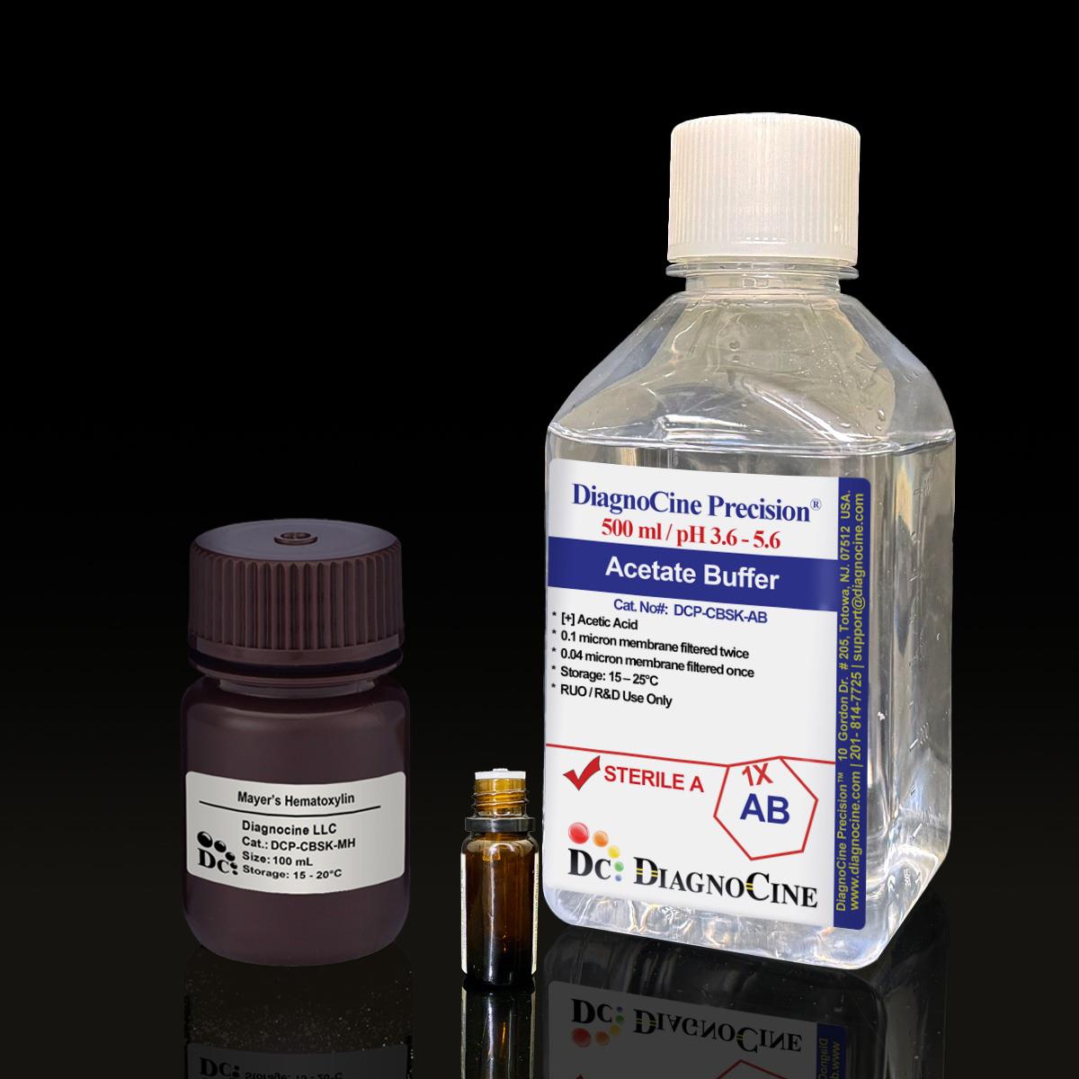
Copper Blood Staining Kit
FluxMPS™ Copper Blood Staining Kit
The FluxMPS™ Copper Blood Staining Kit (DCP-CBSK) delivers a quadruple-stage ultra-filtered rhodanine staining system engineered for reliable histochemical detection of copper deposits in formalin-fixed, paraffin-embedded tissue and forensic bloodstain evidence. Every solution — Rhodanine Solution, Acetate Buffer, and Mayer’s Hematoxylin — is filtered through a 0.1 µm membrane twice and a 0.04 µm membrane twice, delivering microchannel-safe purity for automated stainers and low-background microscopy. Optimized for Wilson’s disease diagnostics, copper metabolism research, and latent bloodstain enhancement, the kit completes a full run in 90 minutes, or under 15 minutes with the microwave-assisted protocol.
- Quadruple-stage ultra-filtration: 0.1 µm membrane twice and 0.04 µm membrane twice for microchannel-safe, low-particulate staining reagents
- Rhodanine-based copper detection sensitive to 0.1–0.5 µg/g tissue
- Pre-mixed Acetate Buffer at pH 8.0 for consistent chelation and reduced preparation time
- 50-test kit compatible with 4 µm FFPE sections and automated stainers
- Standard 90-minute protocol or microwave-accelerated workflow in under 15 minutes
- Confirmed DNase- and RNase-free after 18-hour room-temperature incubation
- Manufactured under ISO 13485-certified, CE-approved facilities in Totowa, New Jersey, USA
- Custom concentrations, pH, and additive modifications available on request
- Format3-solution kit, 50 tests
- Section Compatibility4 µm FFPE sections
- Acetate Buffer pH8.0
- Rhodanine Working Conc.0.2% (alcoholic)
- Detection Sensitivity0.1–0.5 µg/g tissue
- Filtration Architecture0.1 µm x2 + 0.04 µm x2
- DNase / RNaseNone detected (18 hr, RT)
- Rhodanine Storage2–8°C, 6 months
- Buffer / Hematoxylin Storage15–25°C, 12–18 months
- Standard Protocol Time90 min (or <15 min microwave)
Engineered where standard staining reagents fail
Conventional 0.22 µm-filtered staining solutions can carry subvisible particulate, uneven buffering, and undetected nuclease contamination into diagnostic and forensic workflows — obscuring fine collagen and copper-deposit detail and clogging automated stainer lines. DCP-CBSK removes those failure points at the source.
Microchannel-safe purity
Every kit component passes through a 0.1 µm membrane twice and a 0.04 µm membrane twice, removing subvisible particulate that would otherwise accumulate in automated stainer lines and obscure fine tissue detail.
Defined, chelation-ready buffering
The Acetate Buffer ships pre-mixed at pH 8.0, holding the rhodanine chelation reaction at its optimal working point without in-lab pH adjustment.
Ultrapure-grade water
All aqueous components are prepared with Ultrapure Type 1 water (18.2 MΩ·cm), reducing background staining and nonspecific binding.
Low background for diagnostic imaging
Reduced particulate and controlled acetic-acid content keep contrast high for bright-field histology and forensic bloodstain pattern photography alike.
Defined, traceable composition
Rhodanine Solution, Acetate Buffer, and oxidized Mayer’s Hematoxylin are each lot-released, and pre-stained fetal liver control sections (50–100 µg Cu/g) are available to confirm batch-to-batch reliability.
Customization on demand
Alternate concentrations, additional chemicals, compounds, proteins, or supplements, and different pH targets can be arranged — contact support@diagnocine.com.
Quadruple-stage filtration system
DCP-CBSK is manufactured through DiagnoCine Precision’s full quadruple-stage filtration line: a 0.1 µm membrane pass twice and a 0.04 µm membrane pass twice, applied to every solution in the kit before aseptic fill.
-
1
0.1 µm Pre-filtration I
First 0.1 µm membrane pass removes large particulate and aggregates from the Rhodanine Solution, Acetate Buffer, and Mayer’s Hematoxylin, extending the working life of downstream filters.
-
2
0.04 µm Pre-filtration II
First 0.04 µm membrane pass retains fine particulates and bioburden that a 0.1 µm pass alone would allow through.
-
3
0.1 µm Sterile-filtration I
Second 0.1 µm pass adds redundancy ahead of final polishing.
-
4
0.04 µm Sterile-filtration II — Final Polish
Second 0.04 µm pass is the final polish performed under ISO Class 5 (Class 100) conditions before aseptic fill, holding back organisms as small as mycoplasma, the smallest of which are about 0.2 microns in size.
Performance vs. conventional buffer
Sequential 0.1 µm and 0.04 µm filtration, repeated across four total passes, removes finer particulates than a single 0.22 µm pass used in conventional staining reagents, protecting automated stainer valves and preserving clean microscopy backgrounds.

© Diagnocine® — DCP-CBSK
Diagnostic, forensic & research applications
DCP-CBSK supports gold-standard rhodanine histochemistry across diagnostic pathology, forensic science, veterinary medicine, and copper-metabolism research, with protocols compatible with both manual benches and automated stainer platforms.
Automated Bioreactors & Robotics
All DCP-CBSK solutions are compatible with automated slide stainers and robotic liquid handlers. For laboratories running fully automated, high-throughput staining lines, an optional 0.01 µm (10 nm) ultra-filtered variant is available to further protect valves, dispense tips, and optical sensors from subvisible particulate.
- Total Particulate Exclusion: the 0.01 µm grade removes particulate below the resolution of standard 0.1/0.04 µm filtration.
- Valve & Sensor Protection: reduces mechanical wear and optical interference in automated dispensing systems.
- Extended Perfusion Stability: supports longer unattended run times on high-throughput stainer platforms.
Inquiry Required: the 0.01 µm ultra-filtered grade is available upon request — contact support@diagnocine.com for automated-platform formulations.
Wilson’s Disease & Copper Metabolism Diagnostics
Gold-standard detection of hepatic copper in Wilson’s disease, primary biliary cholangitis, and copper toxicosis.
Latent Bloodstain Visualization
Reacts with hemoglobin and serum albumin on porous and non-porous surfaces, producing permanent blue-black patterns while remaining compatible with DNA preservation protocols.
Copper Dysregulation in Neurodegeneration & Oncology
Study copper distribution in neurodegenerative models (Alzheimer’s, Parkinson’s) and oncology research into copper dysregulation in tumors.
Copper Storage Disorder Identification
Identify copper storage disorders in canine and ovine specimens.
Collagen & Connective Tissue Visualization
Binds selectively to collagen fibers in tissue sections, enabling differential visualization of connective tissues in a 10–15 minute protocol.
Automated Stainer & High-Throughput Compatibility
All solutions are compatible with automated stainers, and the microwave-assisted protocol completes staining in under 15 minutes for high-throughput labs.
Staining Protocol Overview
| Step | Standard Method | Microwave-Assisted Method |
|---|---|---|
| Deparaffinization & Hydration | Xylene I/II, 6 min each; 100%/95%/70% ethanol, 2 min steps; distilled water, 2 min | Same as standard method |
| Copper Staining | 50 mL Working Rhodanine Solution, 60°C for 1–2 hours (covered) | 100 mL water heated to 90°C (1–2 min at 700W); 5 drops Working Rhodanine Solution; 700W in 30-sec cycles, 3–4 cycles total, cooling between cycles |
| Rinsing | Acetate Buffer, 1 min x3 | Same as standard method |
| Counterstaining | Mayer’s Hematoxylin, 5–10 seconds | Same as standard method |
| Differentiation | 0.5% HCl, 1–2 dips | Same as standard method |
| Dehydration & Mounting | 70% ethanol 2 min; 95% ethanol 2 min x2; 100% ethanol 2 min x2; xylene 5 min x2; mount with resin medium | Same as standard method |
Technical specifications
Every value below is measured or declared for DCP-CBSK as supplied.
| Parameter | Specification |
|---|---|
| Formulation / Composition | 3-solution kit: Rhodanine Solution, Acetate Buffer, Mayer’s Hematoxylin |
| Acetate Buffer pH USP <791> | 8.0 |
| Rhodanine Working Concentration | 0.2% (alcoholic) |
| Differentiation Reagent | 0.5% HCl |
| Detection Sensitivity | 0.1–0.5 µg/g tissue copper |
| Section Thickness Compatibility | 4 µm |
| Parameter | Specification |
|---|---|
| Filtration System | 0.1 µm membrane twice, 0.04 µm membrane twice |
| DNase Activity | None detected (plasmid DNA, 18 hr, room temperature) |
| RNase Activity | None detected (ribosomal RNA, 18 hr, room temperature) |
| Water Quality | Ultrapure Type 1 water, 18.2 MΩ·cm |
| Manufacturing Standard | ISO 13485-certified, CE-approved |
| Fill Environment | ISO Class 5 (Class 100) |
| Parameter | Specification |
|---|---|
| Rhodanine Solution Storage | 2–8°C, amber glass, 6 month shelf life |
| Acetate Buffer Storage | 15–25°C, 12 month shelf life |
| Mayer’s Hematoxylin Storage | 15–25°C, 18 month shelf life |
| Rhodanine Incubation Handling | Cover to prevent evaporation during 60°C, 1–2 hour incubation |
| Parameter | Specification |
|---|---|
| Raw Material Grade ISO 13485 | Ultrapure Type 1 water, 18.2 MΩ·cm |
| Manufacturing QMS | ISO 13485:2016 |
| Regulatory Alignment | CE-approved manufacturing facility |
| Production Method | Final packaging, QA, and testing at the DiagnoCine R&D and Quality Testing Center, Totowa, New Jersey, USA |
| Intended Use | Research Use Only (RUO) |
Full composition
Each kit solution is prepared under lot-release quality control and supplied ready to use.
| Component | CAS Number | Concentration |
|---|---|---|
| Rhodanine Solution | 5426-90-4 | 30 mL supplied |
| Acetate Buffer (pH 8.0) | 500 mL supplied | |
| Mayer’s Hematoxylin (oxidized) | 517-28-2 | 125 mL supplied |
Manufacturing & compliance
DCP-CBSK is manufactured, filled, and lot-released under a single traceable quality system.
ISO 13485:2016 QMS
Manufactured under an ISO 13485-certified, CE-approved quality management system.
Ultrapure Type 1 Water
Every aqueous component is prepared with 18.2 MΩ·cm Ultrapure Type 1 water.
ISO Class 5 Fill & Finish
Final 0.04 µm filtration and aseptic fill are performed under ISO Class 5 (Class 100) conditions.
Micro-Batch Precision
Final packaging, QA, and customization assembly are completed at DiagnoCine Precision in Totowa, New Jersey, USA.
DNase / RNase Testing
No DNase or RNase activity detected after 18-hour incubation with plasmid DNA and ribosomal RNA, respectively, at room temperature.
Filtration Validation
Every solution is verified through the quadruple-stage 0.1 µm twice / 0.04 µm twice filtration line prior to fill.
Integrated Controls
Pre-stained fetal liver control sections (50–100 µg Cu/g) and spiked positive control preparation guidelines are provided for batch-to-batch verification.
Documentation / CoA
A lot-specific Certificate of Analysis documenting filtration and DNase/RNase testing is available on request.
How DCP-CBSK compares
A side-by-side look at how DCP-CBSK’s filtration and quality controls compare to conventional 0.22 µm-filtered staining reagents.
| Parameter | DCP-CBSK (FluxMPS™) | Conventional Version (0.22 µm filtered) | Standard Alternative (0.22 µm filtered) |
|---|---|---|---|
| Final filtration pore size | 0.04 µm | 0.22 µm | 0.22 µm |
| Number of filtration stages | 4 | 1 | 1 |
| DNase / RNase validated | check_circle | cancel | cancel |
| Water quality | Ultrapure Type 1, 18.2 MΩ·cm | Standard purified water | Standard purified water |
| Manufacturing QMS | ISO 13485:2016 | Standard GMP | Not specified |
| Automated stainer compatibility | check_circle | check_circle | cancel |
| Pre-mixed Acetate Buffer at defined pH | pH 8.0 | Variable | Variable |
| Custom formulation available | check_circle | cancel | cancel |
Frequently asked questions
Answers to the questions we hear most about DCP-CBSK.
Supporting literature
Curated references relevant to rhodanine copper histochemistry, Wilson’s disease diagnostics, and forensic bloodstain enhancement.
- Uzman LL. Histochemical localization of copper with rhodanine. Proc Soc Exp Biol Med. 1956. doi:10.3181/00379727-91-22261
- Sternlieb I, Scheinberg IH. Chronic hepatitis as a first manifestation of Wilson's disease. Ann Intern Med. 1972. doi:10.7326/0003-4819-76-1-59
- Ferenci P. Regional distribution of mutations of the ATP7B gene in patients with Wilson disease. Hum Genet. 2006. doi:10.1007/s00439-006-0202-5
- Bull PC, et al. The Wilson disease gene is a putative copper transporting P-type ATPase. Nat Genet. 1993. doi:10.1038/ng0393-327
- Bevel T, Gardner RM. Bloodstain Pattern Analysis: With an Introduction to Crime Scene Reconstruction. CRC Press. 2008. doi:10.1201/9781420043918
- James SH, Kish PE, Sutton TP. Principles of Bloodstain Pattern Analysis: Theory and Practice. CRC Press. 2005. doi:10.1201/9781420037070
- Barnham KJ, Bush AI. Biological metals and metal-targeting compounds in major neurodegenerative diseases. Chem Soc Rev. 2014. doi:10.1039/C4CS00138A
- Denoyer D, Masaldan S, La Fontaine S, Cater MA. Targeting copper in cancer therapy. Metallomics. 2015. doi:10.1039/C4MT00327F
- Twedt DC, Sternlieb I, Gilbertson SR. Clinical, morphologic, and chemical studies on copper toxicosis of Bedlington Terriers. J Am Vet Med Assoc. 1979.
- Bancroft JD, Gamble M. Theory and Practice of Histological Techniques. Churchill Livingstone. 2008. doi:10.1016/B978-0-443-10279-0.X5001-5