Fast Green FCF Staining Solution
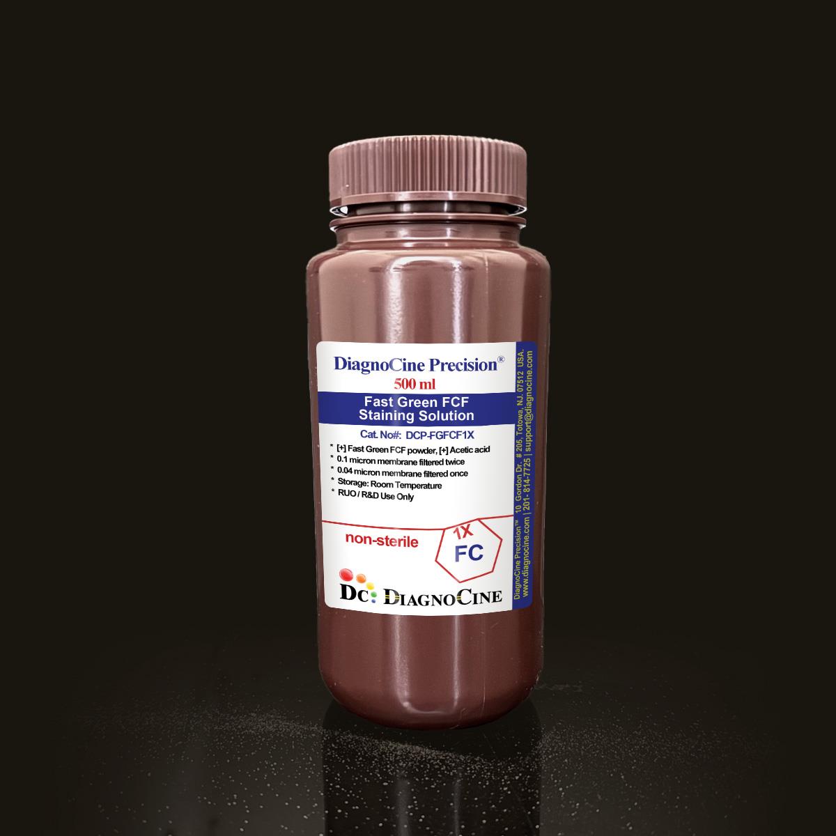
FluxMPS™ Fast Green FCF Staining Solution
An MPS-grade, quadruple-stage filtered Fast Green FCF Staining Solution formulated for brilliant, stable collagen, cytoplasmic, and protein visualization. Filtered 0.1 µm membrane twice and 0.04 µm membrane twice, delivering ultra-clean staining performance for histology, electrophoresis, and imaging workflows built around microfluidic and organ-on-a-chip (OoC) platforms.
- Quadruple-stage filtered — 0.1 µm membrane twice and 0.04 µm membrane twice for ultra-clean staining performance
- Formulated as one of the world’s cleanest reagents for cell and molecular biology experiments
- Deep green, brilliant, and fade-resistant coloration for collagen, cytoplasmic, and protein staining
- Validated free of DNase and RNase activity after 18-hour incubation at room temperature
- Compatible with trichrome histology protocols and SDS-PAGE/IEF protein staining (0.1–20 µg/band)
- Near-infrared fluorescence detection at λmax 624 nm for solvent-free imaging
- Manufactured under ISO 13485-certified, CE-approved quality systems
- Custom concentrations, pH, and additive formulations available on request
- FormulationFast Green FCF 0.1% / Acetic Acid 1%
- AppearanceDeep Green solution
- Fluorescence Emissionλmax 624 nm
- Protein Load Range0.1–20 µg/band
- Filtration0.1 µm x2 + 0.04 µm x2
- DNase ActivityNone detected (18 hr, RT)
- RNase ActivityNone detected (18 hr, RT)
- StorageRoom temperature, protected from light
- Shelf Life6 months
- Size500 mL
Engineered where standard staining reagents fall short
Conventional 0.22 µm-filtered stains carry subvisible particulates that accumulate in microchannels, interfere with fluorescence imaging, and introduce lot-to-lot variability. FluxMPS™ Fast Green FCF Staining Solution is built to eliminate these failure modes at the source.
Microchannel-safe purity
Quadruple-stage filtration (0.1 µm membrane twice and 0.04 µm membrane twice) delivers a final 0.04 µm polish that keeps microchannels and imaging surfaces free of particulate interference.
Precise, reproducible staining
A defined 0.1% Fast Green FCF / 1% acetic acid formulation produces consistent, brilliant green coloration across histology and electrophoresis runs.
Ultrapure-grade water
Prepared with Ultrapure Type 1 water (18.2 MΩ·cm) to minimize background and ionic interference in sensitive staining and imaging protocols.
Low background for imaging & assays
Near-infrared emission at λmax 624 nm supports sensitive fluorescence detection without organic solvent interference.
Defined, traceable composition
Lot-released Fast Green FCF powder and acetic acid content, validated free of DNase and RNase activity after 18-hour incubation at room temperature.
Customization on demand
Alternate concentrations, chemical or compound additions, and pH modifications are available on request.
Quadruple-stage filtration system
Fast Green FCF Staining Solution is processed through a four-stage filtration sequence — 0.1 µm membrane filtration twice, followed by 0.04 µm membrane filtration twice — to remove particulates and bioburden that a single-pass 0.22 µm filter cannot capture.
-
1
0.1 µm Pre-filtration I
Removes large particulate and aggregates, extending the life of downstream filters.
-
2
0.04 µm Pre-filtration II
Retains fine particulates and bioburden ahead of final sterile filtration.
-
3
0.1 µm Sterile-filtration I
A second 0.1 µm pass adds redundancy ahead of final polishing.
-
4
0.04 µm Sterile-filtration II — Final Polish
A second 0.04 µm pass provides the ultimate polish supporting an ISO Class 5 aseptic fill.
Performance vs. conventional buffer
Sequential 0.1 µm and 0.04 µm filtration removes finer particulates than a single 0.22 µm pass used in conventional staining solutions, supporting cleaner backgrounds for microscopy, electrophoresis, and chip-based imaging.

© Diagnocine® — DCP-FGFCF1X
Where Fast Green FCF Staining Solution performs
From classic trichrome histology to quantitative protein electrophoresis, Fast Green FCF Staining Solution delivers brilliant, stable coloration across a wide range of biochemical and histological workflows.
Automated Bioreactors & Robotics
For automated liquid-handling and robotic staining platforms, an optional 0.01 µm (10 nm) ultra-filtered variant is available to further reduce particulate load in fine-bore valves and sensors.
- Total Particulate Exclusion — minimizes subvisible particulate reaching automated dispensing systems
- Valve & Sensor Protection — reduces particulate accumulation in fine-bore microvalves and optical sensors
- Extended Perfusion Stability — supports consistent staining performance across automated, high-throughput runs
Inquiry Required: the 0.01 µm (10 nm) ultra-filtered grade is available by request — contact support@diagnocine.com.
Micro Physiological System (MPS) & Chip
Ultra-clean formulation supports staining of tissue sections and models processed within chip-based and microfluidic platforms.
Wash, Dilution & Reconstitution
Ultrapure, particulate-controlled formulation supports clean handling steps upstream of staining and detection.
iPSC-Derived Model Handling
Low-background formulation supports histological staining of iPSC-derived tissue models.
Endothelial & Primary Cell Perfusion
Compatible with collagen and cytoplasmic staining of vascular and primary cell tissue sections.
ELISA, Blotting & Blocking
Linear, quantitative counterstain compatible with electrophoresis, blotting, and multicolor staining workflows.
Microscopy & Optical Sensing
Near-infrared emission at λmax 624 nm supports sensitive imaging without organic solvent interference.
Full specification sheet
All parameters below reflect values stated for this Fast Green FCF Staining Solution lot.
| Parameter | Specification |
|---|---|
| Formulation / Composition | Fast Green FCF powder 0.1%, Acetic Acid 1% |
| Appearance | Deep Green solution |
| Parameter | Specification |
|---|---|
| Sterility USP <71> | Filtered 0.1 µm membrane twice and 0.04 µm membrane twice |
| DNase Activity | None detected after 18-hour incubation with plasmid DNA at room temperature |
| RNase Activity | None detected after 18-hour incubation with ribosomal RNA at room temperature |
| Water Quality | Ultrapure Type 1 water (18.2 MΩ·cm) |
| Fill Environment | ISO Class 5 (Class 100) |
| Parameter | Specification |
|---|---|
| Storage Temperature | Room temperature, away from bright light |
| Shelf Life | 6 months |
| Parameter | Specification |
|---|---|
| Raw Material Grade Ultrapure | World’s cleanest reagent grade for cell and molecular biology |
| Manufacturing QMS ISO 13485 | ISO 13485-certified, CE-approved facilities |
| Traceability | Final packaging, QA, testing, and customization performed at DiagnoCine Precision, Totowa, New Jersey, USA |
| Production Method | Filtered 0.1 µm membrane twice and 0.04 µm membrane twice |
| Intended Use | Research Use Only (RUO) |
Full composition
Fast Green FCF Staining Solution is a two-component formulation released per lot for consistent, reproducible coloration.
| Component | CAS Number | Concentration |
|---|---|---|
| Fast Green FCF powder | 2353-45-9 | 0.1% |
| Acetic Acid | 64-19-7 | 1% |
Manufacturing & compliance
Fast Green FCF Staining Solution is manufactured, filled, and tested under a certified quality management system.
ISO 13485:2016 QMS
Manufactured under ISO 13485-certified and CE-approved facilities (suppliers of DiagnoCine Precision).
Ultrapure Type 1 Water
Formulated with Ultrapure Type 1 water (18.2 MΩ·cm) to minimize background interference.
ISO Class 5 Fill & Finish
Final packaging, quality assurance, and testing performed at the DiagnoCine R&D and Quality Testing Center.
Micro-Batch Precision
Customization requests and assembly are accomplished at DiagnoCine Precision in Totowa, New Jersey, USA.
DNase / RNase Safety Testing
No DNase or RNase activity detected after 18-hour incubation with plasmid DNA and ribosomal RNA, respectively, at room temperature.
Particulate Control USP <788>
Quadruple-stage 0.1 µm and 0.04 µm membrane filtration supports sub-visible particulate control during fill.
Appearance Verification
Each lot is verified to present as a Deep Green solution prior to release.
Documentation / CoA
A Certificate of Analysis documenting appearance, filtration, and DNase/RNase testing is available on request.
How DCP-FGFCF1X compares
A side-by-side look at filtration architecture and quality controls versus conventional staining solutions.
| Parameter | DCP-FGFCF1X (FluxMPS™) | Conventional 0.22 µm-Filtered Stain | Standard Alternative |
|---|---|---|---|
| Filtration architecture | Quadruple-stage (0.1 µm x2 + 0.04 µm x2) | Single-stage | Single-stage |
| Final filtration pore size | 0.04 µm | 0.22 µm | 0.22 µm |
| Number of filtration stages | 4 | 1 | 1 |
| Water quality | Ultrapure Type 1 (18.2 MΩ·cm) | Standard purified water | Standard purified water |
| DNase / RNase activity testing | check_circle | cancel | cancel |
| Manufacturing QMS | ISO 13485:2016 | cancel | cancel |
| CoA availability | check_circle | cancel | cancel |
| Near-infrared fluorescence compatibility | check_circle | cancel | cancel |
| Custom formulation on request | check_circle | cancel | cancel |
| Microfluidic / chip imaging compatibility | check_circle | cancel | cancel |
Frequently asked questions
Common questions about DCP-FGFCF1X Fast Green FCF Staining Solution.
Supporting literature
Curated literature relevant to Fast Green FCF staining, histology, protein electrophoresis, and reagent purity control.
- Bancroft JD, Layton C. The Hematoxylins and Eosin, in Theory and Practice of Histological Techniques. Churchill Livingstone. doi:10.1016/B978-0-7020-4226-3.00010-X
- Titford M. Progress in the development of microscopical techniques for diagnostic pathology. J Histotechnol. doi:10.1179/his.2009.32.1.9
- Chiarugi V, et al. Masson-Goldner trichrome variants for collagen visualization in fibrotic tissue. Histochem J. doi:10.1023/A:1003682114032
- Steinberg TH. Protein gel staining methods: an introduction and overview. Methods Enzymol. doi:10.1016/S0076-6879(09)63024-3
- Wong C, et al. Quantitative protein staining in SDS-PAGE using anionic dyes. Electrophoresis. doi:10.1002/elps.1150151102
- Neuhoff V, et al. Improved staining of proteins in polyacrylamide gels. Electrophoresis. doi:10.1002/elps.1150090103
- Huh D, et al. Reconstituting organ-level lung functions on a chip. Science. doi:10.1126/science.1188302
- Bhatia SN, Ingber DE. Microfluidic organs-on-chips. Nat Biotechnol. doi:10.1038/nbt.2989
- Sutherland R, et al. Sterile filtration and mycoplasma control in cell culture reagents. In Vitro Cell Dev Biol Anim. doi:10.1007/s11626-014-9803-3