

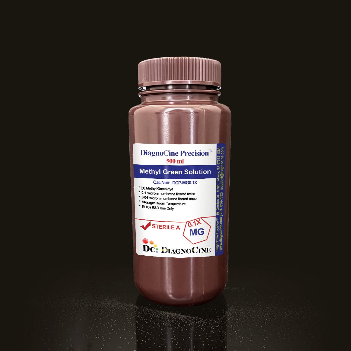
Methyl Green Solution
FluxMPS™ Methyl Green Solution
FluxMPS™ Methyl Green Solution (DCP-MG0.1X) is a ready-to-use, 0.1% w/v aqueous cationic triphenylmethane dye engineered for nuclear counterstaining, DNA/RNA differentiation, and far-red fluorescent DNA imaging across histology, cytology, and molecular biology workflows. The formulation is quadruple-stage ultra-filtered and chloroform-purified to remove crystal violet contaminants, delivering consistent, low-background staining performance for automated, microfluidic, and organ-on-a-chip (OoC) sample-processing platforms.
- Sterile, Ultrapure, quadruple-stage filtered: 0.1-micron membrane twice and 0.04-micron membrane twice
- 0.1% w/v Methyl Green dye, chloroform-extracted to remove crystal violet contaminants
- Far-red fluorescence when bound to DNA (excitation: 633 nm; emission: 677 nm) for confocal imaging
- pH-dependent color transition (yellow at pH 0.1 to blue at pH 2.3) supports precise visual monitoring
- Non-intercalating, AT-rich electrostatic DNA binding preserves nucleic acid integrity
- Compatible with aqueous and non-aqueous (resin-based) mounting media
- Formulated with Ultrapure Type 1 water and manufactured under ISO 13485-certified, CE-approved facilities
- Custom concentrations, additives, and pH modifications available on request
- Formulation0.1% w/v Methyl Green dye, aqueous
- AppearanceBlue-green liquid
- Fluorescence (Ex/Em)633 nm / 677 nm
- pH Color TransitionYellow at pH 0.1 to blue at pH 2.3
- Filtration System0.1 µm x2 + 0.04 µm x2
- DNase ActivityNone detected (18 hr, RT)
- RNase ActivityNone detected (18 hr, RT)
- Water QualityUltrapure Type 1 (18.2 MΩ·cm)
- StorageRoom temperature, protected from light
- Shelf Life24 months unopened
Engineered where standard staining reagents fail
Conventional 0.22 µm-filtered staining solutions can carry subvisible particulates and dye contaminants that accumulate in microchannels, elevate fluorescence background, and compromise nuclear contrast. FluxMPS™ Methyl Green Solution is purified, filtered, and quality-controlled to remove these failure points before they reach the slide or the chip.
Microchannel-safe purity
Quadruple-stage 0.1 µm / 0.04 µm filtration removes particulates that would otherwise obstruct microfluidic channels and automated staining lines.
Purified dye chemistry
Chloroform-extraction removes crystal violet contaminants from the Methyl Green dye, ensuring consistent 0.1% w/v staining quality lot to lot.
Ultrapure-grade water
Formulated with Ultrapure Type 1 water (18.2 MΩ·cm) aligned with USP <85>, reducing background contamination in sensitive assays.
Low background for imaging & assays
Far-red fluorescence (excitation 633 nm; emission 677 nm) when bound to DNA supports high-resolution confocal imaging with minimal spectral overlap.
Defined, traceable composition
Lot-released 0.1% w/v Methyl Green dye content with documented DNase- and RNase-free quality control testing.
Customization on demand
Other concentrations, additions of chemicals, compounds, proteins, or supplements, and different pH formulations are available on inquiry.
Quadruple-stage filtration system
FluxMPS™ Methyl Green Solution is filtered through a four-stage sequence — 0.1-micron membrane filtration twice followed by 0.04-micron membrane filtration twice — to deliver sterile, ultrapure staining performance and guard against mycoplasma contamination.
-
1
0.1 µm Pre-filtration I
Removes large particulates and aggregates from the aqueous Methyl Green formulation, extending the service life of downstream filters.
-
2
0.04 µm Pre-filtration II
Retains fine particulates and bioburden ahead of the final sterile-filtration passes.
-
3
0.1 µm Sterile-filtration I
Provides a second-pass redundancy that reinforces the sterility assurance of the formulation.
-
4
0.04 µm Sterile-filtration II — Final Polish
Delivers the final polish supporting an ISO Class 5 aseptic fill environment and helps prevent mycoplasma contamination; the smallest mycoplasma species measure about 0.2 microns.
Performance vs. conventional buffer
Sequential 0.1 µm and 0.04 µm filtration removes finer particulates than a single 0.22 µm pass used in conventional staining solutions, supporting cleaner microfluidic and automated staining workflows.

© Diagnocine® — DCP-MG0.1X
Nuclear staining, DNA/RNA differentiation & fluorescent imaging
FluxMPS™ Methyl Green Solution supports nuclear counterstaining in histology and cytology, DNA/RNA differentiation, fluorescent imaging, electrophoresis, and educational and diagnostic laboratory use.
Automated Bioreactors & Robotics
Automated staining and dispensing robotics used in microfluidic and organ-on-a-chip workflows are highly sensitive to subvisible particulates that can obstruct valves, sensors, and microchannels. An optional 0.01 µm (10 nm) ultra-filtered variant of this Methyl Green formulation is available for these systems, extending uptime and protecting precision fluid-handling hardware.
- Total Particulate Exclusion — minimizes subvisible particulate load entering automated fluid paths
- Valve & Sensor Protection — reduces the risk of microvalve and optical sensor fouling
- Extended Perfusion Stability — supports longer unattended run times in continuous staining or perfusion setups
Inquiry Required: the 0.01 µm (10 nm) ultra-filtered grade is available upon request; contact support@diagnocine.com to arrange this configuration.
Micro Physiological System (MPS) & Chip
Provides crisp nuclear counterstaining for tissue and cell models processed on microfluidic and organ-on-a-chip platforms.
Wash, Dilution & Reconstitution
Stock solution can be diluted 1:5,000 in PBS for fluorescent whole-mount embryo or tissue staining protocols.
iPSC-Derived Model Handling
Non-intercalating, AT-rich electrostatic DNA binding preserves nucleic acid integrity for nuclear visualization in iPSC-derived models.
Endothelial & Primary Cell Perfusion
Compatible with aqueous and non-aqueous mounting media for nuclear contrast studies in endothelial and primary cell cultures.
ELISA, Blotting & Blocking
Pairs with chromogenic substrates such as DAB in immunohistochemistry (IHC) and in situ hybridization (ISH) workflows.
Microscopy & Optical Sensing
Photostable far-red fluorescence (excitation 633 nm; emission 677 nm) supports live-cell nuclei imaging and thick tissue section confocal microscopy.
Detailed product specifications
Measured and declared values for FluxMPS™ Methyl Green Solution (DCP-MG0.1X).
| Parameter | Specification |
|---|---|
| Formulation / Composition | 0.1% w/v Methyl Green dye, aqueous solution |
| Appearance | Blue-green liquid |
| Dye Concentration | 0.1% |
| pH-Dependent Color Transition | Yellow at pH 0.1 to blue at pH 2.3 |
| Fluorescence Excitation | 633 nm |
| Fluorescence Emission | 677 nm |
| Parameter | Specification |
|---|---|
| Sterility USP <71> | Filtered 0.1-micron membrane twice and 0.04-micron membrane twice |
| DNase Activity | None detected (plasmid DNA, 18 hours, room temperature) |
| RNase Activity | None detected (ribosomal RNA, 18 hours, room temperature) |
| Water Quality | Ultrapure Type 1 water (18.2 MΩ·cm) USP <85> |
| Manufacturing Standard ISO 13485 | ISO 13485-certified, CE-approved facilities |
| Parameter | Specification |
|---|---|
| Storage Temperature | Room temperature, away from bright light |
| Shelf Life | 24 months unopened |
| Usage Note | Use before the expiry date printed on the product label |
| Parameter | Specification |
|---|---|
| Raw Material Grade | Chloroform-extracted, purified Methyl Green dye (crystal violet contaminants removed) |
| Manufacturing QMS ISO 13485 | ISO 13485-certified facilities (suppliers of DiagnoCine Precision) |
| Regulatory Alignment | CE-approved facilities |
| Production Method | Final packaging, QA, and testing at the DiagnoCine R&D and Quality Testing Center; customization and assembly at DiagnoCine Precision, Totowa, New Jersey, USA |
| Intended Use | For Research Use Only (RUO) |
Full composition
Component and concentration values are released per lot as part of the DiagnoCine Precision quality framework.
| Component | CAS Number | Concentration |
|---|---|---|
| Methyl Green dye | 82-94-0 | 0.1% |
Manufacturing & compliance
FluxMPS™ Methyl Green Solution is produced under a documented quality management system with traceable raw materials and controlled fill environments.
ISO 13485:2016 QMS
Manufactured under ISO 13485-certified and CE-approved facilities (suppliers of DiagnoCine Precision).
Ultrapure Type 1 Water
Formulated with Ultrapure Type 1 water (18.2 MΩ·cm) to minimize background contamination in downstream assays.
ISO Class 5 Fill & Finish
Final packaging, quality assurance, and testing are performed at the DiagnoCine R&D and Quality Testing Center.
Micro-Batch Precision
Custom formulation requests and assembly are accomplished at DiagnoCine Precision in Totowa, New Jersey, USA.
Endotoxin USP <85>
Bacterial Endotoxins Test methodology forms part of the routine QC framework for DiagnoCine Precision buffers and staining solutions.
Particulate USP <788> Method 2
Quadruple-stage 0.1 µm / 0.04 µm filtration supports particulate control aligned with Method 2 practices.
Osmolality USP <785>
Osmolality testing methodology is part of the DiagnoCine Precision quality framework where applicable to a given formulation.
Documentation / CoA
Lot-specific documentation is maintained for DNase activity, RNase activity, and appearance testing.
How DCP-MG0.1X compares
A side-by-side look at the FluxMPS™ filtration and quality-control architecture versus conventional staining solutions.
| Parameter | DCP-MG0.1X (FluxMPS™) | Conventional (0.22 µm filtered) | Standard alternative (0.22 µm filtered) |
|---|---|---|---|
| Chloroform-Purified (Crystal Violet Removed) | check_circle | cancel | cancel |
| Final filtration pore size | 0.04 µm | 0.22 µm | 0.22 µm |
| Number of filtration stages | 4 | 1 | 1 |
| DNase / RNase Activity Testing | check_circle | cancel | cancel |
| ISO 13485 / CE-Approved Manufacturing | check_circle | cancel | cancel |
| Water quality | Ultrapure Type 1 (18.2 MΩ·cm) | Standard purified water | Standard purified water |
| Microfluidic / automated staining compatibility | check_circle | cancel | cancel |
| Custom formulation available | check_circle | cancel | cancel |
Frequently asked questions
Common questions about FluxMPS™ Methyl Green Solution (DCP-MG0.1X).
Supporting literature
Curated references relevant to Methyl Green staining chemistry, fluorescence imaging, and microfluidic/organ-on-a-chip applications.
- Kapuscinski, J. "DAPI: a DNA-specific fluorescent probe." Biotechnic & Histochemistry. doi:10.3109/10520299509064050
- Darzynkiewicz, Z. et al. "Methyl green as a fluorescent probe for chromatin condensation." Cytometry. doi:10.1002/cyto.990030109
- Kasten, F. H. "The Chemistry of Schiff's Reagent and related staining methods." The Journal of Histochemistry and Cytochemistry. doi:10.1177/2.6.1974.353
- Bukhari, S. et al. "Methyl green-pyronin staining: a review of applications in cytology." Journal of Cytology. doi:10.4103/0970-9371.126650
- Bhatia, S. N. & Ingber, D. E. "Microfluidic organs-on-chips." Nature Biotechnology. doi:10.1038/nbt.2989
- Huh, D. et al. "From 3D cell culture to organs-on-chips." Trends in Cell Biology. doi:10.1016/j.tcb.2011.09.005
- Zanetti-Domingues, L. C. et al. "Fluorescent nuclear dyes for live-cell imaging." Methods in Molecular Biology. doi:10.1007/978-1-4939-2617-6_9
- Puchtler, H. & Waldrop, F. S. "On the mechanism of Methyl Green staining." Histochemistry. doi:10.1007/BF00493389
- Rasmussen, M. et al. "Filtration and sterility assurance in cell-based assay reagents." SLAS Technology. doi:10.1177/2472630318804090