

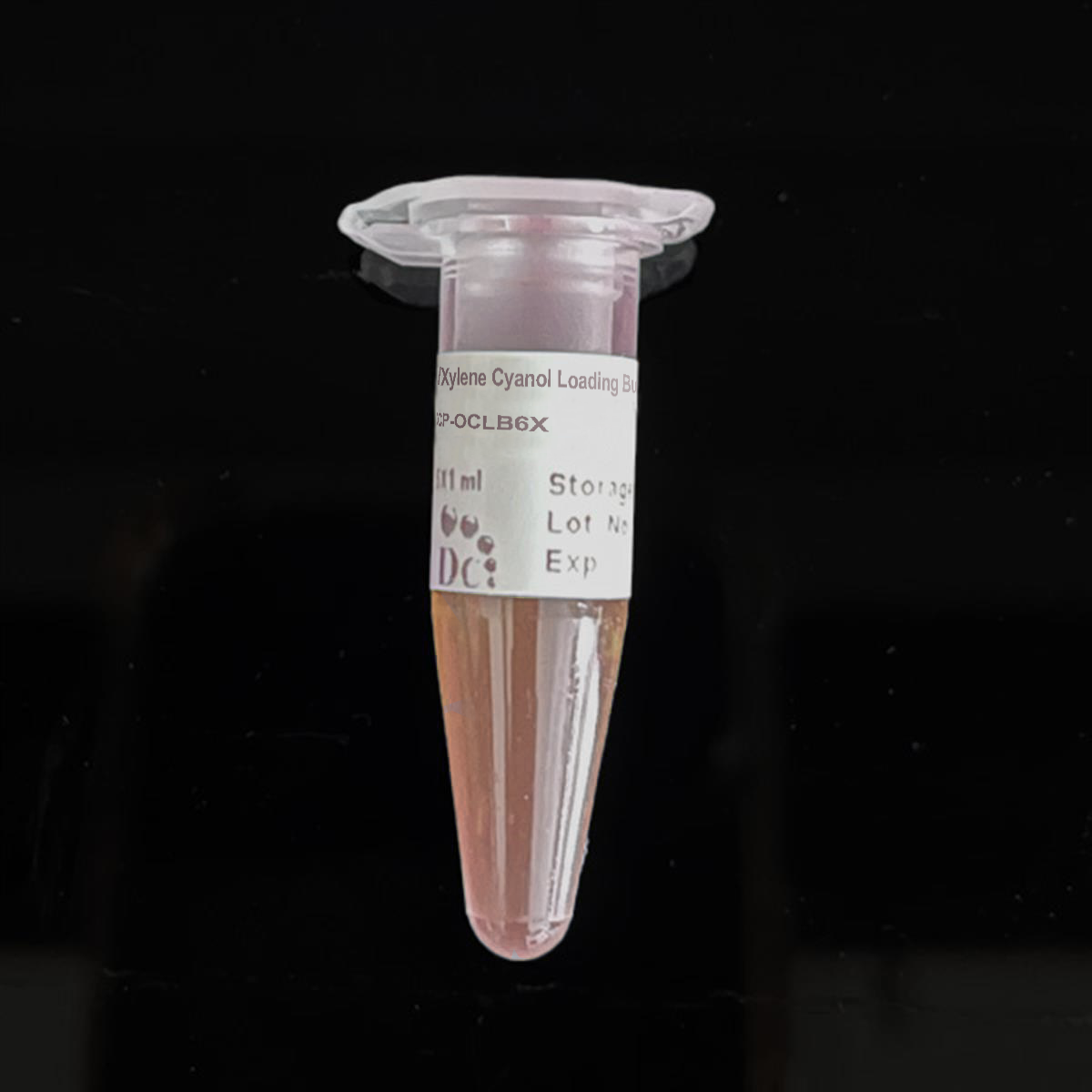
Orange-G/Xylene Cyanol Loading Buffer [6X]
FluxMPS™ Orange-G/Xylene Cyanol Loading Buffer [6X]
FluxMPS™ Orange-G/Xylene Cyanol Loading Buffer [6X] is an ultra-filtered, sterile gel loading dye engineered for DNA and RNA electrophoresis workflows. Formulated at a stable pH of 7.6 with defined Tris-HCl and EDTA concentrations, it pairs dual-dye visual tracking with nuclease-free purity for dependable nucleic acid analysis in molecular biology and microfluidic gel platforms.
- Filtered 0.1 micron membrane twice and 0.04 micron membrane twice for ultra-clean, nuclease-free purity
- Formulated at pH 7.6 with 10 mM Tris-HCl and 60 mM EDTA for stable nucleic acid protection
- Dual tracking dyes, Orange G and Xylene Cyanol FF, span approximately 50 bp to 4000 bp for visual migration monitoring
- 30% glycerol ensures reliable sample density and well loading
- Tested free of DNase and RNase activity after 18-hour room-temperature incubation
- Manufactured under ISO 13485-certified, CE-approved facilities
- Custom pH, dye, and concentration formulations available on request
- pH7.6
- Tris-HCl10 mM
- EDTA60 mM
- Glycerol30%
- Orange G0.36%
- Xylene Cyanol FF0.36%
- Filtration0.1 µm x2 + 0.04 µm x2
- SterilityFiltered in sterile environment
- Storage-20°C
- Shelf Life1 year
Engineered where standard loading buffers fall short
Conventional 0.22 µm-filtered loading buffers can carry residual particulates, nuclease contamination, and inconsistent pH that compromise band resolution and sample integrity. FluxMPS™ Orange-G/Xylene Cyanol Loading Buffer [6X] is built to eliminate those failure modes at the source.
Nuclease-free, ultra-filtered purity
Sequential 0.1 µm and 0.04 µm membrane filtration removes finer particulates than a single-pass 0.22 µm filter, supporting clean, artifact-free electrophoresis.
Precise, stable pH
Formulated at pH 7.6 with 10 mM Tris-HCl and 60 mM EDTA to maintain nucleic acid integrity throughout sample loading and run time.
Ultrapure-grade water
Prepared with Ultrapure Type 1 water (18.2 MΩ·cm), aligned to USP <85> water-quality expectations.
Low background dye tracking
Orange G and Xylene Cyanol FF provide dual-color migration tracking across approximately 50 bp to 4000 bp without obscuring downstream band visualization.
Defined, traceable composition
Every component — glycerol, dyes, Tris-HCl, and EDTA — is present at a stated, lot-consistent concentration.
Customization on demand
Alternate concentrations, tracking dyes, or pH values can be formulated on request.
Quadruple-stage filtration system
DCP-OCLB6X is filtered 0.1-micron membrane twice and 0.04-micron membrane twice, a four-pass architecture that supports the ultra-clean, nuclease-free purity required for reliable nucleic acid electrophoresis.
- 1
0.1 µm Pre-filtration I
Removes large particulates and aggregates, extending downstream filter life.
- 2
0.04 µm Pre-filtration II
Retains fine particulates and bioburden, including mycoplasma-scale contaminants (the smallest mycoplasma types are approximately 0.2 microns).
- 3
0.1 µm Sterile-filtration I
Second-pass redundancy for consistent sterility assurance.
- 4
0.04 µm Sterile-filtration II — Final Polish
Final polish performed in a sterile environment prior to fill.
Performance vs. conventional buffer
Sequential 0.1 µm and 0.04 µm filtration removes finer particulates than a single 0.22 µm pass typical of conventional loading buffers, supporting mycoplasma-scale contamination prevention.

© Diagnocine® — DCP-OCLB6X
Where DCP-OCLB6X performs
Formulated for agarose gel electrophoresis and nucleic acid sample preparation, with purity suited to sensitive downstream molecular biology and microfluidic workflows.
Automated Bioreactors & Robotics
An optional 0.01 µm (10 nm) ultra-filtered variant can be formulated for automated liquid-handling and robotic gel-loading platforms where microvalves and optical sensors are especially sensitive to particulate buildup.
- Total Particulate Exclusion
- Valve & Sensor Protection
- Extended Perfusion Stability
Inquiry Required: contact support@diagnocine.com to request the 0.01 µm ultra-filtered grade.
Micro Physiological System (MPS) & Chip
Supports nucleic acid loading and tracking in microfluidic gel electrophoresis and chip-based analysis platforms.
Wash, Dilution & Reconstitution
Convenient pre-mixed 6X format for dilution and loading of DNA and RNA samples prior to electrophoresis.
iPSC-Derived Model Handling
Used for genotyping and verification of iPSC-derived lines via nucleic acid gel electrophoresis.
Endothelial & Primary Cell Perfusion
Supports plasmid and transfection verification workflows in endothelial and primary cell models.
ELISA, Blotting & Blocking
Applicable to nucleic acid gel electrophoresis and blotting workflows preceding Southern and Northern blot analysis.
Microscopy & Optical Sensing
Dual-dye tracking complements imaging-based nucleic acid quantification and downstream optical analysis.
Full specification sheet
Parameters as measured and declared for DCP-OCLB6X.
| Parameter | Specification |
|---|---|
| Appearance | Blue color |
| pH (USP <791>) | 7.6 |
| Tris-HCl concentration | 10 mM |
| EDTA concentration | 60 mM |
| Glycerol | 30% |
| Orange G | 0.36% |
| Xylene Cyanol FF | 0.36% |
| Parameter | Specification |
|---|---|
| Sterility USP <71> | Filtered 0.1 micron membrane twice and 0.04 micron membrane twice in a sterile environment |
| DNase activity | None detected after incubation with plasmid DNA for 18 hours at room temperature |
| RNase activity | None detected after incubation with ribosomal RNA for 18 hours at room temperature |
| Manufacturing standard ISO 13485 | ISO 13485-certified and CE-approved facilities |
| Parameter | Specification |
|---|---|
| Storage temperature | -20°C |
| Shelf life | 1 year |
| Parameter | Specification |
|---|---|
| Manufacturing QMS ISO 13485 | ISO 13485-certified and CE-approved facilities (Suppliers of DiagnoCine Precision) |
| Regulatory alignment | CE-approved |
| Production method | Final packaging, quality assurance, and testing at the DiagnoCine R&D and Quality Testing Center; customization and assembly at DiagnoCine Precision, Totowa, New Jersey, USA |
| Intended use | Research Use Only (RUO) |
Full composition
Every component of the DCP-OCLB6X 6X loading buffer formulation, listed with concentration as stated in the product record.
| Component | CAS Number | Concentration |
|---|---|---|
| Glycerol | 56-81-5 | 30% |
| Orange G | 1936-15-8 | 0.36% |
| Xylene Cyanol FF | 2650-17-1 | 0.36% |
| Tris-HCl | 1185-53-1 | 10 mM |
| EDTA | 60-00-4 | 60 mM |
Manufacturing & compliance
DCP-OCLB6X is manufactured, filled, and tested under a controlled quality system.
ISO 13485:2016 QMS
Manufactured under ISO 13485-certified, CE-approved facilities (Suppliers of DiagnoCine Precision).
Ultrapure Type 1 Water
Prepared with Ultrapure Type 1 water (18.2 MΩ·cm).
Sterile Fill & Finish
Final packaging, quality assurance, and testing performed at the DiagnoCine R&D and Quality Testing Center.
Custom Assembly Precision
Customization requests and assembly completed at DiagnoCine Precision in Totowa, New Jersey, USA.
DNase/RNase Testing
No DNase activity detected after incubation with plasmid DNA for 18 hours at room temperature; no RNase activity detected after incubation with ribosomal RNA for 18 hours at room temperature.
Sterility USP <71>
Filtered 0.1 micron membrane twice and 0.04 micron membrane twice in a sterile environment.
pH Verification
Formulated and verified at pH 7.6.
Documentation / CoA
Certificate of Analysis available on request, covering appearance, pH, sterility, and DNase/RNase testing.
How DCP-OCLB6X compares
A side-by-side look at filtration architecture, testing, and formulation flexibility.
| Parameter | DCP-OCLB6X (FluxMPS™) | Conventional loading buffer | Standard alternative |
|---|---|---|---|
| Final filtration pore size | 0.04 µm | 0.22 µm | 0.22 µm |
| Number of filtration stages | 4 | 1 | 1 |
| DNase/RNase tested | check_circle | cancel | cancel |
| Sterility tested (USP <71>) | check_circle | cancel | check_circle |
| ISO 13485 manufacturing | check_circle | cancel | cancel |
| Dual-color dye tracking | check_circle | check_circle | cancel |
| Custom formulation available | check_circle | cancel | cancel |
Frequently asked questions
Common questions about DCP-OCLB6X.
Supporting literature
Selected references relevant to nucleic acid gel electrophoresis and loading buffer chemistry.
- Sambrook J, Russell DW. Molecular Cloning: A Laboratory Manual. Agarose gel electrophoresis of DNA. doi:10.1101/pdb.prot4017
- Lee PY, Costumbrado J, Hsu CY, Kim YH. Agarose gel electrophoresis for the separation of DNA fragments. J Vis Exp. 2012. doi:10.3791/3923
- Voytas D. Agarose gel electrophoresis. Curr Protoc Mol Biol. 2000. doi:10.1002/0471142727.mb0205as51
- Ausubel FM, et al. Short Protocols in Molecular Biology. Buffer systems for nucleic acid electrophoresis. doi:10.1002/0471142727
- Green MR, Sambrook J. The basics of agarose gel electrophoresis. Cold Spring Harb Protoc. 2019. doi:10.1101/pdb.top100404
- Nordstrom JL. EDTA as a chelating agent in nuclease inhibition and nucleic acid stabilization. Anal Biochem. doi:10.1016/0003-2697(85)90506-9
- Stellwagen NC. Electrophoresis of DNA in agarose gels, polyacrylamide gels and in free solution. Electrophoresis. 2009. doi:10.1002/elps.200900052
- Rasmussen HB. Restriction fragment analysis and gel electrophoresis. Methods Mol Biol. doi:10.1007/978-1-60327-411-1_18
- Uphoff CC, Drexler HG. Detection of mycoplasma contamination in cell cultures. Methods Mol Biol. 2011. doi:10.1007/978-1-61779-108-6_1
- Kim S, et al. Microfluidic gel electrophoresis for nucleic acid analysis. Lab Chip. doi:10.1039/C8LC00458G