

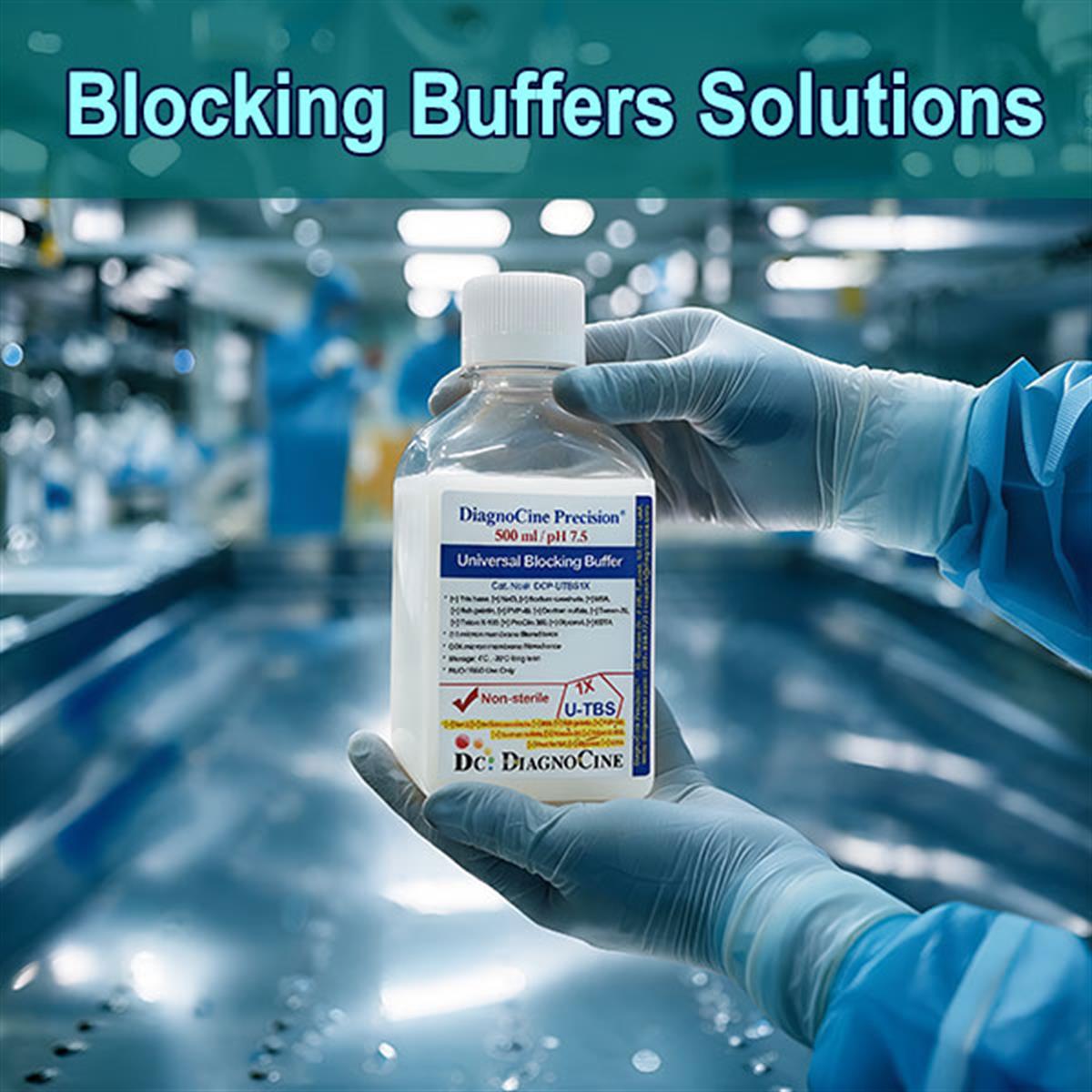
Blocking Buffers — A Selection Guide for the Research Laboratory
Seven blockers, one page. Go straight to the product you already know you need — or read on to match a blocker to your detection chemistry.
| Blocking Buffer | Blocker / Composition | Primary Applications | Product Page |
|---|---|---|---|
| Nonfat Milk Based Blocking Buffer | Milk proteins · not protein-free | Economical, robust general-purpose blocker for standard chemiluminescent Western blots | Viewarrow_forward |
| Casein Based Blocking Buffers | Purified milk protein fraction · not protein-free | Cleaner and more reproducible than whole milk, low particulate background for blots and ELISA | Viewarrow_forward |
| BSA Based Blocking Buffers | Single purified protein (bovine serum albumin) · not protein-free | Standard for phospho-specific detection, gentle, good ELISA and antibody-diluent base | Viewarrow_forward |
| Gelatin Based Blocking Buffers | Denatured collagen, often fish-skin · not protein-free | Non-mammalian option lowers cross-reactivity with anti-mammalian antibodies, displaces weakly bound proteins on membranes | Viewarrow_forward |
| PVP Based Blocking Buffers | Polyvinylpyrrolidone (synthetic) · protein-free | No cross-reactivity with anti-animal antibodies, useful for nucleic acid hybridization and as an additive | Viewarrow_forward |
| FluxMPS™ Universal Blocking Buffer | Optimized blend · protein-free status varies | Broad format compatibility, reduces optimization time, reproducible across blots and plates | Viewarrow_forward |
| Blocking Buffer Test Set | Multi-buffer assessment kit | Side-by-side screening to find the best signal-to-noise for a given antibody pair | Viewarrow_forward |
Not sure which blocker? Match the blocker to your detection system · Compare all seven blocking agents · See the blocking step by assay · Read the FAQ
What a blocking buffer actually does — and why the choice is not interchangeable
A blocking buffer saturates the unoccupied protein-binding sites on a solid phase (nitrocellulose, PVDF, or polystyrene) after the target has been immobilized, so that antibodies and detection reagents bind only their intended target. The goal is signal-to-noise, not blocking for its own sake. The central practical fact is that no single blocker is optimal for every antibody-antigen pair, and the correct choice is driven by the detection chemistry and the target protein, not by cost or habit [4,5].
- Seven options stocked: nonfat milk, casein, BSA, gelatin, PVP, a universal blend, and a Test Set for side-by-side screening.
- Phospho-specific work goes to BSA, because the phosphoproteins in milk and casein directly raise the background.
- Biotin-streptavidin and lectin work avoid milk, because milk contributes endogenous biotin and glycoproteins.
- Fluorescent Western blotting avoids milk for background reasons and favors fish gelatin, BSA, or a purpose-made fluorescent blocker.
- If the secondary antibody is anti-mammalian, a non-mammalian blocker (fish gelatin or protein-free PVP) reduces cross-reactivity.
- On polystyrene ELISA plates, milk and casein are the strongest blockers, while hydrolyzed gelatin performs poorly [5]. On membranes, gelatin and detergent-based approaches are more competitive.
- Pair detergent with protein: adding 0.05 to 0.1 percent Tween 20 to wash and incubation buffers lowers background [2], but detergent alone can produce false positives.
- Do not over-block. Excess blocker or detergent can strip low-affinity antigen and weaken the specific band.
- Blocking options in this category7
- Protein-free optionPVP based
- Anti-phospho (pSer/pThr/pTyr)BSA — avoid milk, casein
- Biotin / streptavidin or avidinBSA, PVP — avoid milk
- Lectin or glycoprotein probingBSA, PVP — avoid milk
- Near-infrared fluorescent Westernfish gelatin, BSA — avoid milk
- Standard chemiluminescent Westernnonfat milk, casein
- ELISA on polystyrenecasein, milk
- Typical ELISA block1–3 % protein, 1 h RT or o/n 4 °C
- Typical Western block5 % milk or 3–5 % BSA in TBST/PBST
The interference modes below are chemical, not preferences
Each failure mode comes from a specific chemical conflict between the blocker and the detection system. Knowing which one applies to your assay is what prevents most failed blots.
Phosphoprotein background
Milk and casein are phosphoproteins and generate high phospho background. Anti-phospho antibodies (pSer / pThr / pTyr) therefore go to BSA.
Endogenous biotin
Milk carries endogenous biotin that saturates streptavidin. Biotin / streptavidin and avidin systems use BSA or PVP instead.
Glycoprotein cross-reaction
Milk glycoproteins react with lectins. Lectin or glycoprotein probing uses BSA or PVP.
Fluorescent background
Milk raises fluorescent background. Near-infrared fluorescent Western blotting uses fish gelatin, BSA, or a dedicated fluorescent blocker.
Secondary-antibody species
Where the secondary antibody is anti-mammalian, a non-mammalian blocker — fish gelatin or protein-free PVP — avoids secondary cross-reactivity.
Surface chemistry
On polystyrene ELISA plates, milk and casein block plastic most effectively while hydrolyzed porcine gelatin blocks poorly [5]. On membranes, gelatin and detergent-based approaches are more competitive.
Each rule on this page traces to a published comparison
The blocker recommendations above are not house preferences. They come from the foundational method papers and reviews listed at the foot of this page, in the order those papers appeared.
-
1
1979 The solid phase that made blocking necessary
Towbin, Staehelin, and Gordon described the electrophoretic transfer of proteins from polyacrylamide gels to nitrocellulose sheets [1]. Once a target is immobilized on a membrane, the unoccupied binding sites around it have to be saturated — which is the job a blocking buffer does.
-
2
1982 Detergent enters the picture
Batteiger, Newhall, and Jones reported the use of Tween 20 as a blocking agent for proteins transferred to nitrocellulose [2]. This is the origin of the 0.05 to 0.1 percent Tween 20 that still appears in wash and incubation buffers today.
-
3
1984 Nonfat dry milk becomes the workhorse
Johnson, Gautsch, Sportsman, and Elder published the improved nonfat dry milk technique for proteins and nucleic acids transferred to nitrocellulose [3]. Milk became the economical general-purpose blocker — and, later, the reason purified casein or BSA is preferred where lot-to-lot reproducibility matters.
-
4
1985 Blockers are shown not to be interchangeable
Spinola and Cannon demonstrated that different blocking agents cause variation in the immunologic detection of proteins on nitrocellulose [4]. This is the direct basis for the rule that no universal best blocker exists and that each antibody-antigen pair must be validated.
-
5
1987 The plate is measured separately from the membrane
Vogt and colleagues quantified how various proteins perform as blocking agents on ELISA microtiter plates [5]. Their results are why milk and casein are named for polystyrene, and why hydrolyzed gelatin is called out as a poor plate blocker.
-
6
2017–2020 Blocking as one controlled variable
Contemporary reviews place blocking inside a validated end-to-end workflow rather than treating it as a step to optimize in isolation [6,7,8]. For quantitative fluorescent Western blotting in particular, follow the validated workflow rather than tuning the block alone [7].
All seven options, side by side
Composition, whether the blocker is protein-free, what it is good at, and — the column most often skipped — what it must not be paired with.
| Blocking agent | Composition / mechanism | Protein-free | Strengths | Avoid with |
|---|---|---|---|---|
| Nonfat milk (“Blotto”) | Complex mixture of milk proteins | No | Economical, robust general-purpose blocker for standard chemiluminescent Western blots | Anti-phospho antibodies, biotin-streptavidin systems, lectin / glycoprotein probes, fluorescent detection |
| Casein based | Purified milk protein fraction | No | Cleaner and more reproducible than whole milk, low particulate background for blots and ELISA | Anti-phospho antibodies (casein is a phosphoprotein), biotin systems |
| BSA based | Single purified protein (bovine serum albumin) | No | Standard for phospho-specific detection, gentle, good ELISA and antibody-diluent base | Anti-BSA cross-reactivity; verify BSA grade for residual IgG / contaminants |
| Gelatin based | Denatured collagen, often fish-skin | No | Non-mammalian option lowers cross-reactivity with anti-mammalian antibodies, displaces weakly bound proteins on membranes | Polystyrene ELISA plates, where hydrolyzed gelatin blocks poorly [5] |
| PVP based | Polyvinylpyrrolidone (synthetic) | Yes | No cross-reactivity with anti-animal antibodies, useful for nucleic acid hybridization and as an additive | Standalone use where strong blocking is needed; usually combined with detergent or protein |
| Universal blocking buffer | Optimized blend | Varies | Broad format compatibility, reduces optimization time, reproducible across blots and plates | Applications needing a defined single-component background |
| Blocking Buffer Test Set | Multi-buffer assessment kit | n/a | Side-by-side screening to find the best signal-to-noise for a given antibody pair | Not applicable; this is the optimization tool itself |
Where blocking sits in each workflow
Select an assay format to see where the blocking step falls and the conditions the source specifies for it.
- Block after antigen coating
- 1 to 3 percent protein blocker
- 1 h at room temperature or overnight at 4 °C
- Milk and casein block plastic most effectively; avoid hydrolyzed porcine gelatin [5]
- Block membrane after transfer
- Typically 5 percent milk or 3 to 5 percent BSA in TBST or PBST
- 1 h at room temperature
- Nonfat milk or casein for standard chemiluminescent detection; fish gelatin, BSA, or a dedicated fluorescent blocker for near-infrared fluorescent detection
- Block after antigen retrieval
- Normal serum matched to the secondary antibody host species, plus BSA
- Block endogenous biotin and peroxidase separately when relevant
- Pretreat conjugate pad and membrane to control nonspecific capture
The table that prevents most failed blots
Find your detection system in the first column, then use the blocker named in “Use” and keep the one in “Avoid” out of the workflow.
| Detection system | Use | Avoid | Reason |
|---|---|---|---|
| Anti-phospho antibodies (pSer / pThr / pTyr) | BSA | Milk, casein | Milk and casein are phosphoproteins and generate high phospho background |
| Biotin / streptavidin or avidin | BSA, PVP | Milk | Milk carries endogenous biotin that saturates streptavidin |
| Lectin or glycoprotein probing | BSA, PVP | Milk | Milk glycoproteins react with lectins |
| Near-infrared fluorescent Western | Fish gelatin, BSA, dedicated fluorescent blocker | Milk | Milk raises fluorescent background |
| Standard chemiluminescent Western | Nonfat milk, casein | (none specific) | Economical and effective for routine targets |
| Anti-mammalian secondary antibody | Fish gelatin, PVP | Cross-reactive mammalian-protein blockers | Non-mammalian blocker avoids secondary cross-reactivity |
| ELISA on polystyrene | Casein, milk | Hydrolyzed porcine gelatin | Milk and casein block plastic most effectively [5] |
Frequently asked questions
The questions that come up most often when a blocking protocol meets a new antibody.
Foundational papers and reviews
The published record behind every recommendation on this page, reproduced as supplied with the source description.
Foundational
- Towbin H, Staehelin T, Gordon J (1979). Electrophoretic transfer of proteins from polyacrylamide gels to nitrocellulose sheets: procedure and some applications. Proc Natl Acad Sci USA 76:4350–4354.
- Batteiger B, Newhall WJ, Jones RB (1982). The use of Tween 20 as a blocking agent in the immunological detection of proteins transferred to nitrocellulose membranes. J Immunol Methods 55:297–307.
- Johnson DA, Gautsch JW, Sportsman JR, Elder JH (1984). Improved technique utilizing nonfat dry milk for analysis of proteins and nucleic acids transferred to nitrocellulose. Gene Anal Tech 1:3–8.
- Spinola SM, Cannon JG (1985). Different blocking agents cause variation in the immunologic detection of proteins transferred to nitrocellulose membranes. J Immunol Methods 81:161–165.
- Vogt RF Jr, Phillips DL, Henderson LO, Whitfield W, Spierto FW (1987). Quantitative differences among various proteins as blocking agents for ELISA microtiter plates. J Immunol Methods 101:43–50.
Reviews
- Bass JJ, Wilkinson DJ, Rankin D, Phillips BE, Szewczyk NJ, Smith K, Atherton PJ (2017). An overview of technical considerations for Western blotting applications to physiological research. Scand J Med Sci Sports 27:4–25.
- Pillai-Kastoori L, Schutz-Geschwender AR, Harford JA (2020). A systematic approach to quantitative Western blot analysis. Anal Biochem 593:113608.
- Pillai-Kastoori L, Heaton S, Shiflett SD, Roberts AC, Solache A, Schutz-Geschwender AR (2020). Antibody validation for Western blot: by the user, for the user. J Biol Chem 295:926–939.