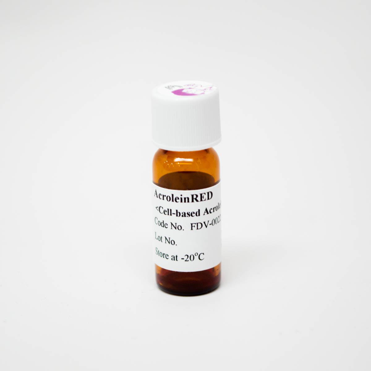
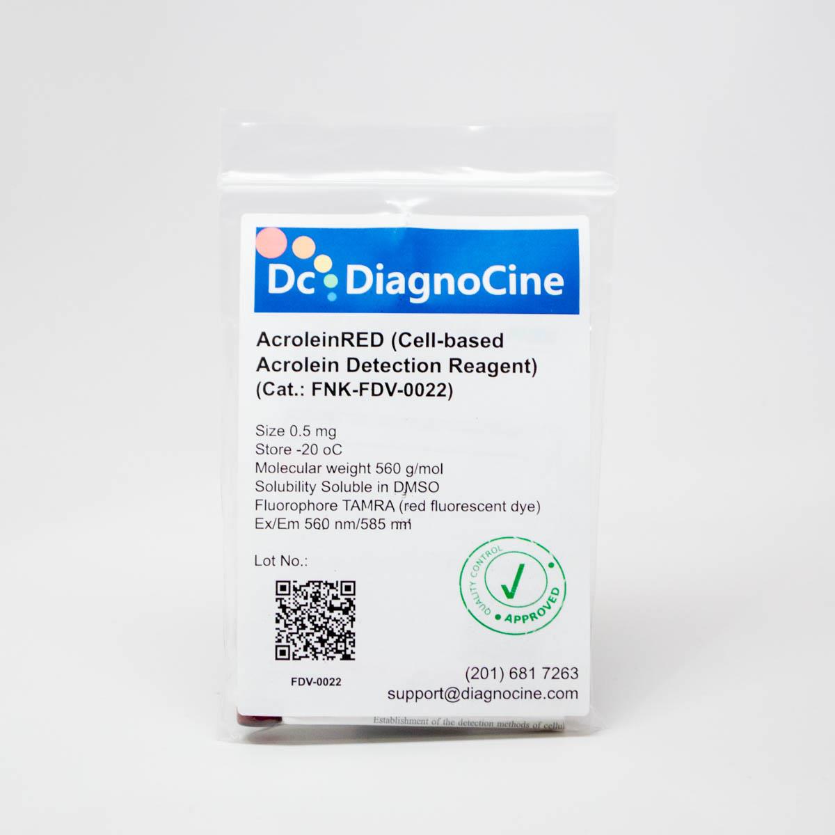
AcroleinRED (Cell-based Acrolein Detection Reagent)
AcroleinRED (Cell-based Acrolein Detection Reagent)
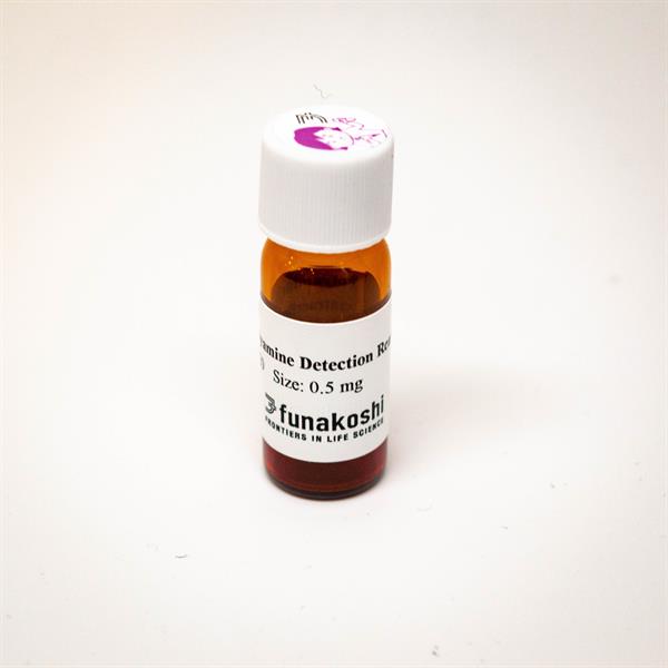
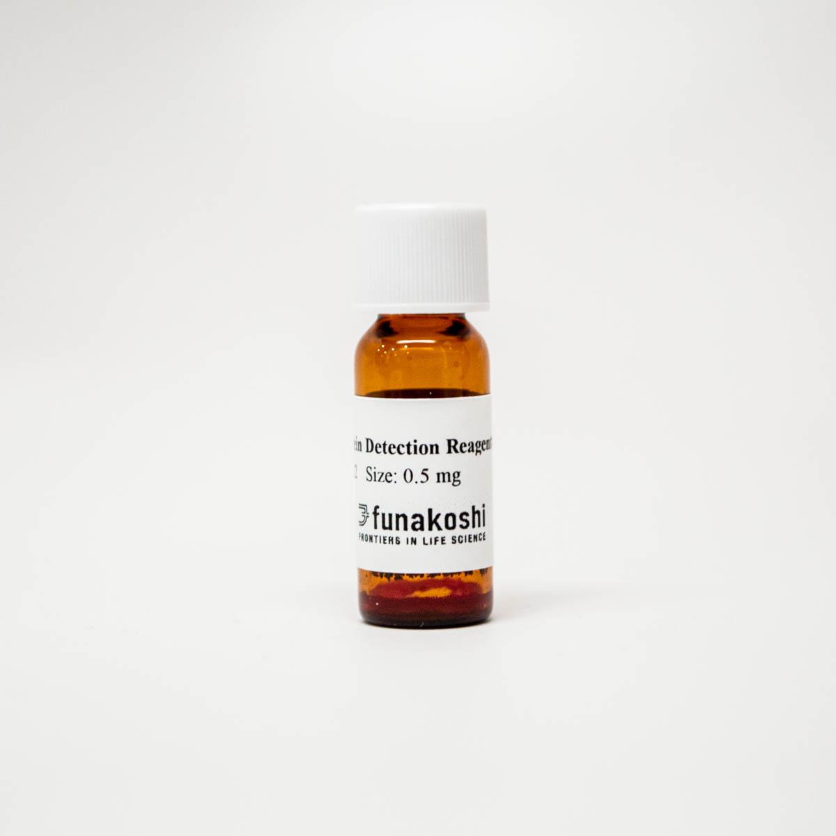
Cat.No. FNK-FDV-0022
Size 0.5 mg
Store -20 oC
Catalog Number FDV-0022
Molecular weight 560 g/mol
Solubility Soluble in DMSO
Fluorophore TAMRA (red fluorescent dye)
Ex/Em 560 nm/585 nm
Description
The world's first Acrolein Detection Reagent in Live Cells

Acrolein, which is found in a common dietary and environmental pollutant, is known as a highly toxic metabolite for cells. Not only in outer environment, but also acrolein is endogenously gererated in cells, especially under the oxidative stress condition.
Acrolein has being researched for more than half-century and many detection methods were developed, such as fluorometric method, mass spectromy or antibody based detection method, etc.
However, conventional methods are not appropriate for the detection in live cells and show poor selectivity and sensitivity.
Our AcroleinRED is the world first cell-based acrolein-detection reagent in live cells condition without any pre-treatment and cell lysis.
This reagent is based on novel phenylazide-acrolein click chemistry discovered by Drs. Katsunori Tanaka and Ambara Pradipta (Ref.1).
AcroleinRED specifically react with either extracellular acrolein conjugate released from cell surface lipids or intracellular acrolein generated via enzymatic pathway and label acrolein with TAMRA fluorophore.
In case of extracellular labeling, TAMRA-labeled acrolein is immediately incorporated into cells through the endocytosis pathway. Subsequently, the TAMRA-labeled acrolein conjugate reacts with biomolecules such as proteins and remain in the cells.


This product has been commercialized with the support of Biofunctional Synthetic Chemistry Laboratory, Cluster for Pioneering Research, RIKEN.

Application
- Semi-quantification of cellular acrolein
- Estimation of effects of any drugs on cellular acrolein production
- Staining of ex vivo tissues under non-fixed condition
NOTE AcroleinRED cannot be applied into localization study of intracellular acrolein.
Reconstitution and Storage
Reconstitution:
stock solution in 100% DMSO.
Storage (solution) :
Store powder at -20oC. After reconstitution in DMSO, aliquot and store at -20 °C. Avoid repeated freeze-thaw cycles. Protect from light.
Features
- Specifically labels acrolein
- High sensitivity
Can be detected by filter set for Rhodamine (Ex./Em. = 560 / 585 nm)
Cell permeable : compatible with live cell imaging
No pretreatments : Just add, incubate and wash.
Can do semi-quantification of total acrolein amount by intracellular fluorescence intensity.
Chemical Information
- Molecular weight : 560 g/mol
- Solubility : Soluble in DMSO
- Fluorophore : TAMRA (red fluorescent dye)

Example Data
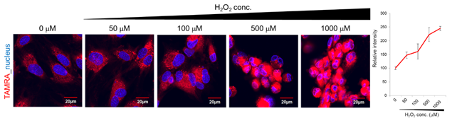
HUVECs were pretreated with 0-1000 μM H2O2 for 2 hours and subsequently treated with 10 μM AcroleinRED for 30 min. Right after labeling, cells were washed, stained with hoechest and observed under live cell condition. In the absence of H2O2, the acrolein endogenously produced by HUVECs was observed.
Intracellular TAMRA signals were increased in H2O2 dose-dependent manner compared with the endogenous acrolein level.
Note: Ref.1 confirmed AcroleinRED did not react with H2O2 directly. Please refer to Ref.1 for the detail of the experiments.

Fig.2 Observation of reactive oxygen species (ROS)-induced acrolein production
HUVECs were treated with 25 μM menadione, an inducer of reactive oxygen species, for 0-60 min and subsequently treated with Total ROS detection dye (Enzo Life Science) and AcroleinRED for 60 min. After labeling, fluorescent signal of ROS (green) or acrolein (red) was observed.
By the addition of menadione, ROS were immediately increased. In contrast, the acrolein level started to increase 60 min after menadione treatment. Namely, the late stage production of acrolein through ROS-initiated process was clearly imaged by using AcroleinRED.
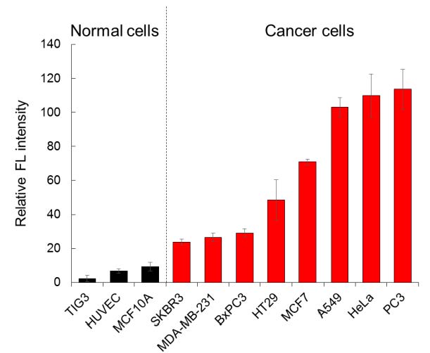
Comparison of acrolein-production levels of various cell lines by AcroleinRED
Three non-cancer cells (TIG4, HUVEC and MCF10A) and eight cancer cell lines were treated with 22.5 ?M of AcroleinRED for 30 min at 37oC. Red fluorescent signals of cancer cells were much higher than that of normal cells. Furthermore, AcroleinRED revealed that acrolein-production levels of eight cancer cell lines are significantly different from each other.
|
|
|

Visualization of tumors from breast gland tissues
Surgical tissues derived from breast cancer patients or a healthy control were immediately incubated with AcroleinRED (20 ?M)/Hoechst mixed solution under non-fixed condition for 5 min. After washing tissues by PBS, double-stained tissues were observed by fluorescent microscopy. AcroleinRED visualized tumor region of patient-derived breast gland, but not derived from healthy control.
IDC; invasive ductal carcinoma,
DCIS; ductal carcinoma in situ, NBG; normal breast gland

* Above two data are cited from 2
Reference
- A.R. Paradipt, M. Taichi, I. Nakase, E. Saigitbatalova, A. Kurbangalieva, S. Kitazume, N. Taniguchi, K. Tanaka, ACS Sens., 1, 623-632 (2016) Uncatalyzed Click Reaction between Phenyl Azides and Acrolein: 4-Formyl-1,2,3-Triazolines as “Clicked” Markers for Visualizations of Extracellular Acrolein Released from Oxidatively Stressed Cells.
- T. Tanei, A. R. Pradipta, K. Morimoto, M. Fujii, M. Arata, A. Ito, M. Yoshida, E. Saigitbatalova, A.
Kurbangalieva, J. Ikeda, E. Morii, S. Noguchi, and K. Tanaka, Adv. Sci., 1801479 (2018) Cascade reaction in
human live tissue allows clinically applicable diagnosis of breast cancer morphology. - Miyamoto H.D., Ikeda M., Ide T., Tadokoro T., Furusawa S., Abe K., Ishimaru K., Enzan N., Sada M., Yamamoto T., Matsushima S., Koumura T., Yamada K., Imai H., Tsutsui H., (2022) Iron Overload via Heme Degradation in the Endoplasmic Reticulum Triggers Ferroptosis in Myocardial Ischemia-Reperfusion Injury. JACC Basic Transl Sci. 7(8):800-819.
- Mao Z. er al., (2024) Hydrogen sulfide protects against toxicant acrolein-induced ferroptotic cell death in Sertoli cells. Front Pharmacol. 15:1440147.
- Lai S. et al., (2024) Effect of nonsense-mediated mRNA decay factor SMG9 deficiency on premature aging in zebrafish. Commun Biol. 7(1):654.
- McNamara, K. M., Sierra, J. C., Latour, Y. L., Hawkins, C. V., Asim, M., Williams, K. J., ... & Wilson, K. T. (2024). Spermine oxidase promotes Helicobacter pylori-mediated gastric carcinogenesis through acrolein production. Oncogene, 44(5), 296–306.
- Gobert, A. P., Hawkins, C. V., McNamara, K. M., & Wilson, K. T. (2025). Alterations in polyamine metabolism induced by the pathogen Helicobacter pylori: implications for gastric inflammation and carcinogenesis. Methods in Enzymology, 715, 137–153.
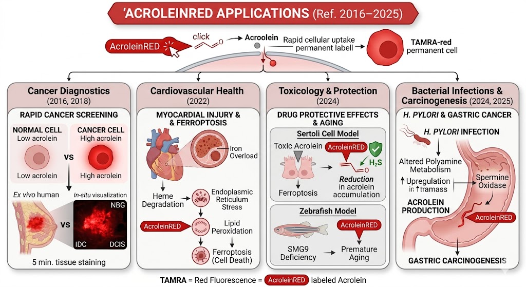
Here is a summary of how AcroleinRED was applied in the cited research papers:
1. Discovery and Visualization of Oxidative Stress (2016, 2018)
-
Visualizing Extracellular Acrolein: The initial study established a "click chemistry" method to label acrolein with a TAMRA fluorophore. This allowed researchers to visualize acrolein released from cell surface lipids during oxidative stress.
-
Cancer Diagnosis: It was discovered that cancer cells produce significantly higher levels of acrolein than normal cells. This property was applied to breast cancer diagnosis, where AcroleinRED could distinguish tumor regions in fresh human tissue within just 5 minutes.
2. Ferroptosis and Myocardial Injury (2022)
-
Myocardial Ischemia-Reperfusion: Researchers used the reagent to detect acrolein as a marker of oxidative damage and ferroptosis (a type of programmed cell death) triggered by iron overload in heart tissues.
3. Protective Mechanism and Toxicity Studies (2024)
-
Protective Effects of $H_2S$: In studies involving Sertoli cells, AcroleinRED was used to measure how Hydrogen Sulfide ($H_2S$) reduces the accumulation of toxic acrolein, thereby protecting cells from ferroptotic death.
-
Aging Models: It served as a tool to monitor oxidative stress levels in zebrafish models to study the effects of SMG9 deficiency on premature aging.
4. Gastric Carcinogenesis and Bacterial Infection (2024, 2025)
-
H. pylori Infection: The reagent was instrumental in proving that Helicobacter pylori induces gastric cancer through the production of acrolein via the spermine oxidase pathway.
-
Polyamine Metabolism: It was applied to map the spatial distribution and concentration of acrolein resulting from altered polyamine metabolism during chronic gastric inflammation and tumor development.