TBS Blocking Buffer with PVP
FluxMPS™ TBS Blocking Buffer with PVP
FluxMPS™ TBS Blocking Buffer with PVP is a sterile, ultrapure, 0.1 µm membrane-filtered blocking buffer built on a defined 50 mM Tris-Base / 150 mM Sodium Chloride system with 1% PVP-40. It delivers a non-protein, low-autofluorescence block for Western blotting, ELISA, and IHC workflows, including microfluidic and organ-on-a-chip (OoC) sample handling where a clean, particulate-controlled buffer matters.
- Sterile, ultrapure, filtered through a 0.1 µm membrane once for particulate and bioburden control
- Formulated with 1% PVP-40, 50 mM Tris-Base, and 150 mM Sodium Chloride at pH 7.5
- Non-protein PVP-40 blocking agent avoids protein-protein interference and shows low autofluorescence
- Tested with no detectable DNase or RNase activity after 18 hr incubation at room temperature
- Tris-based formulation is compatible with alkaline phosphatase detection systems where phosphate-containing PBS would interfere
- Custom concentrations, additives, pH, and formulation modifications available on inquiry
- pH7.5
- Tris-Base50 mM
- Sodium Chloride150 mM
- PVP-401%
- SterilityFiltered 0.1 µm membrane once
- AppearanceClear, colorless liquid
- DNase ActivityNone detected
- RNase ActivityNone detected
- Storage4°C
- Shelf Life9 months
Engineered where standard blocking buffers fall short
Conventional blocking buffers filtered only through a 0.22 µm membrane, or based on protein blockers like milk or BSA, can carry particulates, introduce interfering proteins, or add autofluorescent background. FluxMPS™ TBS Blocking Buffer with PVP is built to remove those variables from sensitive immunoassay and microfluidic workflows.
Microchannel-safe purity
A single 0.1 µm membrane filtration pass, performed in a sterile environment, yields a clear, colorless buffer suited to particulate-sensitive microfluidic channels.
Precise, stable pH
A defined pH 7.5 buffering system built on 50 mM Tris-Base and 150 mM Sodium Chloride supports consistent blocking conditions across incubation steps.
Ultrapure-grade water
Manufactured with Ultrapure Type 1 water (18.2 MΩ·cm) consistent with USP <85> laboratory water quality expectations.
Low background for imaging & assays
PVP-40 exhibits very low autofluorescence, minimizing background noise in fluorescence detection channels used in confocal and biosensor assays.
Defined, traceable composition
Every lot is formulated to the same 1% PVP-40 / 50 mM Tris-Base / 150 mM Sodium Chloride specification with documented pH, DNase, and RNase results.
Customization on demand
Alternate concentrations, additional chemicals, compounds, proteins, supplements, or a different pH can be produced on inquiry.
Single-stage 0.1 µm filtration
DCP-PVPTBS1X is finished with a single 0.1 µm membrane filtration pass in a sterile environment, giving this TBS blocking buffer with PVP a clear, colorless, sterile finish suited to immunoassay and microfluidic sample handling.
-
1
0.1 µmSterile-filtration
A single pass through a 0.1 µm membrane, performed in a sterile environment, retains particulates and bioburden before final packaging.
Performance vs. conventional buffer
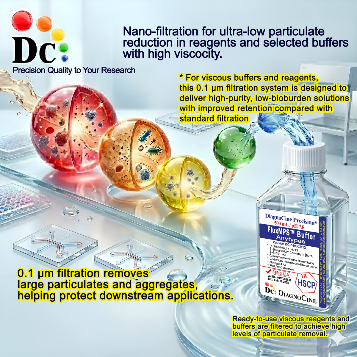
A 0.1 µm membrane pass retains particulates larger than 0.1 µm — finer than the 0.22 µm membranes commonly used for laboratory blocking buffers — supporting lower particulate carryover into sensitive immunoassay and microchannel workflows.
© Diagnocine® — DCP-PVPTBS1X
Where DCP-PVPTBS1X is used
TBS Blocking Buffer with PVP reduces non-specific binding and background staining across Western blotting, ELISA, and immunohistochemistry, and its non-protein, low-autofluorescence chemistry extends its usefulness into imaging and microfluidic sample workflows.
Automated Bioreactors & Robotics
For automated bioreactor and robotics-integrated workflows, an optional ultra-filtered 0.01 µm (10 nm) variant of this buffer can be produced on request for facilities requiring the finest available particulate exclusion ahead of valves, sensors, and perfusion lines.
- Total Particulate Exclusion: finer membrane options for automated fluidic systems
- Valve & Sensor Protection: reduced particulate load protects precision fluidic components
- Extended Perfusion Stability: cleaner input buffer supports longer unattended run times
Inquiry Required: the 0.01 µm ultra-filtered grade is produced to order — contact support@diagnocine.com to request this configuration.
Micro Physiological System (MPS) & Chip
A clear, sterile, particulate-controlled blocking buffer suited to immunostaining steps within microfluidic chip-based platforms.
Wash & Blocking Steps
The Tris-buffered saline base supports use as a wash buffer between blocking and antibody incubation steps in immunodetection protocols.
iPSC-Derived Model Immunostaining
PVP-40's non-protein chemistry blocks non-specific binding during immunostaining of iPSC-derived cell models without adding interfering protein background.
Endothelial & Primary Cell Immunodetection
Blocks non-specific antibody binding sites in endothelial and primary cell cultures ahead of marker staining.
ELISA, Blotting & Blocking
Used to block non-specific binding sites on membranes and microplates, improving specificity in Western blotting, ELISA, and IHC, and is particularly suited to detecting small proteins where protein-based blockers can interfere.
Microscopy & Optical Sensing
PVP-40's very low autofluorescence minimizes background noise in fluorescence detection channels used in confocal microscopy and biosensor assays.
Full specification sheet
Measured and declared values for DCP-PVPTBS1X, as reported by DiagnoCine Quality Control.
| Parameter | Specification |
|---|---|
| Formulation | 1% PVP-40, 50 mM Tris-Base, 150 mM Sodium Chloride |
| Appearance | Clear, colorless liquid |
| pH (USP <791>) | 7.5 |
| Tris-Base concentration | 50 mM |
| Sodium Chloride concentration | 150 mM |
| PVP-40 concentration | 1% |
| Parameter | Specification |
|---|---|
| Sterility (USP <71>) | Filtered 0.1 µm membrane once in a sterile environment |
| DNase Activity | None detected (18 hr incubation, room temperature) |
| RNase Activity | None detected (18 hr incubation, room temperature) |
| Water Quality | Ultrapure Type 1 water (18.2 MΩ·cm) |
| Manufacturing Standard ISO | ISO 13485-certified, CE-approved facilities |
| Parameter | Specification |
|---|---|
| Storage temperature | 4°C |
| Shelf life | 9 months |
| Parameter | Specification |
|---|---|
| Raw material grade | Ultrapure |
| Manufacturing QMS ISO | ISO 13485-certified and CE-approved (DiagnoCine Precision suppliers) |
| Production & assembly location | Final packaging, QA, and testing at the DiagnoCine R&D and Quality Testing Center; customization and assembly at DiagnoCine Precision, Totowa, New Jersey, USA |
| Intended use | Research Use Only (RUO) |
Full composition
Every lot of DCP-PVPTBS1X is formulated to the ingredient and concentration specification below.
| Component | CAS Number | Concentration |
|---|---|---|
| PVP-40 (Polyvinylpyrrolidone) | 9003-39-8 | 1% |
| Tris-Base | 77-86-1 | 50 mM |
| Sodium Chloride | 7647-14-5 | 150 mM |
Manufacturing & compliance
DCP-PVPTBS1X is manufactured, packaged, and quality tested under a controlled quality system.
ISO 13485:2016 QMS
Manufactured under ISO 13485-certified and CE-approved facilities (Suppliers of DiagnoCine Precision).
Ultrapure Type 1 Water
Formulated with Ultrapure Type 1 water (18.2 MΩ·cm).
Sterile, Filtered Fill
Filtered through a 0.1 µm membrane once in a sterile environment prior to fill.
Micro-Batch Precision
Final packaging, QA, and testing performed at the DiagnoCine R&D and Quality Testing Center; customization and assembly completed at DiagnoCine Precision in Totowa, New Jersey, USA.
DNase & RNase Testing
No DNase or RNase activity was detected after 18 hr incubation of plasmid DNA or ribosomal RNA with this product at room temperature.
Sterility (USP <71>)
Filtered 0.1 µm membrane once in a sterile environment.
Manufacturing Platform
DiagnoCine Precision's sterile buffer manufacturing platform is equipped to apply 0.1 µm filtration twice and 0.04 µm filtration twice across its buffer catalog to help exclude mycoplasma contamination, the smallest species of which measures about 0.2 µm, with filtration architecture configured per product specification.
Documentation / CoA
A Certificate of Analysis reflecting lot-specific appearance, pH, sterility, and DNase/RNase results is available on request.
How DCP-PVPTBS1X compares
A side-by-side look at DCP-PVPTBS1X against conventional TBS buffer and protein-based blocking buffers.
| Parameter | DCP-PVPTBS1X (FluxMPS™) | Conventional 0.22 µm-filtered TBS buffer (no PVP) | Protein-based blocking buffer (milk/BSA) |
|---|---|---|---|
| Non-protein PVP-40 blocking agent | check_circle | cancel | cancel |
| Final filtration pore size | 0.1 µm | 0.22 µm | Not typically filtered |
| Number of filtration stages | 1 | 1 | Not applicable |
| Low autofluorescence | check_circle | cancel | cancel |
| Water quality | Ultrapure Type 1 (18.2 MΩ·cm) | Standard purified water | Standard purified water |
| Manufacturing QMS | ISO 13485:2016 | Not typically certified | Not typically certified |
| Microfluidic / OoC channel compatibility | check_circle | cancel | cancel |
| Custom formulation available | check_circle | cancel | cancel |
Frequently asked questions
Common questions about DCP-PVPTBS1X, TBS Blocking Buffer with PVP.
Supporting literature
Curated literature relevant to TBS/PVP blocking chemistry, immunoassay technique, and microfluidic/organ-on-a-chip applications.
- Mahmood, T. & Yang, P.C. Western blot: technique, theory, and trouble shooting. N Am J Med Sci. 2012. doi:10.4103/1947-2714.100998
- Kurien, B.T. & Scofield, R.H. Western blotting: an introduction. Methods Mol Biol. 2015. doi:10.1007/978-1-4939-2694-7_3
- Hnasko, T.S. & Hnasko, R.M. The Western blot. Methods Mol Biol. 2015. doi:10.1007/978-1-4939-2694-7_1
- Sompuram, S.R. et al. A novel blocking chemistry for reducing nonspecific background in immunohistochemistry. J Histochem Cytochem. 2004. doi:10.1177/002215540405200613
- Huh, D. et al. Reconstituting organ-level lung functions on a chip. Science. 2010. doi:10.1126/science.1188302
- Ronaldson-Bouchard, K. & Vunjak-Novakovic, G. Organs-on-a-chip: a fast track for engineered human tissues in drug development. Cell Stem Cell. 2018. doi:10.1016/j.stem.2018.02.011
- Bhatia, S.N. & Ingber, D.E. Microfluidic organs-on-chips. Nat Biotechnol. 2014. doi:10.1038/nbt.2989
- Lin, J.S. & Lai, E.M. Protein-protein interactions: co-immunoprecipitation. Methods Mol Biol. 2017. doi:10.1007/978-1-4939-7033-9_18