

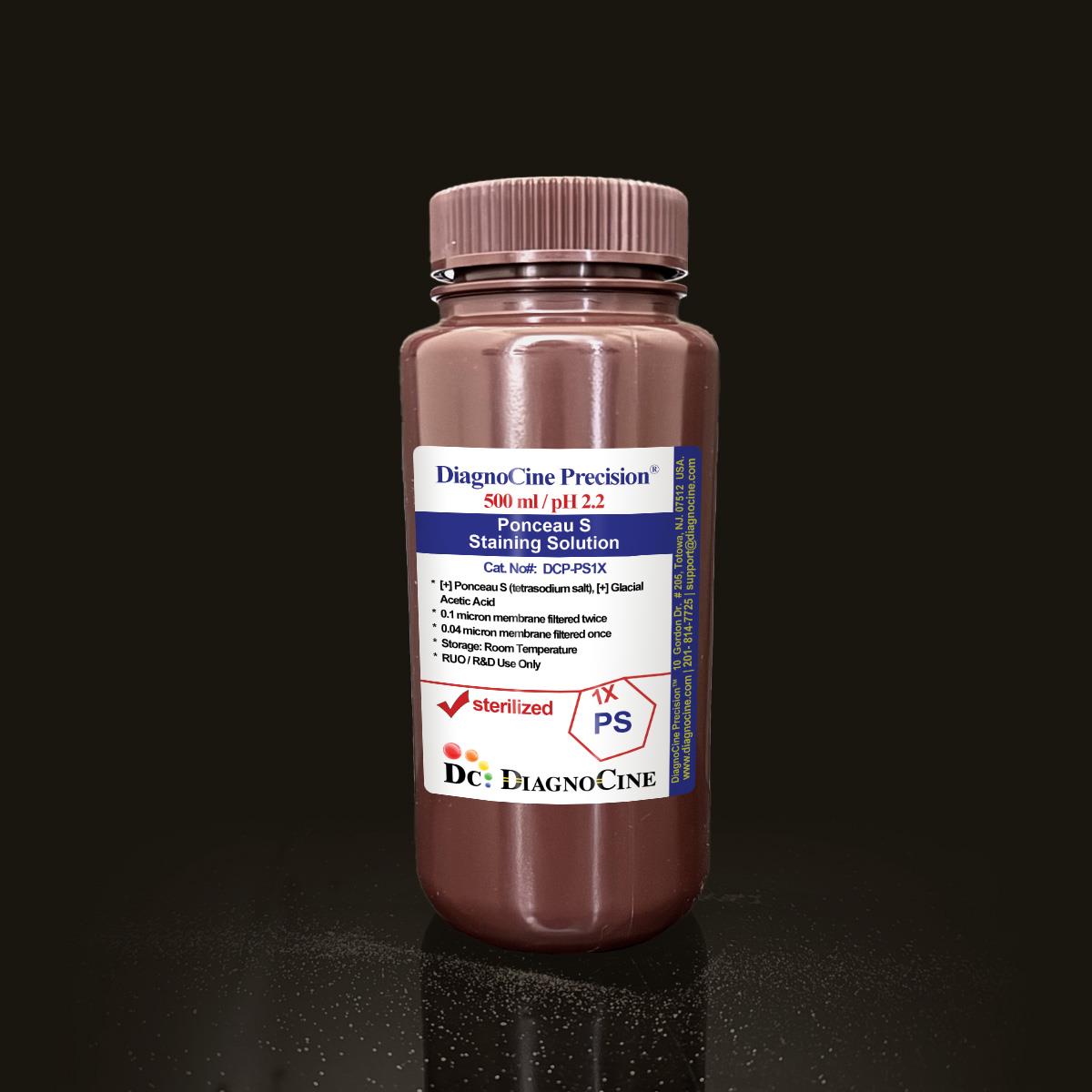
Ponceau S Staining Solution
FluxMPS™ Ponceau S Staining Solution
MPS-grade Ponceau S Staining Solution formulated for rapid, reversible total-protein detection on Western blot membranes. Sterile-filtered through a quadruple-stage 0.1 µm and 0.04 µm membrane architecture and manufactured in an ISO 13485-certified, CE-approved facility, it delivers a consistently clean, low-background stain for protein transfer verification and loading normalization.
- Quadruple-stage filtration: 0.1 µm membrane twice and 0.04 µm membrane twice
- Filtered and finished in a sterile environment to help exclude mycoplasma-scale contaminants (about 0.2 microns)
- pH 2.2, formulated with 0.1% Ponceau S (tetrasodium salt) and 5% glacial acetic acid
- No DNase activity and no RNase activity detected in QC testing
- Ultrapure Type 1 water (18.2 MΩ·cm) base for a clean, low-background working solution
- Compatible with nitrocellulose and PVDF membranes for Western blot workflows
- Rapid, reversible stain removable with water or mild acetic acid for downstream immunoblotting
- Custom concentrations, additives, and pH available on request
- pH2.2
- Formulation0.1% Ponceau S / 5% Acetic Acid
- Filtration0.1 µm x2 + 0.04 µm x2
- SterilityFiltered in sterile environment
- DNase ActivityNone Detected
- RNase ActivityNone Detected
- AppearanceRed Liquid
- StorageRoom Temperature
- Shelf Life12 Months
- Intended UseRUO
Engineered where standard Ponceau S falls short
Conventional single-pass, 0.22 µm-filtered Ponceau S stains can carry residual particulates and inconsistent bioburden control, contributing to background artifacts and membrane-to-membrane variability. FluxMPS™ DCP-PS1X is built for a clean, reproducible working solution from the first use to the last.
Microchannel-safe purity
Quadruple-stage 0.1 µm and 0.04 µm filtration reduces particulate carryover for a cleaner, more consistent working stain.
Defined, stable staining chemistry
A fixed 0.1% Ponceau S (tetrasodium salt) and 5% glacial acetic acid formulation at pH 2.2 supports reproducible band visualization.
Ultrapure-grade water
Formulated with Ultrapure Type 1 water (18.2 MΩ·cm), consistent with USP <85> water-quality expectations.
Low background for imaging & blotting
A clean particulate profile supports crisp, low-background bands against a clear membrane background.
Defined, traceable composition
Every component is disclosed with concentration, and DNase/RNase activity is verified negative on each lot.
Customization on demand
Alternate concentrations, additives, and pH are available — contact support@diagnocine.com.
Quadruple-stage filtration system
DCP-PS1X is filtered 0.1-micron membrane twice and 0.04-micron membrane twice, then filled in a sterile environment — a four-stage architecture designed to keep the working solution clean pass after pass.
-
1
0.1 µm Pre-filtration I
Removes larger particulates and aggregates from the staining solution, protecting downstream filters and membranes.
-
2
0.04 µm Pre-filtration II
Retains finer particulates and bioburden ahead of the final polishing passes.
-
3
0.1 µm Sterile-filtration I
A second 0.1 µm pass provides redundancy in particulate and bioburden reduction.
-
4
0.04 µm Sterile-filtration II — Final Polish
A second 0.04 µm pass performed in a sterile environment provides the final polish, helping exclude mycoplasma-scale contaminants — the smallest mycoplasma types are about 0.2 microns.
Performance vs. conventional staining solution
Sequential 0.1 µm and 0.04 µm filtration removes finer particulates than a single-pass 0.22 µm filtration typical of standard Ponceau S stains, supporting a cleaner working solution for sensitive membrane-based protein detection.

© Diagnocine® — DCP-PS1X
Where DCP-PS1X fits in your workflow
Ponceau S Staining Solution provides a rapid, reversible view of protein transfer quality before committing a membrane to antibody-based detection, and its clean, ultrafiltered formulation supports use across cell and molecular biology workflows, including microfluidic and organ-on-a-chip sample handling.
Automated Bioreactors & Robotics
For automated liquid-handling and robotic Western blot platforms, an optional 0.01 µm (10 nm) ultra-filtered variant is available to further protect fluidic valves and sensors.
- Total Particulate Exclusion for sensitive automated optics and valves
- Valve & Sensor Protection in robotic blot-processing systems
- Extended Perfusion Stability for continuous-flow staining/destaining modules
Inquiry Required: the 0.01 µm ultra-filtered grade is available on request — contact support@diagnocine.com.
Western Blot Transfer Verification
Rapid, reversible staining to visually confirm complete and even protein transfer before immunoblotting.
Protein Sample Quality Assessment
Quick visual check of sample integrity and banding pattern ahead of downstream analysis.
Micro Physiological System (MPS) & Chip Sample Handling
Ultraclean formulation supports low-background staining of protein samples derived from chip-based culture systems.
Protein Loading Normalization
Used to normalize protein loading across lanes prior to detection of the target of interest.
iPSC-Derived Model Protein Screening
Supports quick membrane-level protein checks from iPSC-derived model lysates before targeted detection.
Membrane Orientation & Marker Visualization
Visualizes protein markers to confirm membrane orientation in vascular and primary cell studies.
Detailed product specifications
Measured and declared values for DCP-PS1X, verified as part of routine lot release.
| Parameter | Specification |
|---|---|
| Formulation / Composition | Ponceau S (tetrasodium salt) 0.1%; Glacial Acetic Acid 5% |
| Appearance | Red Liquid |
| pH USP <791> | 2.2 |
| Parameter | Specification |
|---|---|
| Sterility USP <71> | Filtered 0.1 micron membrane twice and 0.04 micron membrane twice in a sterile environment |
| DNase Activity | None detected after incubation of plasmid DNA with this product for 18 hr at room temperature |
| RNase Activity | None detected after incubation of ribosomal RNA with this product for 18 hr at room temperature |
| Water Quality | Ultrapure Type 1 water (18.2 MΩ·cm) |
| Fill Environment | ISO Class 5 (Class 100) |
| Parameter | Specification |
|---|---|
| Storage Temperature | Room temperature |
| Shelf Life | 12 months |
| Parameter | Specification |
|---|---|
| Manufacturing QMS ISO 13485 | ISO 13485-certified and CE-approved facilities (Suppliers of DiagnoCine Precision) |
| Manufacturing Location | Final packaging, quality assurance, and testing at the DiagnoCine R&D and Quality Testing Center; customization and assembly at DiagnoCine Precision, Totowa, New Jersey, USA |
| Regulatory Alignment | ISO 13485:2016 |
| Production Method | Filtered 0.1 micron membrane twice and 0.04 micron membrane twice |
| Intended Use | For Research Use Only (RUO) |
Full composition
Component concentrations for DCP-PS1X, expressed as declared in the formulation record.
| Component | CAS Number | Concentration |
|---|---|---|
| Ponceau S (tetrasodium salt) | 6226-79-5 | 0.1% |
| Glacial Acetic Acid | 64-19-7 | 5% |
Manufacturing & compliance
DCP-PS1X is produced and released under a documented quality system, from raw material control through final fill.
ISO 13485:2016 QMS
Manufactured under ISO 13485-certified and CE-approved facilities (Suppliers of DiagnoCine Precision).
Ultrapure Type 1 Water
Formulated using Ultrapure Type 1 water (18.2 MΩ·cm).
ISO Class 5 Fill & Finish
Final filtration and fill are carried out in a sterile, ISO Class 5 (Class 100) environment.
Traceable Micro-Batch Precision
All final packaging, quality assurance, and testing are completed at the DiagnoCine R&D and Quality Testing Center, with customization and assembly at DiagnoCine Precision, Totowa, New Jersey, USA.
DNase / RNase Verified
No DNase activity detected after 18 hr incubation with plasmid DNA; no RNase activity detected after 18 hr incubation with ribosomal RNA, both at room temperature.
Sterility USP <71>
Filtered 0.1 micron membrane twice and 0.04 micron membrane twice in a sterile environment.
pH Control
Formulated and verified to pH 2.2.
Documentation / CoA
A Certificate of Analysis is available for this lot — contact support@diagnocine.com.
How DCP-PS1X compares
A side-by-side look at DCP-PS1X against a standard 0.22 µm-filtered Ponceau S stain and a Coomassie Blue stain.
| Parameter | DCP-PS1X (FluxMPS™) | Standard Ponceau S (0.22 µm Filtered) | Coomassie Blue Stain |
|---|---|---|---|
| Sensitivity | ~1-10 µg | ~1-10 µg | ~0.1-1 µg |
| Reversibility | check_circle | check_circle | cancel |
| Processing Time | 5-10 minutes | 5-10 minutes | 30+ minutes |
| Detection Limit | 200+ ng | 200+ ng | 50+ ng |
| Final Filtration Pore Size | 0.04 µm | 0.22 µm | Not applicable |
| Number of Filtration Stages | 4 | 1 | Not applicable |
| DNase / RNase Verified | check_circle | cancel | cancel |
| Manufacturing QMS | ISO 13485 & CE | Variable | Variable |
| Custom Formulation Available | check_circle | cancel | cancel |
Frequently asked questions
Common questions about DCP-PS1X Ponceau S Staining Solution.
Supporting literature
Curated references on protein blotting, membrane staining, and microfluidic/organ-on-a-chip sample handling relevant to DCP-PS1X.
- Salinovich O, Montelaro RC. Reversible staining and peptide mapping of proteins transferred to nitrocellulose membranes after separation by sodium dodecylsulfate-polyacrylamide gel electrophoresis. Anal Biochem. 1986. doi:10.1016/0003-2697(86)90600-3
- Romero-Calvo I, et al. Reversible Ponceau staining as a loading control alternative to actin in Western blots. Anal Biochem. 2010. doi:10.1016/j.ab.2010.02.036
- Kurien BT, Scofield RH. Protein blotting: a review. J Immunol Methods. 2003. doi:10.1016/S0022-1759(02)00516-3
- Bass JJ, et al. An overview of technical considerations for Western blotting applications to physiological research. Scand J Med Sci Sports. 2017. doi:10.1111/sms.12702
- Bhattacharya S, et al. Organs-on-chips: into the next decade. Nat Rev Drug Discov. 2021. doi:10.1038/s41573-020-0079-3
- Low LA, et al. Organs-on-chips: into the next decade. Nat Rev Drug Discov. 2021. doi:10.1038/s41573-020-0079-3
- Kurien BT, Scofield RH. Western blotting: an introduction. Methods Mol Biol. 2015. doi:10.1007/978-1-4939-2694-7_17
- Razo-Mejia M, et al. Determination of protein concentration and loading normalization in microfluidic protein assays. Lab Chip. 2018. doi:10.1039/C8LC00393B