No More Sales-NpFlamma® MMP-2,9 675
Packing Unit
-
10 tests

-
50 tests

-
100 tests

NpFlamma® MMP-2,9 675
Cat. No. List below
Description
Specifications
- Fluorophore: Flamma® Fluors 675
- Probe type: Activatable by MMP-2, -9
- Application: In vivo imaging
- Particle size: 250 ± 50 nm
- Excitation/Emission Max.(nm): 683/694
- Appearance: Blue Solid
- Storage conditions: 4 ℃, protect from light
Table 1. NpFlamma® MMP product list
| Quick link (Cat.#) | Series | Ex * (nm) | Em* (nm) | Common filter set |
Excitation source |
| PNM0103 | NpFlamma® MMP-2,9 648 | 648 | 670 | Cy 5 | 594, 633 nm |
| PNM0104 | NpFlamma® MMP-2,9 675 | 683 | 694 | Cy 5.5 | 633, 680 nm |
| PNM0105 | NpFlamma® MMP-2,9 749 | 760 | 778 | Cy 7 | 785 nm |
| PNM0106 | NpFlamma® MMP-2,9 774 | 793 | 810 | Cy 7.5 | 785 nm |
| PNM0101 | NpFlamma® MMP-2,9 ICG | 798 | 835 | Cy 7.5 | 785 nm |
| PNM0203 | NpFlamma® MMP-3,7 648 | 648 | 670 | Cy 5 | 594, 633 nm |
| PNM0204 | NpFlamma® MMP-3,7 675 | 683 | 694 | Cy 5.5 | 633, 680 nm |
| PNM0205 | NpFlamma® MMP-3,7 749 | 760 | 778 | Cy 7 | 785 nm |
| PNM0206 | NpFlamma® MMP-3,7 774 | 793 | 810 | Cy 7.5 | 785 nm |
| PNM0201 | NpFlamma® MMP-3,7 ICG | 798 | 835 | Cy 7.5 | 785 nm |
| PNM0303 | NpFlamma® MMP-13 648 | 648 | 670 | Cy 5 | 594, 633 nm |
| PNM0304 | NpFlamma® MMP-13 675 | 683 | 694 | Cy 5.5 | 633, 680 nm |
| PNM0305 | NpFlamma® MMP-13 749 | 760 | 778 | Cy 7 | 785 nm |
| PNM0306 | NpFlamma® MMP-13 774 | 793 | 810 | Cy 7.5 | 785 nm |
| PNM0301 | NpFlamma® MMP-13 ICG | 798 | 835 | Cy 7.5 | 785 nm |
Overview
BioActs developed NpFlamma® MMP series, MMP-activatable polymeric in vivo fluorescent probes, for early diagnosis and for visualization of overexpressed MMPs related diseases. The probes consist of a fluorescent dye that connected to a quencher through a MMP-cleavable peptide, and a chitosan based nanoparticle (CNP). The one end of peptide is chemically conjugated to a chitosan based nanoparticle. NpFlamma® MMPs are optically silent in their inactivated state yet would be highly fluorescent following MMP-cleaved activation. CNP can selectively accumulate in cancer tissues due to high permeability for loose new blood vessels around cancer tissues and retention effect. The polymeric nanoparticles form self-aggregates size of several hundred nanometers in the aqueous system accumulate only in vicinity of cancer tissues. Thus, CNP enables to bring up the probe to tumor cells, and the cleavage of the peptide by MMPs allows to selective detection of tumor by realizing fluorescence imaging. NpFlamma® MMP series equipped with several different MMPs (MMP-2, -3, -7, -9, -13) cleavable peptides that enable to detect a wide range of diseases. Since self-assembled CNPs have already been used as vehicles for hydrophobic drug delivery, and have shown therapeutic efficacy for mouse tumors. Therefore, the role of
NpFlamma® MMP series might be extended as theranostic agents, which enabling simultaneously monitoring therapeutic responses and delivering therapy. BioActs offers NpFlamma® MMP series as smart fluorescent probes for monitoring MMP-related diseases such as cancer progression, invasion and metastasis, rheumatoid arthritis, pulmonary diseases and areas of cardiovascular disease, and also for evaluating the potential therapeutic efficacy of drugs targeting for these diseases.
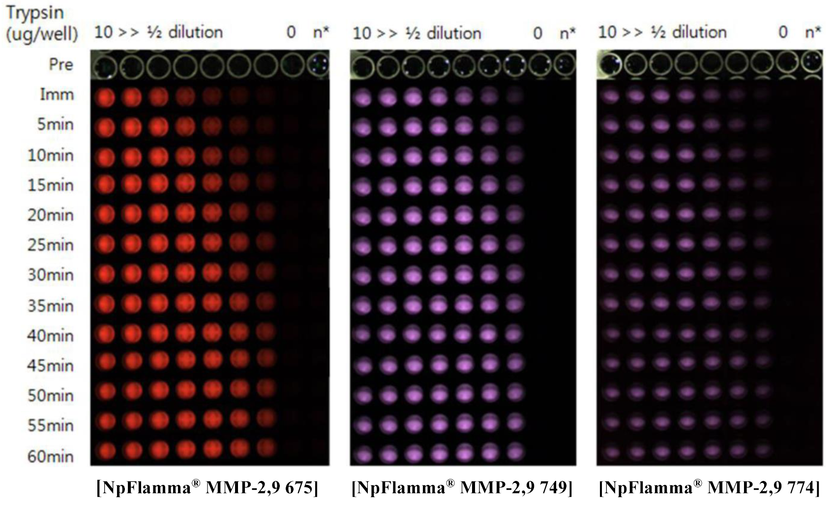
Figure 1. Activation of NpFlamma® MMP series with Trypsin
 |
 |
 |
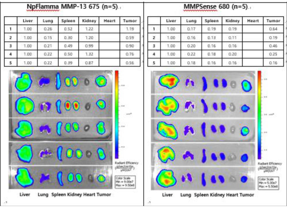 |
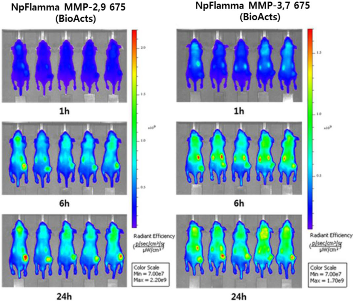
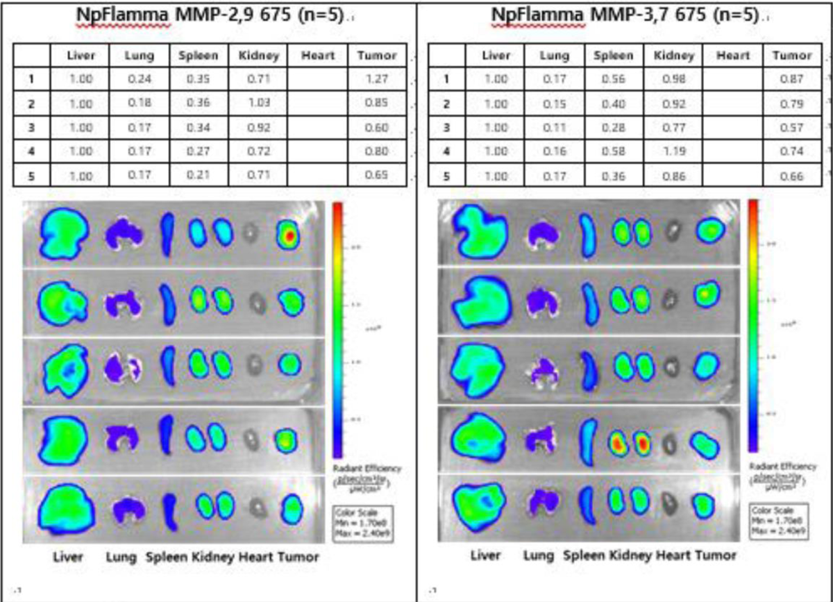
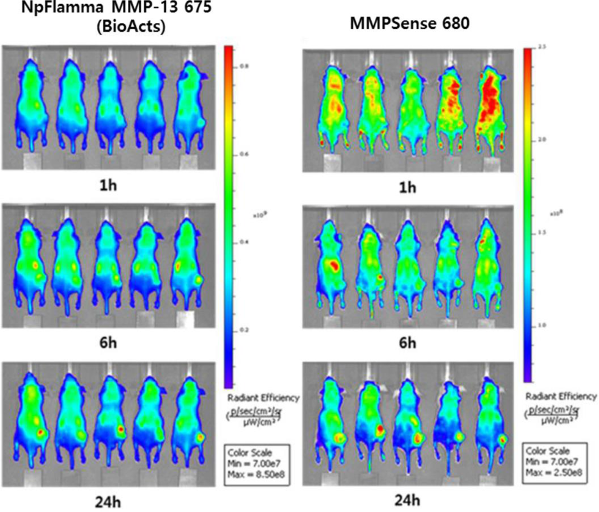
| Figure 2. Comparison of in vivo imaging of three NpFlamma® MMPs and MMPSense 680 | Figure 3. Comparison of ex-vivo imaging of three of NpFlamma® MMPs and MMPSense 680 NpFlamma® MMP series displays higher tumor cell accumulation than that of MMPSense 680 |
In vivo Imaging Protocol
General
- Since fluorescent substances are unstable under light, they should be stored in the dark.
- Mouse fur may cause scattering or absorption of excitation of light during optical imaging process. Use nude mouse or remove the mouse fur in advance.
- It is recommended to use 31 G syringe needle.
- Prepare 5 week-old male Balb/c-nude mouse.
Typical procedure for mouse model tumor imaging with NpFlamma® MMP
- When the volume of tumor cell reaches to 60~80 mm3, take the zero time image of each subject.
- Inject NpFlamma® MMP series (120 μg per 100 μL) intravenously to mouse.
- The optimal interval for fluorescence imaging is 1 h, 3 h, 6 h, 9 h, and 24 h after injection.
- After take the 24 h imaging, extract major organs (liver, lung, spleen, kidney, heart) and tumor cell, and perform the ex-vivo imaging process.
Citation & Reference
1.Optimization of matrix metalloproteinase fluorogenic probes for osteoarthritis imaging (Ju Hee Ryu, Aeju Lee, Jin Hee Na, Seulki Lee, Hyung Jun Ahn, Jong Woong Park, Cheol-Hee Ahn, Byung-Soo Kim, Ick Chan Kwon, Kuiwon Choi, Inchan Youn, Kwangmeyung Kim, Amino Acids, 2011, Volume 41, Issue 5, pp 1113–1122)
2.Dark Quenched Matrix Metalloproteinase Fluorogenic Probe for Imaging Osteoarthritis Development in Vivo (Seulki Lee, Kyeongsoon Park, Seung-Young Lee, Ju Hee Ryu, Jong Woong Park, Hyung Jun Ahn, Ick Chan Kwon, In-Chan Youn, Kwangmeyung Kim, Kuiwon Choi, Bioconjugate Chem., 2008, 19 (9), pp 1743–1747
3.Early Diagnosis of Arthritis in Mice With Collagen-Induced Arthritis, Using a Fluorogenic Matrix Metalloproteinase 3–Specific Polymeric Probe (Ju Hee Ryu, Aeju Lee, Jun-Uk Chu, Heebeom Koo, ChangYong Ko, Han Sung Kim, Soo-Young Yoon, Byung-Soo Kim, Kuiwon Choi, Ick Chan Kwon, Kwangmeyung Kim, Inchan Youn, ARTHRITIS & RHEUMATISM, 2011, Vol. 63, No. 12, pp 3824–3832)
4.Polymeric Nanoparticle-Based Activatable Near-Infrared Nanosensor for Protease Determination In Vivo (Seulki Lee, Ju Hee Ryu, Kyeongsoon Park, Aeju Lee, Seung-Young Lee, In-Chan Youn, Cheol-Hee Ahn, Soon Man Yoon, Seung-Jae Myung, Dae Hyuk Moon, Xiaoyuan Chen, Kuiwon Choi, Ick Chan Kwon, Kwangmeyung Kim, Nano Lett., 2009, Vol. 9, No. 12, pp 4412-4416)