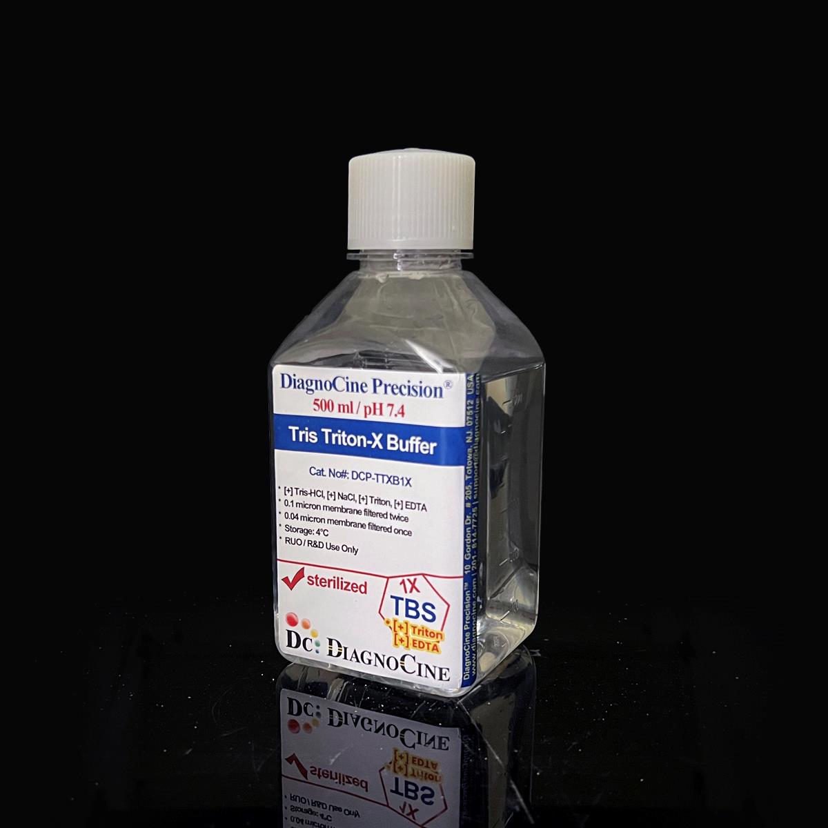
Tris Triton-X Buffer
FluxMPS™ Tris Triton-X Buffer
FluxMPS™ Tris Triton-X Buffer is an MPS-grade, non-denaturing cell lysis buffer formulated at pH 7.4 with a defined 50 mM Tris-HCl, 150 mM NaCl, 1% Triton X-100, and 5 mM EDTA composition. Manufactured with quadruple-stage filtration and ultrapure Type 1 water, it delivers the low-particulate, sterile purity required for microfluidic channels, organ-on-a-chip (OoC) systems, and sensitive downstream protein assays.
- Quadruple-stage filtered: 0.1 µm membrane twice and 0.04 µm membrane twice
- Precise, defined pH of 7.4 for near-physiological, non-denaturing protein extraction
- Defined formulation: 50 mM Tris-HCl, 150 mM NaCl, 1% Triton X-100, 5 mM EDTA
- Sterile, DNase- and RNase-tested (18-hour incubation, negative activity detected)
- Non-denaturing lysis chemistry preserves native protein conformation and complexes
- Manufactured in ISO 13485-certified, CE-approved facilities
- Compatible with SDS-PAGE, Western blotting, immunoprecipitation, and enzyme assays
- Custom concentrations, pH, and additives available on request
- pH7.4
- Tris-HCl50 mM
- NaCl150 mM
- Triton X-1001%
- EDTA5 mM
- AppearanceClear, Colorless Liquid
- Filtration0.1 µm x2, 0.04 µm x2
- DNase ActivityNone Detected
- Storage4°C or Room Temperature
- Shelf Life1 Year
Engineered where standard lysis buffers fail
Conventional 0.22 µm-filtered lysis buffers can carry subvisible particulates, inconsistent ionic strength, and unverified bioburden into sensitive protein workflows and microfluidic channels. FluxMPS™ Tris Triton-X Buffer is built to remove those failure points at the source.
Microchannel-safe purity
Quadruple-stage filtration (0.1 µm membrane twice, 0.04 µm membrane twice) reduces particulate load that could obstruct fine microfluidic channels or interfere with sensitive assays.
Precise, stable pH
Formulated at pH 7.4 using a Tris-HCl system, which provides high buffering capacity, especially between pH 7.5-9.0, to help stabilize the pH of downstream reaction systems.[3]
Ultrapure-grade water
Manufactured using ultrapure water within an ISO 13485-certified, CE-approved facility for a clean, consistent aqueous background.
Low background for imaging & assays
EDTA chelates divalent cations, minimizing interference with downstream biochemical processes and avoiding precipitation with calcium, magnesium, or heavy metal ions.
Defined, traceable composition
Every lot contains a fixed 50 mM Tris-HCl, 150 mM NaCl, 1% Triton X-100, 5 mM EDTA formulation, tested negative for DNase and RNase activity.
Customization on demand
Alternate concentrations, additional chemicals, compounds, proteins, supplements, and pH values are available — contact support@diagnocine.com.
Quadruple-stage filtration system
Every batch of FluxMPS™ Tris Triton-X Buffer passes through four sequential membrane filtration stages — 0.1 µm membrane twice followed by 0.04 µm membrane twice — in a sterile environment, delivering an ultra-clean, sterile lysis buffer suited to microfluidic channels and sensitive protein workflows.
-
1
0.1 µm Pre-filtration I
Removes large particulates and aggregates, extending the life of downstream filters.
-
2
0.04 µm Pre-filtration II
Retains fine particulates and bioburden ahead of the final sterile-filtration passes.
-
3
0.1 µm Sterile-filtration I
Provides second-pass redundancy in a sterile environment.
-
4
0.04 µm Sterile-filtration II — Final Polish
Delivers the final polish and helps prevent mycoplasma contamination, since even the smallest mycoplasma species measure approximately 0.2 micron.
Performance vs. conventional buffer
Four sequential filtration passes — 0.1 µm membrane twice and 0.04 µm membrane twice — provide additional particulate and bioburden reduction beyond a single-pass 0.22 µm filtered buffer, while preserving the sterile, low-particulate profile needed for microfluidic and organ-on-a-chip workflows.

© Diagnocine® — DCP-TTXB1X
Where FluxMPS™ Tris Triton-X Buffer is used
A gentle, non-denaturing lysis buffer designed to solubilize and extract cytoskeletal and membrane-bound proteins while maintaining protein-protein interactions and native conformation.
Automated Bioreactors & Robotics
An optional 0.01 µm (10 nm) ultra-filtered variant is available for automated bioreactor and robotic liquid-handling platforms where valve and sensor protection is critical.
- Total Particulate Exclusion for closed-system automation
- Valve & Sensor Protection in robotic fluid paths
- Extended Perfusion Stability across long-duration runs
Inquiry Required: Contact support@diagnocine.com to request the 0.01 µm ultra-filtered grade.
Micro Physiological System (MPS) & Chip
Ultra-clean buffer background for chip-based cell and tissue models.
Wash, Dilution & Reconstitution
Supports lysis, dilution, reconstitution, and rinse steps in protein workflows.
iPSC-Derived Model Handling
Gentle, non-denaturing lysis suitable for iPSC-derived cell models.
Endothelial & Primary Cell Perfusion
Compatible with perfusion-based protein extraction from primary and endothelial cells.
ELISA, Blotting & Blocking
Versatile buffer compatible with SDS-PAGE, Western blotting, IHC, and IF workflows.
Microscopy & Optical Sensing
Low-interference extraction buffer suited to downstream optical and biosensor readouts.
Full technical profile
All parameters below reflect the values stated for this specific FluxMPS™ Tris Triton-X Buffer lot formulation.
| Parameter | Specification |
|---|---|
| Formulation | Tris-HCl, NaCl, Triton X-100, EDTA |
| Appearance | Clear, Colorless Liquid |
| pH | 7.4 |
| Tris-HCl Concentration | 50 mM |
| NaCl Concentration | 150 mM |
| Triton X-100 Concentration | 1% |
| EDTA Concentration | 5 mM |
| Parameter | Specification |
|---|---|
| Sterility USP | Filtered 0.1 µm membrane twice and 0.04 µm membrane twice in a sterile environment |
| DNase Activity | None detected (18 hr incubation, plasmid DNA, room temperature) |
| RNase Activity | None detected (18 hr incubation, ribosomal RNA, room temperature) |
| Water Quality | Ultrapure Type 1 water (18.2 MΩ·cm) |
| Fill Environment | Sterile environment |
| Parameter | Specification |
|---|---|
| Storage Temperature | 4°C or room temperature |
| Shelf Life | 1 year |
| Parameter | Specification |
|---|---|
| Manufacturing QMS ISO | ISO 13485-certified facilities |
| Regulatory Alignment | CE-approved facilities |
| Production Method | Final packaging, quality assurance, and testing performed at the DiagnoCine R&D and Quality Testing Center; customization completed at DiagnoCine Precision, Totowa, New Jersey, USA |
| Intended Use | Research Use Only (RUO) |
Full composition
Every lot of FluxMPS™ Tris Triton-X Buffer is manufactured to the fixed formulation below.
| Component | CAS Number | Concentration |
|---|---|---|
| Tris-HCl | 1185-53-1 | 50 mM |
| NaCl | 7647-14-5 | 150 mM |
| Triton X-100 | 9002-93-1 | 1% |
| EDTA | 60-00-4 | 5 mM |
Manufacturing & compliance
FluxMPS™ Tris Triton-X Buffer is manufactured, filtered, and packaged under a controlled quality system.
ISO 13485:2016 QMS
Manufactured under ISO 13485-certified and CE-approved facilities (Suppliers of DiagnoCine Precision).
Ultrapure Type 1 Water
Formulated using ultrapure water for a clean, consistent aqueous background.
Sterile Fill & Finish
Filtered and packaged in a sterile environment to help prevent mycoplasma contamination.
Micro-Batch Precision
Final packaging, quality assurance, and testing are completed at the DiagnoCine R&D and Quality Testing Center.
DNase & RNase Testing
No DNase or RNase activity detected after 18-hour incubation with plasmid DNA and ribosomal RNA, respectively, at room temperature.
Filtration Validation USP
Filtered through 0.1 µm membrane twice and 0.04 µm membrane twice in a sterile environment.
Regulatory Facilities ISO
Manufactured under ISO 13485-certified and CE-approved supplier facilities; customization performed at DiagnoCine Precision, Totowa, New Jersey, USA.
Documentation / CoA
A Certificate of Analysis summarizing appearance, pH, sterility/filtration, and DNase/RNase results is available on request.
How DCP-TTXB1X compares
A side-by-side look at FluxMPS™ Tris Triton-X Buffer against typical conventional lysis buffers.
| Parameter | DCP-TTXB1X (FluxMPS™) | Conventional Tris-Triton Buffer | Standard Alternative |
|---|---|---|---|
| Defined pH & Fixed Formulation | check_circle | cancel | cancel |
| Final Filtration Pore Size | 0.04 µm | 0.22 µm | 0.22 µm |
| Number of Filtration Stages | 4 | 1 | 1 |
| DNase & RNase Tested Negative | check_circle | cancel | cancel |
| Water Quality | Ultrapure Type 1 water | Purified water | Purified water |
| Manufacturing QMS | ISO 13485 / CE | Unspecified | Unspecified |
| Microfluidic Channel Compatibility | check_circle | cancel | cancel |
| Custom Formulation Available | check_circle | cancel | cancel |
Frequently asked questions
Common questions about FluxMPS™ Tris Triton-X Buffer (DCP-TTXB1X).
Supporting literature
Curated literature relevant to Tris-Triton lysis chemistry, filtration, and microphysiological system applications.
- Ronaldson-Bouchard K, Vunjak-Novakovic G. Organs-on-a-chip: a fast track for engineered human tissues in drug development. Cell Stem Cell. 2018.doi:10.1016/j.stem.2018.02.011
- Bhatia SN, Ingber DE. Microfluidic organs-on-chips. Nat Biotechnol. 2014.doi:10.1038/nbt.2989
- Ellis KJ, Morrison JF. Buffers of constant ionic strength for studying pH-dependent processes. Methods Enzymol. 1982.doi:10.1016/S0076-6879(82)87007-1
- Koley D, Bard AJ. Triton X-100 concentration effects on membrane permeability. Anal Chem. 2010.doi:10.1021/ac1006547
- Ausubel FM, et al. Current Protocols in Molecular Biology: Cell Lysis Methods. Wiley. 2003.doi:10.1002/0471142727
- Nagatomo H, et al. EDTA chelation and its effect on metalloenzyme activity in cell extracts. J Biochem. 2005.doi:10.1093/jb/mvi048
- Uphoff CC, Drexler HG. Detection of mycoplasma contamination in cell cultures. Methods Mol Biol. 2011.doi:10.1007/978-1-61779-108-6_1
- Williams DC, et al. Endotoxin and particulate control in cell culture perfusion systems. Biotechnol Prog. 2009.doi:10.1002/btpr.208
- Mahmoudi M, et al. Protein extraction methods and downstream compatibility with SDS-PAGE and Western blotting. Nat Protoc. 2015.doi:10.1038/nprot.2015.024
- van Meer BJ, et al. Microfluidic biosensors for organ-on-chip monitoring. Lab Chip. 2019.doi:10.1039/C9LC00190E