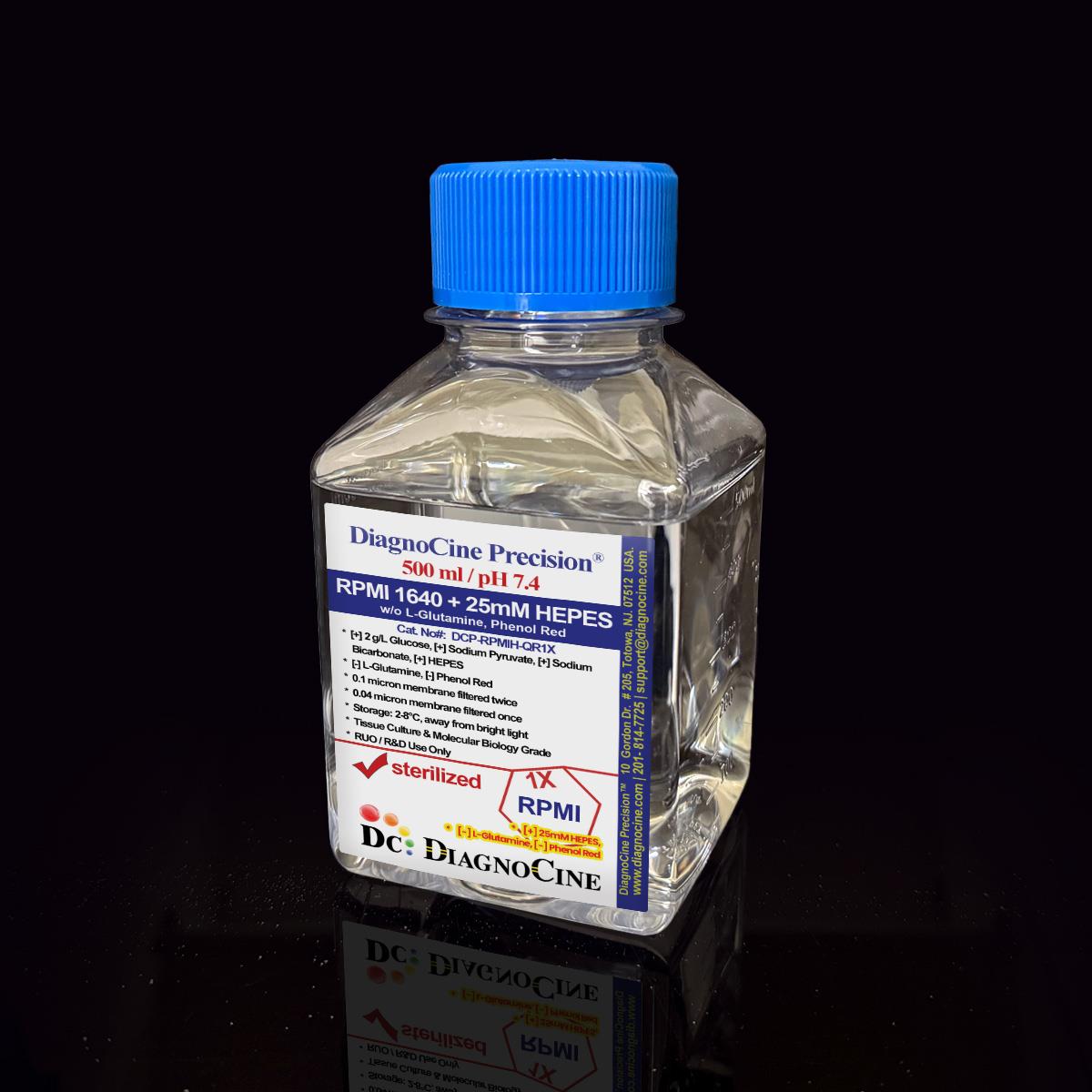
FluxMPS™ RPMI 1640 Medium with 25mM HEPES w/o L-Glutamine, Phenol Red: 1X Liquid
FluxMPS™ RPMI 1640 + 25mM HEPES
FluxMPS™ DCP-RPMIH-QR1X is a Microfluidics Suitable, quadruple-stage ultra-filtered (0.1 µm ×2 + 0.04 µm ×2) RPMI 1640 + 25 mM HEPES formulation engineered for hematopoietic and lymphocyte-based cell models on organ-on-a-chip (OoC) and microphysiological system (MPS) platforms. The train reaches a 0.04 µm final cut-off — five times finer than the 0.22 µm membranes used for conventional sterile filtration. HEPES (25 mM, pKa 7.3 at 37°C) supplements the sodium bicarbonate buffering system for pH stability during bench handling and flow cytometry preparation. Formulation: [+] Sodium Bicarbonate, [+] HEPES (25 mM), [+] Calcium, [+] Magnesium, [+] Glucose (2.0 g/L), [+] Sodium Pyruvate | [-] L-Glutamine, [-] Phenol Red.
- Glucose source: 2000 mg/L (2.0 g/L) D-glucose for standard glycolytic metabolism in RPMI-adapted cell lines
- 25 mM HEPES (pKa 7.3 at 37°C) supplements bicarbonate buffering for pH stability during bench handling and flow cytometry prep
- Glutathione (reduced, 1.0 mg/L) included — antioxidant support for redox-sensitive lymphocyte and hematopoietic cell types
- Quadruple-stage filtration train: 0.1 µm ×2 + 0.04 µm ×2, reaching a 0.04 µm final cut-off
- Endotoxin release specification: < 0.05 EU/mL (LAL, USP <85>) — relevant for TLR4-sensitive immune cell assays
- Phenol red–free and L-glutamine–free formulation — supports reduced-background imaging and researcher-controlled glutamine supplementation
- Manufactured under an ISO 13485:2016 quality management system; final QC at Diagnocine, Totowa, NJ
- pH 7.4; osmolality 280–320 mOsm/kg H2O — formulated for RPMI-based lymphocyte and hematopoietic culture
- Media familyRPMI 1640 + 25mM HEPES
- Formulation[+] Sodium Bicarbonate, [+] HEPES 25mM, [+] Calcium, [+] Magnesium, [+] Glucose 2.0g/L, [+] Sodium Pyruvate | [-] L-Glutamine, [-] Phenol Red
- AppearancePale yellow to colorless, clear solution
- pH (USP <791>)7.4
- Osmolality (USP <785>)280–320 mOsm/kg H2O
- Endotoxin (USP <85>)< 0.05 EU/mL
- Filtration0.1 µm ×2 + 0.04 µm ×2 (Quadruple-stage)
- Storage2–8°C, protect from light
- Shelf Life12 months from date of manufacture, unopened
- ShippingCold pack, temperature-monitored
Engineered where standard media fails
Conventional 0.22 µm–filtered RPMI passes mycoplasma-scale organisms, subvisible particulates, and endotoxin fragments that can activate TLR4 signaling and confound immune cell assays. FluxMPS™ is built to reduce these risks at the filtration stage.
Microchannel-safe purity
0.04 µm final filtration with USP <788> Method 1 particulate compliance supports suspension immune cell OoC platforms and flow cytometry workflows sensitive to particulate interference.
Researcher-controlled metabolic inputs
L-glutamine is excluded, allowing precise, researcher-controlled nitrogen and glutamine-analog supplementation for T cell activation and metabolic flux studies. Sodium pyruvate (110 mg/L) and D-glucose (2.0 g/L) are provided as base carbon sources.
Ultrapure-grade water
Formulated with Type 1 water (18.2 MΩ·cm) under trace-metal and organic-carbon control, reducing lot-to-lot variability contributed by feed water in sensitive lymphocyte culture systems.
Low background for imaging
Phenol red is excluded, reducing spectral interference for flow cytometry and confocal fluorescence microscopy. Riboflavin, present in all RPMI-based formulations, contributes intrinsic background fluorescence common to this medium class — plan filter sets accordingly.
Rich, stable nutrient profile
RPMI's 19-amino-acid and 10-vitamin profile, plus reduced glutathione for redox support, is optimized for lymphocyte, monocyte, and hematopoietic cell maintenance, released per-lot against the full specification matrix.
Customization on demand
pH, nutrient concentrations, HEPES level, and component modifications available on request. Contact support@diagnocine.com.
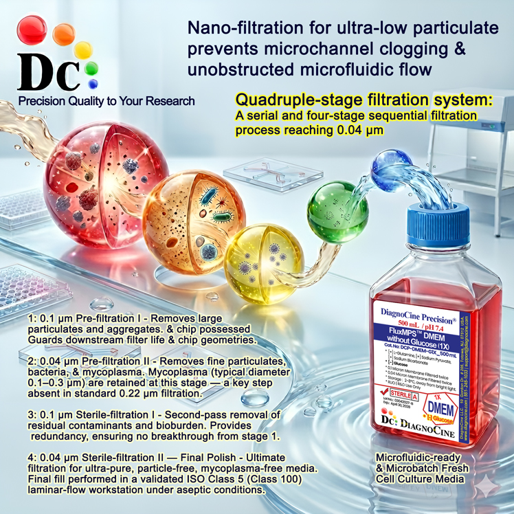
Quadruple-stage filtration system
Four serial filtration passes — two dedicated 0.1 µm/0.04 µm prefilter-and-final-filter pairs — reach a final 0.04 µm polish under aseptic fill conditions.
-
1
0.1 µm Prefiltration I
Removes large particulate, cell debris, and protein aggregates; protects the first 0.04 µm cartridge.
-
2
0.04 µm Final filtration I — Mycoplasma Barrier
Retains organisms in the 0.2–0.3 µm mycoplasma size range — a pore size below what standard 0.22 µm filtration retains.
-
3
0.1 µm Prefiltration II
Second dedicated prefilter, protecting the second 0.04 µm final-filter cartridge.
-
4
0.04 µm Final filtration II — Polish
Ultimate polishing filter ahead of aseptic fill & finish in ISO Class 5 (Class 100) workstations.
Performance vs. conventional media
A quadruple-stage train (0.1 µm ×2 + 0.04 µm ×2) reaches a 0.04 µm final cut-off, five times finer than the 0.22 µm membranes used for conventional sterile filtration.
© Diagnocine® — DCP-RPMIH-QR1X
Immune cell OoC and hematopoietic applications
FluxMPS™ DCP-RPMIH-QR1X — RPMI 1640 + 25mM HEPES — delivers 0.04 µm filtered purity for hematopoietic cells and related OoC applications.
Automated Bioreactors & Robotics
An optional 0.01 µm (10 nm) MPS Grade variant of this formulation — a separate six-stage ultra nano-filtered line beyond the Microfluidics Suitable 0.04 µm tier described above — is available on request for automated bioreactor and robotic perfusion systems.
- Total Particulate Exclusion: 0.01 µm filtration targets nanoparticulate aggregates beyond the 0.04 µm tier
- Valve & Sensor Protection: Reduces micro-fouling risk in automated perfusion systems
- Extended Perfusion Stability: Consistent nutrient delivery over long-duration culture
Inquiry Required: Contact support@diagnocine.com for the 0.01 µm MPS Grade variant.
T Cell & Lymphocyte Culture
RPMI 1640 is the standard base for primary T cells, B cells, NK cells, and monocytes. FluxMPS™ 0.04 µm filtration reduces particulate load that can confound immune assays.
Leukemia & Lymphoma Lines
RPMI supports NCI-60 hematologic cancer lines, Jurkat, Raji, K562, and HL-60, where DMEM would alter proliferation and signaling behavior.
Immune Cell OoC
0.04 µm filtered RPMI supports tumor-immune interaction chips, vascular-immune OoC, and lymph node-on-chip models sensitive to particulate load.
CAR-T & TIL Expansion
The <0.05 EU/mL endotoxin release specification supports CAR-T manufacturing and TIL expansion protocols where endotoxin-driven T cell activation artefacts are a concern.
Immune Cell Metabolic Flux
Defined RPMI base for glycolysis stress assays and ¹³C isotope tracing of lymphocyte activation states. Not compatible with Agilent Seahorse XF assays, which require bicarbonate-free, phenol red–free medium.
Flow Cytometry & Confocal
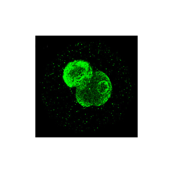
Ultra-low particulate and phenol red–free formulation reduce spectral background for PE-channel flow cytometry and immune cell confocal imaging.
Analytical release specifications
Every lot released against the full specification matrix. CoA: support@diagnocine.com.
| Parameter | Specification |
|---|---|
| Formulation | [+] Sodium Bicarbonate, [+] HEPES 25mM, [+] Calcium, [+] Magnesium, [+] Glucose 2.0 g/L, [+] Sodium Pyruvate | [-] L-Glutamine, [-] Phenol Red |
| Appearance | Pale yellow to colorless, clear solution |
| Glucose | 2000 mg/L (2.0 g/L) |
| HEPES | 25 mM (pKa 7.3 at 37°C) |
| pH USP <791> | 7.4 |
| Osmolality USP <785> | 280–320 mOsm/kg H2O |
| Total ingredients | 40 components across 3 categories |
| Parameter | Specification |
|---|---|
| Endotoxin USP <85> BET | < 0.05 EU/mL (batch release spec, see Manufacturing & Compliance) |
| Sterility USP <71> | No growth / 14 days |
| Mycoplasma | 0.1 µm mycoplasma-retentive filtration (not tested per lot) |
| Particulate ≥10 µm USP <788> Method 1 | NMT 25/mL |
| Particulate ≥25 µm USP <788> Method 1 | NMT 3/mL |
| Water purity | Type 1, 18.2 MΩ·cm |
| Manufacturing std. | ISO 13485:2016 |
| Fill environment | ISO Class 5 (Class 100) |
| Parameter | Specification |
|---|---|
| Storage temperature | 2–8°C, away from light |
| Freeze-thaw | Do not freeze |
| Shelf life | 12 months from date of manufacture, unopened |
| Shipping condition | Cold pack, temperature-monitored |
| CO₂ requirement | 5% CO2 recommended; sodium bicarbonate (2.0 g/L) is the primary buffer, with 25 mM HEPES as supplemental buffering during bench handling |
| Parameter | Specification |
|---|---|
| Raw material grade | Reagent / cell culture grade |
| Traceability | Full lot traceability per ISO 13485 |
| Manufacturing QMS ISO | ISO 13485:2016 certified |
| UNSPSC | 41116155 — Molecular biology and cell culture growth media (UNv260801) |
| Grade | Microfluidics Suitable (0.04 µm final cut-off) |
| Regulatory alignment | 21 CFR Part 820 (QMSR) aligned |
| Production method | Micro-batch, per-lot QC release |
| Intended use | Research Use Only (RUO) |
Full composition (mg/L)
RPMI 1640 + 25mM HEPES: 40 ingredients verified per lot with CAS numbers for raw-material traceability. This formulation includes reduced glutathione and a balanced amino acid and vitamin profile optimized for lymphocyte and hematopoietic cell culture.
| Component | CAS Number | mg/L |
|---|---|---|
| INORGANIC SALTS | ||
| Calcium nitrate tetrahydrate | 13477-34-4 | 100.000 |
| Magnesium sulfate anhydrous | 7487-88-9 | 48.840 |
| Potassium chloride | 7447-40-7 | 400.000 |
| Sodium bicarbonate | 144-55-8 | 2000.000 |
| Sodium chloride | 7647-14-5 | 6000.00 |
| Sodium phosphate dibasic anhydrous | 7558-79-4 | 800.000 |
| Component | CAS Number | mg/L |
|---|---|---|
| AMINO ACIDS | ||
| Glycine | 56-40-6 | 10.000 |
| L-Arginine hydrochloride | 1119-34-2 | 241.000 |
| L-Asparagine | 70-47-3 | 50.000 |
| L-Aspartic acid | 56-84-8 | 20.000 |
| L-Cystine dihydrochloride | 30925-07-6 | 65.200 |
| L-Glutamic acid | 56-86-0 | 20.000 |
| L-Histidine hydrochloride monohydrate | 5934-29-2 | 20.960 |
| L-Hydroxyproline | 51-35-4 | 20.000 |
| L-Isoleucine | 73-32-5 | 50.000 |
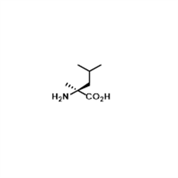
| L-Leucine | 61-90-5 | 50.000 |
| L-Lysine hydrochloride | 657-27-2 | 40.000 |
| L-Methionine | 63-68-3 | 15.000 |
| L-Phenylalanine | 63-91-2 | 15.000 |
| L-Proline | 147-85-3 | 20.000 |
| L-Serine | 56-45-1 | 30.000 |
| L-Threonine | 72-19-5 | 20.000 |
| L-Tryptophan | 73-22-3 | 5.000 |
| L-Tyrosine Disodium Salt | 69847-45-6 | 28.830 |
| L-Valine | 72-18-4 | 20.000 |
| Component | CAS Number | mg/L |
|---|---|---|
| VITAMINS | ||
| Choline chloride | 67-48-1 | 3.000 |
| D-Biotin | 58-85-5 | 0.200 |
| D-Ca-Pantothenate | 137-08-6 | 0.250 |
| Folic acid | 59-30-3 | 1.000 |
| Niacinamide | 98-92-0 | 1.000 |
| Pyridoxine hydrochloride | 58-56-0 | 1.000 |
| Riboflavin | 83-88-5 | 0.200 |
| Thiamine hydrochloride | 67-03-8 | 1.000 |
| Vitamin B12 | 68-19-9 | 0.005 |
| p-Amino benzoic acid (PABA) | 150-13-0 | 1.000 |
| OTHERS | ||
| D-Glucose | 50-99-7 | 2000.000 |
| Glutathione reduced | 70-18-8 | 1.000 |
| HEPES | 7365-45-9 | 5958.00 |
| Sodium Pyruvate | 113-24-6 | 110.000 |
| i-Inositol | 87-89-8 | 35.000 |
Manufacturing & compliance
Every FluxMPS™ product is manufactured and released under a multi-layer quality system.
ISO 13485:2016 Quality Management
Manufactured under an ISO 13485:2016-certified quality management system. Final QC at Diagnocine's R&D Center, Totowa, NJ, USA.
Ultrapure Type 1 Water
18.2 MΩ·cm Type 1 water processed under trace-metal and organic-carbon control.
ISO Class 5 Fill & Finish
Aseptic fill in validated ISO Class 5 (Class 100) laminar-flow workstations.
Micro-Batch Precision
Small-batch, per-lot tested — no blending; Certificate of Analysis issued for every lot.
Endotoxin — USP <85> BET
LAL assay; release specification < 0.05 EU/mL per batch.
Particulate — USP <788> Method 1
≤25/mL (≥10 µm), ≤3/mL (≥25 µm), light obscuration.
Osmolality — USP <785>
Target: 280–320 mOsm/kg H2O.
Documentation & CoA
Full CoA with raw-material traceability available on request.
- Endotoxin — LAL assay, USP <85> Bacterial Endotoxins Test; assay sensitivity 0.005 EU/mL; release specification < 0.05 EU/mL
- pH, osmolality, conductivity, appearance and clarity
- Sterility
How DCP-RPMIH-QR1X compares
FluxMPS™ DCP-RPMIH-QR1X vs. conventional 0.22 µm–filtered RPMI formulations.
| Parameter | DCP-RPMIH-QR1X (FluxMPS™) | Conventional RPMI 1640 + HEPES (0.22 µm filtered) |
Standard DMEM/RPMI (0.22 µm filtered) |
|---|---|---|---|
| Grade | Microfluidics Suitable | Not specified | Not specified |
| RPMI 1640 + 25mM HEPES — no L-Glutamine, no Phenol Red; pH-stable imaging-clean immune base | check_circle Yes | cancel No | cancel No |
| Final filtration pore size | 0.04 µm | 0.22 µm | 0.22 µm |
| Number of filtration stages | 4 (Quadruple) | 1 | 1 |
| Mycoplasma-retentive filtration | check_circle Yes (0.1 µm stage) | cancel No | cancel No |
| Endotoxin (release specification) | FluxMPS™ — < 0.05 EU/mL | Corning classical liquid media — < 0.25 EU/mL Sigma-Aldrich DMEM complete medium — ≤ 2 EU/mL Gibco classical DMEM — Not specified (recorded per lot) |
|
| USP <788> particulate tested | check_circle Yes | cancel No | cancel No |
| Water quality | Type 1, 18.2 MΩ·cm | Purified water | Purified water |
| Manufacturing QMS | ISO 13485:2016 | ISO 9001 or none | ISO 9001 or none |
| Microfluidic channel compatibility | check_circle Yes (Microfluidics Suitable) | cancel Risk of clogging | cancel Risk of clogging |
| Custom formulation available | check_circle Yes | cancel No | cancel No |
Comparison figures from published supplier specifications, accessed 2026-09-02. Suppliers that publish no numeric endotoxin specification are shown as "Not specified".
Frequently asked questions
Common questions about FluxMPS™ DCP-RPMIH-QR1X — RPMI 1640 + 25mM HEPES.
Supporting literature
Key publications supporting RPMI 1640 + 25mM HEPES in immune cell culture and OoC applications.
- Moore GE, et al. Culture of normal human leukocytes. JAMA. 1967;199:519–524. doi:10.1001/jama.1967.03120080053007
- Huh D, et al. Reconstituting organ-level lung functions on a chip. Science. 2010;328:1662–1668. doi:10.1126/science.1188302
- Bhatia SN, Ingber DE. Microfluidic organs-on-chips. Nat Biotechnol. 2014;32:760–772. doi:10.1038/nbt.2989
- Novak R, et al. Robotic fluidic coupling and interrogation of multiple vascularized organ chips. Nat Biomed Eng. 2020;4:407–420. doi:10.1038/s41551-019-0497-x
- Jang KJ, et al. Human kidney proximal tubule-on-a-chip. Integr Biol. 2013;5:1119–1129. doi:10.1039/c3ib40049b
- Schimek K, et al. Integrating biological vasculature into a multi-organ-chip microsystem. Lab Chip. 2013;13:3588–3598. doi:10.1039/c3lc50217a
- Boussommier-Calleja A, et al. Microfluidics: A new tool for modeling cancer-immune interactions. Trends Cancer. 2016;2:6–19. doi:10.1016/j.trecan.2015.12.003
- Sung JH, et al. Microfabricated mammalian organ systems. Lab Chip. 2013;13:1201–1212. doi:10.1039/c3lc41017j