No More Sales-NpFlamma® HGC 675
Packing Unit
-
10 tests

-
50 tests

-
250 tests

NpFlamma® HGC 675
Cat. No. List below
Description
Specifications
- Fluorophore: Flamma® Fluors 675
- Application: In vivo imaging
- Particle size: ~250 nm
- Excitation/Emission Max.(nm): 675/698
- Appearance: Blue Solid
- Storage conditions: 4 ℃, protect from light
Table 1. NpFlamma® HGC series list
| Quick link (Cat.#) | Series | Ex * (nm) | Em* (nm) | Common filter set |
Excitation source |
| PNC1201 | NpFlamma® HGC 648 | 648 | 675 | Cy 5 | 594, 633 nm |
| PNC1401 | NpFlamma® HGC 675 | 675 | 698 | Cy 5.5 | 633, 680 nm |
| PNC1301 | NpFlamma® HGC 749 | 750 | 782 | Cy 7 | 680 nm |
| PNC1601 | NpFlamma® HGC 774 | 777 | 802 | Cy 7.5 | 785 nm |
| PNC1501 | NpFlamma® HGC ICG | 785 | 821 | Cy 7.5 | 785 nm |
Overview
NpFlamma® HGC series is a fluorescent dye incorporated chitosan based amphiphilic nanoparticles that enables to selectively detect tumor cells. Chitosan nanoparticles can selectively accumulate in cancer tissues due to high permeability for new blood vessels in cancer tissues. Chitosan particles display low toxicity along with absence of noticeable side effect in vivo, yet they exhibit a long half-life, high stability and aqueous solubility. Thus, NpFlamma® HGC series is an ideal fluorescence agent for in vivo imaging of angiography and tumor progression. The hydrophobic nature of NpFlamma® HGC series can embed hydrophobic materials, thus they can be utilized as a selective carrier for hydrophobic cancer drugs such as doxorubicin and paclitaxel, etc.
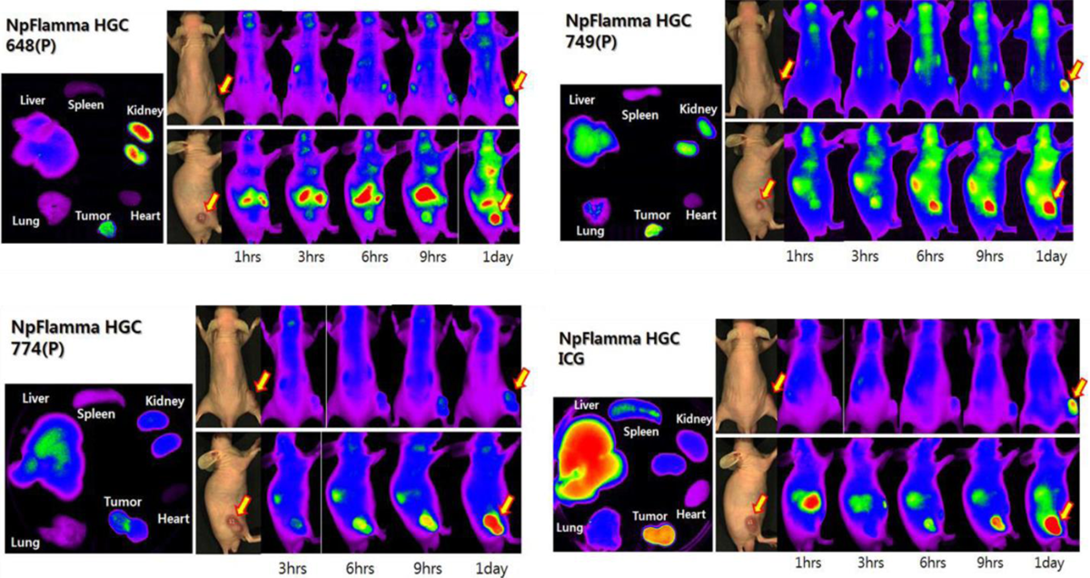
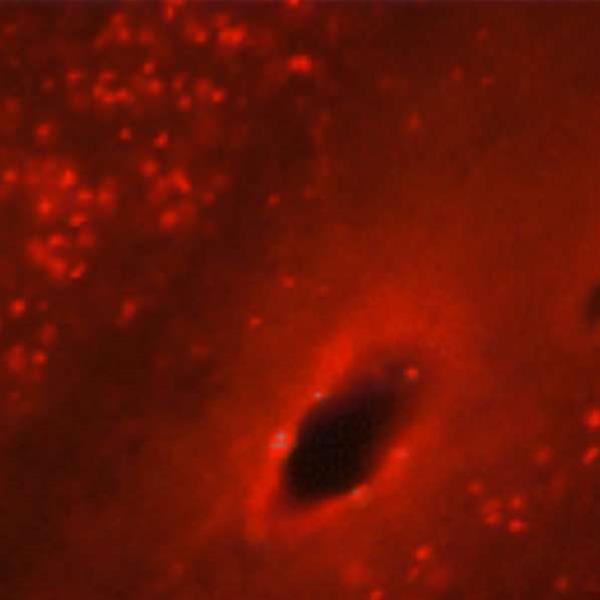
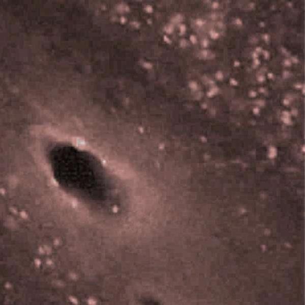
Figure 1. In vivo tumor imaging of NpFlamma® HGC series
 |
 |
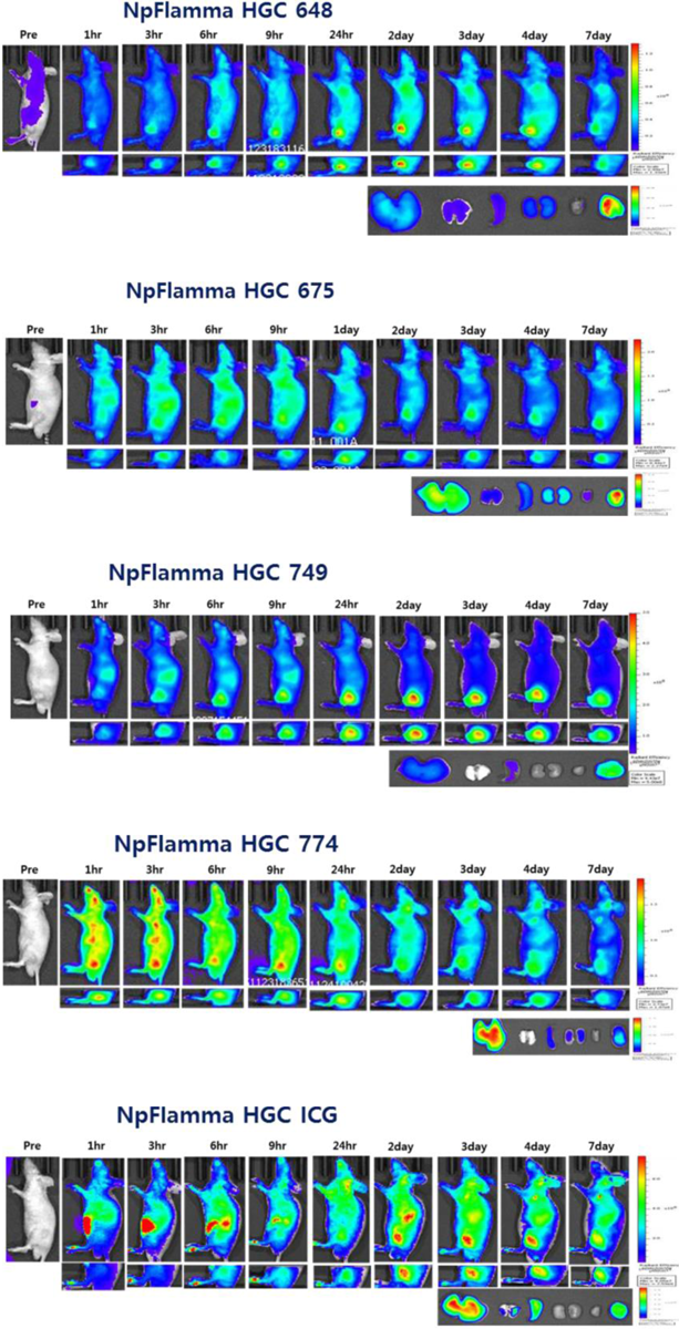
| Figure 2. In vivo imaging of five NpFlamma® HGC series over seven days Fluorescence from cancer cell continued after 7 days injection, but fluorescence from other organs disappeared. |
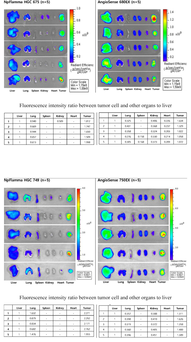
Figure 3. Comparison of ex-vivo imaging of NpFlamma® HGC and another imaging agent NpFlamma® HGC series displays higher tumor cell accumulation than that of AngioSense |
In vivo Imaging Protocol
General
- Since fluorescent substances are unstable under light, they should be stored in the dark.
- Mouse fur may cause scattering or absorption of excitation of light during optical imaging process. Use nude mouse or remove the mouse fur in advance.
- It is recommended to use 31 G syringe needle.
- Prepare 5 week-old male Balb/c-nude mouse.
Typical procedure for mouse model tumor imaging with NpFlamma® HGC
- When the volume of tumor cell reaches to 60~80 mm3, take the zero time image of each subject.
- Inject NpFlamma® HGC series (120 μg per 100 μL) intravenously to mouse.
- The optimal interval for fluorescence imaging is 1 h, 3 h, 6 h, 9 h, and 24 h after injection.
- After take the 24 h imaging, extract major organs (liver, lung, spleen, kidney, heart) and tumor cell, and perform the ex-vivo imaging process.
Citation & Reference
1. New generation of multifunctional nanoparticles for cancer imaging and therapy (Kyeongsoon Park, Seulki Lee, Eunah Kang, Kwangmeyung Kim, Kuiwon Choi, Ick Chan Kwon, Advanced Functional Materials, 2009, Volume 19, Issue 10, Pages 1553–1566)
2. Tumor-homing multifunctional nanoparticles for cancer theragnosis: Simultaneous diagnosis, drug delivery, and therapeutic monitoring (Kwangmeyung Kim, Jong Ho Kim, Hyungkyu Park, Yoo-Shin Kim, Kyeongsoon Park, Heayun Nam, Seulki Lee, Jae Hyung Park, Rang-Woon Park, In-San Kim, Kuiwon Choi, Sang Yoon Kim, Kinam Park, Ick Chan Kwon, Journal of Controlled Release, 2010, Volume 146, Issue 2, Pages 219–227)
3. Glycol chitosan nanoparticles as specialized cancer therapeutic vehicles: Sequential delivery of doxorubicin and Bcl-2 siRNA (Hong Yeol Yoon, Sejin Son, So Jin Lee, Dong Gil You, Ji Young Yhee, Jae Hyung Park, Maggie Swierczewska, Seulki Lee, Ick Chan Kwon, Sun Hwa Kim, Kwangmeyung Kim & Martin G. Pomper, Scientific Reports, 2014, 4, Articlenumber:6878)