LipiORDER
LipiORDER
Membrane Lipid Order Imaging DyeSize: 0.1 mg
Description
LipiORDER is a novel solvatochromic dye for membrane lipid order imaging
Principle and Reference data
Sensing of lipid order by using LipiORDER is based on the following two unique properties.
1) LipiORDER is a pyren-based solvatochromic fluorescent dye which changes fluorescent property in response to their solvent environment (Figure P1). In low polaric solvents such as toluene, LipiORDER shows green fluorescence. On the other hand, in highly polaric solvents such as DMSO and methanol, this dye changes color to orange or red.
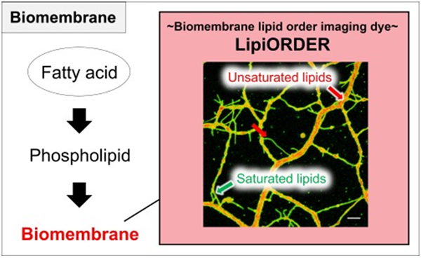
2) LipiORDER is a highly hydrophobic compound and quickly accumulates in the various biological membranes. Combining the two features above, LipiORDER can sense the local environment in a lipid bilayer. Generally, Lo is a high packing lipid bilayer and shows lower polarity, whereas Ld is a sparse packing lipid bilayer and shows high polarity. Based on polarity of lipid bilayer derived from lipid order, LipiORDER will change fluorescent color, from green on Lo membrane to red on Ld membrane (Figure P2).
3) Ratiometric fluorescent value (FR/FG ) is correlated to lipid order (Lo and Ld). Actually, in sphingomyeline/cholesterol (SM/Chol) liposome, one of the model Lo, LipiORDER emits green fluorescence and in 1,2-dioleoyl-sn-glucero-3-phosphocholine (DOPC) liposome, a model Ld, shows red fluorescence. In DOPC/Chol, an intermediate model, the reagent show yellow to orange. The ratiometric values (F575/F510) clearly depend on lipid order, SM/Chol (Lo) is low and DOPC (Ld) is high (Figure P3).
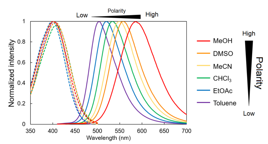
Figure P1 Absorption and fluorescent spectrum of LipiORDER in various solvent
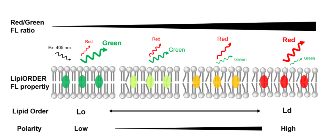
Figure P2 Graphical overview of lipid order-dependent fluorescent change of LipiORDER

Figure P3 Fluorescent spectrum of LipiORDER in model liposomes
Specification
Formulation: C23H21NO
Molecular weight: 327.4 g/mol
Solubility: Soluble in DMSO
Fluorescent characteristics: Ex. 405 nm/Em. 450-650 nm (dependent on solvents)
Application Data
Ratiometric imaging of COS7 cells
COS7 cells were treated with 300 nM LipiORDER in HBSS for 10 min and observed by confocal laser microscopy (Ex. 405 nm, Em 470-550 nm for Green channel and >550 nm for Red channel). Ratiometric analysis was performed with ImageJ using green and red channel data and lipid order was shown by green-to-red pseudocolor
 . Plasma membrane and intramembranes are shown Lo and Ld, respectively.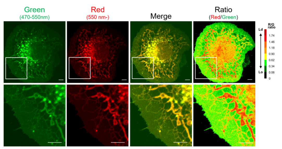
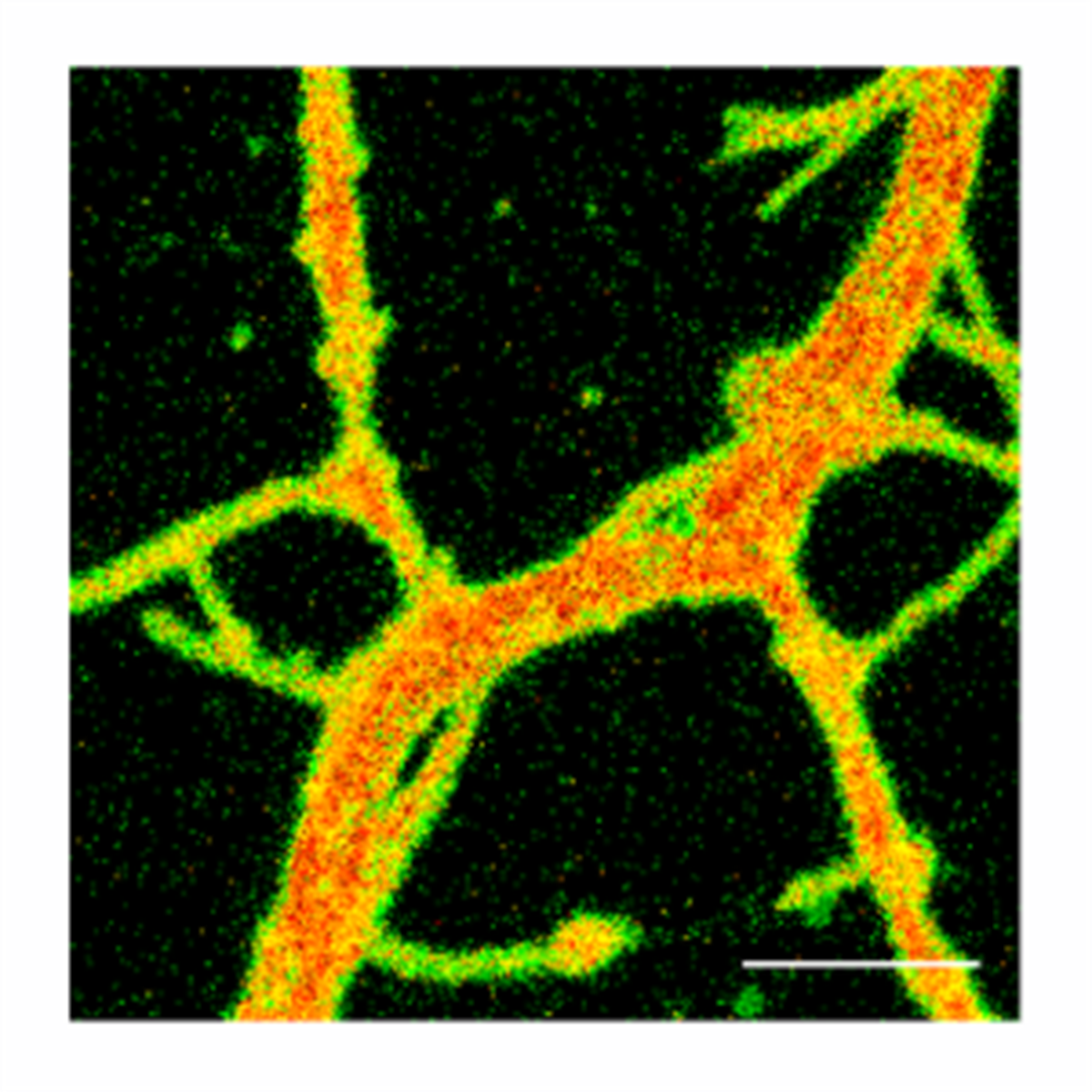
Primary cultured hippocampal neurons (DIV 3 or DIV 12) from E17.5 mice were stained with 300 nM LipiORDER in HBSS for 10 min and observed by confocal laser microscopy (Ex. 405 nm, Em. 470-550 nm for Green channel and >550 nm for Red channel). Ratiometric analysis was performed with ImageJ using green and red channel data and lipid order was shown by green-to-red pseudocolor
.
Drug-induced cellular lipid order changes
COS7 cells were treated with 15 mM beta-cyclodextrin (beta-CD), a membrane-disrupting chemical via removing endogenous cholesterol, for 4 hours. After beta-CD treatment, cells were washed and stained with 300 nM LipiORDER in HBSS for 10 min. The cells were observed by confocal laser microscopy (Ex. 405 nm, Em. 470-550 nm for Green channel and >550 nm for Red channel). Ratiometric analysis was performed with ImageJ using green and red channel data and lipid order is shown by green-to-red pseudocolor
. The cell structure was dramatically changed by beta-CD and at the same time, the distribution of Lo phase (■) clearly changed.
Photostability of LipiORDER
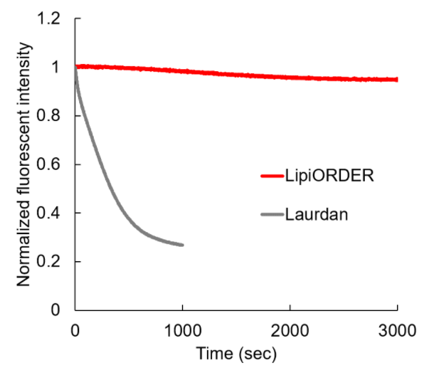
LipiORDER and Laurdan, a conventional membrane lipid order imaging dye in lipid vesicles composed of 0.2 mM DOPC in 20 mM HEPES (pH 7.4) were irradiated with Xe lamp. LipiORDER and Laurdan were excited at 405 nm and 360 nm, respectively and fluorescent intensity was measured. Laurdan was quickly photodegraded, whereas LipiORDER maintains fluorescent intensity for at least 1 hour. LipiORDER is highly stable compared to Laurdan.
Reference
1. Valanciunaite et al., Polarity Mapping of Cells and Embryos by Improved Fluorescent Solvatochromic Pyrene Probe. Anal. Chem., 92, 6512-6520 (2020)
2. Momma Y., et al. The Curcumin Derivative GT863 Protects Cell Membranes in Cytotoxicity by Aβ Oligomers. Int J Mol Sci. 2023 Feb; 24(4): 3089.
3. Okamoto Y., et al. Characterization of Phase Separated Planar Lipid Bilayer Membrane by Fluorescence Ratio Imaging and Scanning Probe Microscope. Membranes (Basel). 2022 Aug 9;12(8):770. doi: 10.3390/membranes12080770.
4. Yatsuzuka K., et al. A fluorescence imaging technique suggests that sweat leakage in the epidermis contributes to the pathomechanism of palmoplantar pustulosis. Sci Rep. 2024 Jan 3;14(1):378. doi: 10.1038/s41598-023-50875-x.
5. Hayakawa E., et al. Budding pouches and associated bubbles: 3D visualization of exo-membrane structures in plasmodium falciparum gametocytes. Front Cell Infect Microbiol. 2022 Aug 22:12:962495. doi: 10.3389/fcimb.2022.962495. eCollection 2022.
6. Suito T. et al. Ether phospholipids modulate somatosensory responses by tuning multiple receptor functions in Drosophila. bioRxiv. https://doi.org/10.1101/2023.09.12.556286 (2023)
7. Honda A., et al., Very-long-chain fatty acids are crucial to neuronal polarity by providing sphingolipids to lipid rafts. Cell Rep. 2023 42(10):113195.
8. Kumar GA., et al., Membrane order regulates clathrin-coated pit dynamics but not initiation. Mol Biol Cell. 2025 36(7):br17.
9. Han, Z., Yan, Z., Ma, Z., Wang, Y., Beus, M., Lu, J., ... & Nikiforov, M. A. (2025). Targeting ABCD1-ACOX1-MET/IGF1R axis suppresses multiple myeloma. Leukemia, 39(3), 720–733.
10. Houser, M. C. Q., Mitchell, S. P. C., Sinha, P., Lundin, B., Berezovska, O., & Maesako, M. (2023). Endosome and Lysosome Membrane Properties Functionally Link to γ-Secretase in Live/Intact Cells. Sensors, 23(5), 2651.
11. Morita, S., Kondo, T., & Inoue, H. (2026). Protocol for measuring lipid membrane fluidity in human iPSC-derived neural cells. STAR Protocols, 7(2), 104440. https://doi.org/10.1016/j.xpro.2026.104440
12. Rui, X., Okamoto, Y., Fukushima, S., Watanabe, N. M., & Umakoshi, H. (2024). Investigating the impact of 2-OHOA-embedded liposomes on biophysical properties of cancer cell membranes via Laurdan two-photon microscopy imaging. Scientific Reports, 14(1), 15831. https://doi.org/10.1038/s41598-024-65812-9
13. Morita, S., Kondo, T., Tokuda, H., Kaneda, Y., Izumo, T., Nakao, Y., & Inoue, H. (2025). Polyunsaturated fatty acids in lipid membranes regulate human neuronal function and amyloid-β production. iScience, 28(6), 112557. https://doi.org/10.1016/j.isci.2025.112557
14. Saito, S., Okumura, Y., Kataoka, S., Fukaya, K., Urabe, D., & Arai, M. A. (2025). The heat-shock metabolite streptolactam D, produced by high-temperature culture of Streptomyces sp. JA74, promotes thermotolerance via self-membrane stabilization. Journal of the American Chemical Society, 147(18), 15676–15685. https://doi.org/10.1021/jacs.5c03026
15. Hasan, M., Kristof, J., Sadiq, A. H., Alam, M. J., Rimi, S. A., Begum, F., & Shimizu, K. (2025). Microplasma-mediated enhancement of FD-150 uptake in HL-60 cells. Membranes, 15(5), 156. https://doi.org/10.3390/membranes15050156
16. Yatsuzuka, K., Kawakami, R., Niko, Y., Tsuda, T., Kameda, K., Kohri, N., Yoshida, S., Shiraishi, K., Muto, J., Mori, H., Fujisawa, Y., Imamura, T., & Murakami, M. (2024). A fluorescence imaging technique suggests that sweat leakage in the epidermis contributes to the pathomechanism of palmoplantar pustulosis. Scientific Reports, 14(1), 378. https://doi.org/10.1038/s41598-023-50875-x
17. Kanamori, T., Yasuda, S., Duan, R., Ohashi, M., Amou, M., Hori, K., Tsuda, R., Fujimoto, T., Higashi, K., Xu, W., Niidome, T., & Hatakeyama, H. (2025). Cholesterol depletion suppresses thermal necrosis resistance by alleviating an increase in membrane fluidity. Scientific Reports, 15(1), 10133. https://doi.org/10.1038/s41598-025-92232-0
18. Yoshida, S., Kawakami, R., Niko, Y., Fujisawa, Y., & Murakami, M. (2025). New characteristics of eccrine sweat glands in acquired idiopathic generalised anhidrosis as determined via three-dimensional fluorescence imaging of cleared skin tissue. Experimental Dermatology, 34(3), e70038. https://doi.org/10.1111/exd.70038
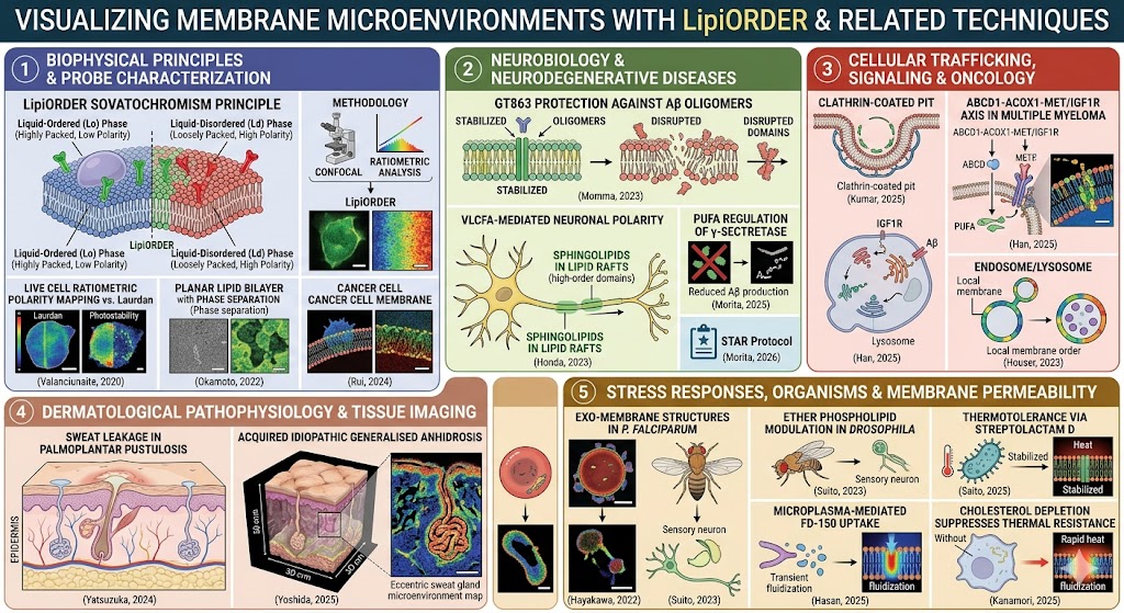
Here is a summarized format of the selected publications detailing how the solvatochromic dye LipiORDER (or related pyrene-based/Laurdan membrane order imaging techniques) was utilized to investigate lipid packing, membrane fluidity, and microenvironments across various biological systems.
1. Biophysical Principles & Probe Characterization
-
Valanciunaite et al. (2020) | Anal. Chem.
-
Application: Developed and characterized the improved fluorescent solvatochromic pyrene probe (the basis of LipiORDER).
-
LipiORDER Role: Demonstrated high photostability and a color shift (green in low-polarity, highly-packed $L_o$ phases; red in high-polarity, loosely-packed $L_d$ phases) to achieve high-resolution ratiometric polarity mapping in live cells and tissue embryos.
-
-
Okamoto et al. (2022) | Membranes
-
Application: Characterization of phase-separated planar lipid bilayer membranes.
-
LipiORDER Role: Utilized via fluorescence ratio imaging alongside scanning probe microscopy to distinguish and map physical variations between liquid-ordered ($L_o$) and liquid-disordered ($L_d$) domains in artificial model membranes.
-
-
Rui et al. (2024) | Sci. Rep.
-
Application: Investigation of 2-OHOA-embedded liposomes on cancer cell membranes.
-
LipiORDER/Laurdan Context: Used advanced two-photon microscopy imaging to determine how specialized therapeutic liposomes alter the rigid biophysical packing and fluidity of cancerous cell membranes.
-
2. Neurobiology & Neurodegenerative Diseases
-
Momma Y., et al. (2023) | Int J Mol Sci.
-
Application: Protective effect of curcumin derivative GT863 against Amyloid-$\beta$ ($A\beta$) oligomer cytotoxicity.
-
LipiORDER Role: Used to visualize and confirm that GT863 physically protects and stabilizes the cell membrane against disruption and fluidization caused by toxic $A\beta$ aggregates.
-
-
Honda A., et al. (2023) | Cell Rep.
-
Application: Role of very-long-chain fatty acids (VLCFAs) in neuronal polarity.
-
LipiORDER Role: Monitored how the supply of sphingolipids to lipid rafts maintains highly ordered, rigid domains crucial for proper neuronal polarization and axonal development.
-
-
Morita, Kondo, & Inoue (2026) | STAR Protocols
-
Application: Protocol for measuring lipid membrane fluidity in human iPSC-derived neural cells.
-
LipiORDER Role: Establishes a standardized method utilizing LipiORDER for high-throughput, ratiometric imaging to assess baseline membrane fluidity shifts in human stem-cell-derived neurons.
-
-
Morita et al. (2025) | iScience
-
Application: Polyunsaturated fatty acids (PUFAs) regulation of human neuronal function and $A\beta$ production.
-
LipiORDER Role: Visualized how PUFA incorporation dynamically increases membrane fluidity (loosens lipid packing), shifting $\gamma$-secretase processing to minimize toxic $A\beta$ generation.
-
3. Cellular Trafficking, Signaling & Oncology
-
Kumar GA., et al. (2025) | Mol Biol Cell
-
Application: Membrane order regulation of clathrin-coated pit dynamics.
-
LipiORDER Role: Probed localized microdomains to reveal that highly ordered lipid packing controls the downstream dynamics and maturation of clathrin pits, rather than their initial formation.
-
-
Han et al. (2025) | Leukemia
-
Application: Targeting the ABCD1-ACOX1-MET/IGF1R axis in multiple myeloma.
-
LipiORDER Role: Measured shifts in the lipid order of cancer cells following metabolic disruption, linking fatty acid oxidation pathways to structural oncogenic signaling on the plasma membrane.
-
-
Houser et al. (2023) | Sensors
-
Application: Endosome and lysosome membrane link to $\gamma$-secretase in intact cells.
-
LipiORDER Role: Directly imaged internal organelle membranes to map how the localized physical order/fluidity within endosomes and lysosomes alters intramembrane enzymatic cleavages.
-
4. Dermatological Pathophysiology & Tissue Imaging
-
Yatsuzuka K., et al. (2024) | Sci Rep. (References 4 & 16)
-
Application: Epidermal sweat leakage pathomechanisms in palmoplantar pustulosis.
-
LipiORDER Role: Provided detailed tissue-level fluorescence ratio imaging to determine how sweat leakage alters the surrounding epidermal lipid matrix packing and barrier function.
-
-
Yoshida S., et al. (2025) | Exp Dermatol.
-
Application: 3D fluorescence imaging of cleared skin tissue in acquired idiopathic generalized anhidrosis.
-
LipiORDER Role: Allowed 3D ratiometric mapping of eccentric sweat gland microenvironments inside optically cleared tissue to identify structural and lipid-level gland abnormalities.
-
5. Stress Responses, Organisms & Membrane Permeability
-
Hayakawa E., et al. (2022) | Front Cell Infect Microbiol.
-
Application: 3D visualization of exo-membrane structures in Plasmodium falciparum gametocytes.
-
LipiORDER Role: Leveraged to visually discriminate the unique lipid packing order between the parasite's internal budding pouches, associated bubbles, and host erythrocyte membranes.
-
-
Suito T. et al. (2023) | bioRxiv
-
Application: Ether phospholipid modulation of somatosensory responses in Drosophila.
-
LipiORDER Role: Imaged mechanosensory membranes to reveal how ether lipids fine-tune the stiffness/fluidity of lipid domains around sensory receptors to modulate physical responsiveness.
-
-
Saito et al. (2025) | J Am Chem Soc.
-
Application: Thermotolerance via self-membrane stabilization by the metabolite streptolactam D.
-
LipiORDER Role: Demonstrated that streptolactam D acts as a physical membrane stabilizer, preventing lethal heat-induced hyperfluidization by preserving tight lipid ordering at high temperatures.
-
-
Hasan et al. (2025) | Membranes
-
Application: Microplasma-mediated enhancement of molecule (FD-150) uptake in HL-60 cells.
-
LipiORDER Role: Monitored transient physical membrane perturbations and localized decrease in lipid order (fluidization) immediately following microplasma treatment to facilitate cellular drug uptake.
-
-
Kanamori et al. (2025) | Sci Rep.
-
Application: Cholesterol depletion suppression of thermal necrosis resistance.
-
LipiORDER Role: Illustrated that pulling cholesterol from the cell membrane alters the baseline lipid order, causing rapid heat-induced fluidization that destroys the cell's natural resistance to thermal necrosis.
-