

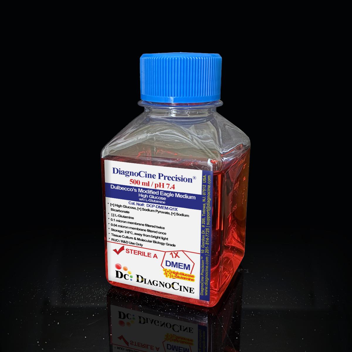
FluxMPS™ Dulbecco’s Modified Eagle Medium (DMEM), High Glucose w/o L-Glutamine: 1X Liquid
FluxMPS™ DMEM, High Glucose, w/o L-Glutamine — 1X Liquid Cell Culture Medium
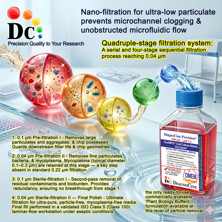
FluxMPS™ DCP-DMEM-Q1X is a Microfluidics Suitable, quadruple-stage ultra-filtered (0.1 µm ×2 + 0.04 µm ×2) Dulbecco’s Modified Eagle Medium (DMEM) formulated with 4,500 mg/L high glucose, sodium bicarbonate, sodium pyruvate and phenol red, without L-Glutamine — engineered specifically for microfluidic channels, organ-on-a-chip (OoC), and microphysiological system (MPS) platforms. A quadruple-stage train (0.1 µm ×2 + 0.04 µm ×2) reaches a 0.04 µm final cut-off, five times finer than the 0.22 µm membranes used for conventional sterile filtration.
- Quadruple-stage nano-filtration: 0.1 µm ×2 + 0.04 µm ×2 for sub-mycoplasma polishing and ultra-low particulate load
- Endotoxin release specification < 0.05 EU/mL, verified per batch by LAL assay (USP <85>)
- High glucose (4,500 mg/L), sodium bicarbonate, sodium pyruvate and phenol red included; formulated without L-Glutamine so you control nitrogen source at time of use
- 4× BME amino acid & vitamin concentration — rich, stable nutrient base for demanding cell types
- Manufactured under an ISO 13485:2016 quality management system with full lot traceability; final QC and fill at Diagnocine, Totowa NJ
- Formulated with Type 1 Ultrapure water (18.2 MΩ·cm) for tight trace-metal and total organic carbon (TOC) control
- Customizable: pH, glucose, HEPES, salts, and nutrient composition available on request — support@diagnocine.com
- Glucose4,500 mg/L (High Glucose)
- L-GlutamineNot included — add exogenously
- Sodium Pyruvate110.0 mg/L
- Sodium Bicarbonate3,700 mg/L
- pH (USP <791>)7.0 - 7.4
- Osmolality (USP <785>)300 - 330 mOsm/kg
- Endotoxin< 0.05 EU/mL (USP <85> BET)
- Filtration System0.1 µm ×2 + 0.04 µm ×2 (Quadruple-stage)
- Storage2–8°C, protect from light
- Shelf Life12 months from date of manufacture, unopened
Engineered where standard DMEM fails
Conventional 0.22 µm-filtered DMEM is designed for well-plate culture, not for the microscale geometries of organ-on-a-chip devices. Residual mycoplasma-sized particles accumulate in microchannels, corrupt biosensor baselines, and shorten perfusion device lifespan. FluxMPS™ DCP-DMEM-Q1X uses a Quadruple-stage filtration architecture engineered to reduce this failure mode while preserving the rich 4× BME amino acid and vitamin profile that DMEM is valued for.[1,2]
Microchannel-Safe Purity
0.04 µm final-pass filtration targets sub-mycoplasma particulates; USP <788> Method 1 (light obscuration) particle testing confirms < 25 particles ≥10 µm per mL — suitable for channels as narrow as 50 µm.
Total Metabolic Control
L-Glutamine-free base lets you define the exact nitrogen source — add stable GlutaMAX™ or glutamine analogues at time of use, or omit entirely for Warburg-effect or amino-acid limitation studies; nitrogen input remains fully user-defined.
Ultrapure-Grade Water
Formulated with Type 1 Ultrapure water (18.2 MΩ·cm, ASTM D1193 / ISO 3696) for tight trace-metal and total organic carbon (TOC) control in microphysiological system applications.
Low Background for Imaging
Quadruple-stage 0.04 µm filtration provides an ultra-low particulate baseline for confocal, two-photon, and brightfield microscopy, supporting long-duration live-cell imaging on OoC platforms. Note: this formulation contains phenol red; select a phenol-red-free FluxMPS™ DMEM variant if autofluorescence-free imaging is required.
Rich, Stable Nutrient Profile
DMEM’s 4× BME amino acid and vitamin concentrations sustain demanding cell types including HeLa, MCF-7, HEK293, primary neurons, and iPSC-derived organoids through extended perfusion runs.[3]
Customization on Demand
pH, glucose concentration, HEPES buffer, sodium bicarbonate, and individual nutrient levels can be adjusted to your exact protocol. Contact support@diagnocine.com for custom formulation inquiries.
Quadruple-stage filtration system
FluxMPS™ DCP-DMEM-Q1X is a Microfluidics Suitable, ready-to-use 0.04 µm-final-filtered DMEM.
-
1
0.1 µm Prefiltration I
First 0.1 µm membrane pass captures large aggregates, cell debris, and coarse particulates, protecting the first 0.04 µm cartridge and establishing a clean base stream for downstream polishing.
-
2
0.04 µm Final filtration I
The first 0.04 µm (40 nm) membrane retains organisms in the 0.2–0.3 µm mycoplasma size range and fine particulates that pass a 0.22 µm filter.
-
3
0.1 µm Prefiltration II
A second, independent 0.1 µm membrane protects the second 0.04 µm cartridge, catching any upstream shed particles before the final polishing stage.
-
4
0.04 µm Final filtration II — Polish
The final 0.04 µm membrane delivers ultimate particulate polishing. Media passes directly into ISO Class 5 (Class 100) laminar-flow fill suites, with particle counts confirmed per USP <788> Method 1 on every lot before release.
Performance vs. conventional media
FluxMPS™ DCP-DMEM-Q1X targets substantially lower subvisible particulate burdens compared with standard 0.22 µm-filtered DMEM, reducing the risk of channel occlusion and biosensor baseline drift in microfluidic perfusion systems.
© Diagnocine® — DCP-DMEM-Q1X
Purpose-built for microphysiological & metabolic research
FluxMPS™ DCP-DMEM-Q1X is a clean, L-Glutamine-free DMEM base for organ-on-a-chip (OoC), microfluidic perfusion, and Warburg-effect metabolomics applications where a user-defined nitrogen source is required.[4,5]
Automated Bioreactors & Robotics
For automated perfusion bioreactors and robotic liquid-handling systems with sub-10 µm valve tolerances, Diagnocine offers an optional ultra-filtered 0.01 µm (10 nm) MPS Grade variant of FluxMPS™ DMEM — a separate product line from the Microfluidics Suitable 0.04 µm medium described on this page.
- Total Particulate Exclusion: 10 nm final filtration targets virtually all particle species — protecting microvalves and sensors in automated organ-on-a-chip workstations
- Valve & Sensor Protection: Reduces actuator wear in PDMS and thermoplastic chip platforms with channel cross-sections below 50 µm
- Extended Perfusion Stability: Maintains defined nutrient composition across 7–21-day continuous perfusion runs
Inquiry Required: The 0.01 µm (10 nm) MPS Grade format is available on special order. Contact support@diagnocine.com to request this format for your automated system.
Microphysiological System (MPS) & Chip
Quadruple-stage filtered, particle-reduced DMEM base for perfused OoC gut, lung, liver, kidney, and multi-organ platforms. L-Glutamine-free to avoid ammonia accumulation during extended perfusion.
Warburg Effect & Metabolic Research
High-glucose (4.5 g/L), glutamine-free base for glycolytic flux studies and isotope tracing where nitrogen source must be precisely controlled.
iPSC-Derived Models
Controlled nutrient base for iPSC differentiation protocols where L-Glutamine is titrated independently. Compatible with GlutaMAX™ supplementation for stable glutamine delivery to fragile iPSC-derived neurons, cardiomyocytes, and hepatocytes.
Endothelial & Primary Cells
Ultra-low particulate DMEM is compatible with primary endothelial cell culture and co-culture vascular-on-chip models.
Metabolic Flux Analysis
Defined, L-Glutamine-free background for 13C glucose isotope tracing and NMR-based metabolomics — nitrogen source remains fully user-defined. Not compatible with Agilent Seahorse XF assays, which require bicarbonate-free, phenol-red-free medium.
Microscopy & Optical Sensing
Quadruple-stage filtration provides an ultra-low particulate baseline for confocal and two-photon live-cell imaging, electrochemical biosensors, and transepithelial electrical resistance (TEER) measurement on chip. Contains phenol red; select a phenol-red-free FluxMPS™ variant for autofluorescence-sensitive assays.
Full QC specification suite
Every lot of FluxMPS™ DCP-DMEM-Q1X is tested against the full specification suite below before release. Certificate of Analysis (CoA) is available on request. Available pack sizes: 500 mL, 1000 mL.
| Parameter | Specification |
|---|---|
| Formulation | DMEM High Glucose (4,500 mg/L), + NaHCO3 (3,700 mg/L), + Sodium Pyruvate (110 mg/L), + Phenol Red (15.9 mg/L), − L-Glutamine |
| Appearance | Red/orange-colored, clear solution (phenol red indicator present) |
| pH USP <791> | 7.0 - 7.4 |
| Osmolality USP <785> | 300 - 330 mOsm/kg H2O |
| Glucose | 4,500 mg/L (High Glucose, 4.5 g/L) |
| L-Glutamine | Not included — add 2 mM – 4 mM exogenously |
| Sodium Pyruvate | 110.00 mg/L |
| Phenol Red | 15.90 mg/L |
| Parameter | Specification |
|---|---|
| Endotoxin USP <85> BET | < 0.05 EU/mL (batch release specification — see Manufacturing & Quality) |
| Sterility USP <71> | No growth at 14 days (aerobic + anaerobic) |
| Mycoplasma | 0.1 µm mycoplasma-retentive filtration (not tested per lot) |
| Particulate ≥10 µm USP <788> M1 | < 25 per mL |
| Particulate ≥25 µm USP <788> M1 | < 3 per mL |
| Water Purity | Ultrapure Type 1, 18.2 MΩ·cm (ASTM D1193 / ISO 3696) |
| Manufacturing Std. | ISO 13485:2016 QMS; 21 CFR Part 820 (QMSR) aligned |
| Fill Environment | ISO Class 5 (Class 100) laminar-flow |
| Parameter | Specification |
|---|---|
| Storage Temperature | 2–8°C, protect from light |
| Freeze-Thaw | Do not freeze |
| Shelf Life | 12 months from date of manufacture, unopened |
| Shipping Condition | Shipped on ice packs (2–8°C); ambient for international orders with express courier |
| CO2 Requirement | ~10% CO2 atmosphere to maintain pH 7.0–7.4 with 3,700 mg/L sodium bicarbonate buffering |
| Parameter | Specification |
|---|---|
| Raw Material Grade | Reagent-grade to USP-grade salts, vitamins, amino acids |
| Traceability | Full lot traceability per ISO 13485 DMR/DHR |
| Manufacturing QMS ISO 13485 | ISO 13485:2016 certified; Totowa, NJ, USA |
| UNSPSC | 41116155 — Molecular biology and cell culture growth media (UNv260801) |
| Regulatory Alignment | ISO 13485:2016; 21 CFR Part 820 (QMSR) aligned |
| Production Method | Micro-batch compounding — lot sizes ≤10 L for traceability |
| Intended Use | For Research Use Only (RUO) |
Full composition (mg/L)
FluxMPS™ DCP-DMEM-Q1X is based on the classical high-glucose DMEM formulation featuring 4× BME amino acid and vitamin concentrations. The composition below reflects the per-lot release basis. L-Glutamine is excluded; add 2–4 mM L-Glutamine or equivalent GlutaMAX™ per your protocol. Sodium Pyruvate and Phenol Red are included as shown below.
| Component | CAS Number | mg/L |
|---|---|---|
| INORGANIC SALTS | ||
| Calcium Chloride (dihydrate) | 10035-04-8 | 265.00 |
| Ferric Nitrate Nonahydrate | 7782-61-8 | 0.100 |
| Magnesium Sulfate (MgSO4) (anhydrous) | 7487-88-9 | 97.720 |
| Potassium Chloride (KCl) | 7447-40-7 | 400.00 |
| Sodium Bicarbonate (NaHCO3) | 144-55-8 | 3700.00 |
| Sodium Chloride (NaCl) | 7647-14-5 | 6400.00 |
| Sodium Phosphate Monobasic (NaH2PO4) | 7558-80-7 | 109.00 |
| Component | CAS Number | mg/L |
|---|---|---|
| AMINO ACIDS | ||
| Glycine | 56-40-6 | 30.00 |
| L-Arginine Hydrochloride | 1119-34-2 | 84.00 |
| L-Cystine ·2HCl | 30925-07-6 | 62.57 |
| L-Histidine Hydrochloride ·H2O | 5934-29-2 | 42.00 |
| L-Isoleucine | 73-32-5 | 105.00 |
| L-Leucine | 61-90-5 | 105.00 |
| L-Lysine Hydrochloride | 657-27-2 | 146.20 |
| L-Methionine | 63-68-3 | 30.00 |
| L-Phenylalanine | 63-91-2 | 66.00 |
| L-Serine | 56-45-1 | 42.00 |
| L-Threonine | 72-19-5 | 95.00 |
| L-Tryptophan | 73-22-3 | 16.00 |
| L-Tyrosine Disodium Salt Dihydrate | 69847-44-5 | 103.79 |
| L-Valine | 72-18-4 | 94.00 |
| Component | CAS Number | mg/L |
|---|---|---|
| VITAMINS | ||
| Choline Chloride | 67-48-1 | 4.00 |
| D-Calcium Pantothenate | 137-08-6 | 4.00 |
| Folic Acid | 59-30-3 | 4.00 |
| Niacinamide (Nicotinamide) | 98-92-0 | 4.00 |
| Pyridoxal hydrochloride | 58-56-0 | 4.00 |
| Riboflavin | 83-88-5 | 0.40 |
| Thiamine Hydrochloride | 67-03-8 | 4.00 |
| i-Inositol | 87-89-8 | 7.20 |
| OTHERS | ||
| Phenol Red | 143-74-8 | 15.900 |
| D-Glucose | 50-99-7 | 4500.00 |
| Sodium Pyruvate | 113-24-6 | 110.00 |
ISO 13485-certified manufacturing & compliance
FluxMPS™ DCP-DMEM-Q1X is manufactured in Totowa, NJ, USA under an ISO 13485:2016-certified Quality Management System with full micro-batch traceability and multi-standard QC release testing.
ISO 13485:2016 Quality Management System
Full QMS certification covering raw material qualification, in-process controls, final product release, and post-market surveillance. Every batch carries a traceable Device History Record (DHR).
Ultrapure Type 1 Water
All formulations use freshly generated Ultrapure Type 1 water (ASTM D1193 / ISO 3696) — controlled for trace metals and total organic carbon (TOC), supporting sensitive biosensor and omics readouts.
ISO Class 5 Fill & Finish
Media is filled aseptically under ISO Class 5 (Class 100) laminar-flow conditions. Particle counts confirmed by USP <788> Method 1 on every lot before release.
Micro-Batch Precision
Production in micro-batches (≤10 L) ensures raw material homogeneity, minimizes lot-to-lot variability, and enables full component traceability — critical for reproducible MPS and OoC experiments.
Endotoxin — USP <85> BET
LAL assay; release specification < 0.05 EU/mL per batch. Full endotoxin data reported on CoA.
Particulate Matter — USP <788> Method 1
Light-obscuration particle counting for particles ≥10 µm and ≥25 µm per mL. Results reported on lot CoA.
Osmolality — USP <785>
Freezing-point osmometry per USP <785>. Target 300–330 mOsm/kg H2O. Actual lot value reported on CoA.
Documentation — Certificate of Analysis
Full CoA available for every lot including: appearance, pH, osmolality, endotoxin, sterility, and particulate data. Request at support@diagnocine.com.
- Endotoxin — LAL assay, USP <85> Bacterial Endotoxins Test; assay sensitivity 0.005 EU/mL; release specification < 0.05 EU/mL
- pH, osmolality, conductivity, appearance and clarity
- Sterility
How DCP-DMEM-Q1X compares
FluxMPS™ DCP-DMEM-Q1X vs. conventional 0.22 µm-filtered DMEM variants and published supplier specifications — across the parameters that matter most for microphysiological systems and microfluidic applications.
| Parameter | DCP-DMEM-Q1X (FluxMPS™) | Standard DMEM (0.22 µm filtered) | Typical Competitor DMEM |
|---|---|---|---|
| Grade | Microfluidics Suitable (0.04 µm final cut-off) | Not specified | Not specified |
| L-Glutamine status | Not included — user-defined nitrogen source | Often included (unstable, degrades to NH&sub3;) | Often included or as GlutaMAX™ |
| Final filtration pore size | 0.04 µm (40 nm) | 0.22 µm | 0.22 µm |
| Number of filtration stages | 4 (Quadruple-stage) | 1 | 1–2 |
| Mycoplasma barrier filtration | check_circle Yes (0.04 µm ×2) | cancel No | cancel No |
| Endotoxin (release specification) | < 0.05 EU/mL | Corning classical liquid media — < 0.25 EU/mL Sigma-Aldrich DMEM complete medium — ≤ 2 EU/mL Gibco classical DMEM — Not specified (recorded per lot) |
|
| USP particulate compliance | check_circle USP <788> Method 1 tested | cancel Not routinely tested | cancel Not routinely tested |
| Water quality | Type 1 Ultrapure 18.2 MΩ·cm | WFI or purified water (variable) | Purified water (variable) |
| Manufacturing QMS | check_circle ISO 13485:2016 | ISO 9001 or not specified | ISO 9001 or not specified |
| Microfluidic channel compatibility | check_circle Validated for ≥50 µm channels | cancel Not validated | cancel Not validated |
| Custom formulation | check_circle Yes — pH, glucose, salts, HEPES | cancel No | cancel Rarely |
Comparison figures from published supplier specifications, accessed 2026-09-02. Suppliers that publish no numeric endotoxin specification are shown as "Not specified".
Frequently asked questions
Common questions about FluxMPS™ DCP-DMEM-Q1X and its use in microphysiological, microfluidic, and metabolic research.
Supporting literature
Key peer-reviewed references supporting the use of ultra-filtered DMEM in organ-on-a-chip, microfluidic perfusion, and metabolic research applications.
- Bhatia, S.N. & Ingber, D.E. Microfluidic organs-on-chips. Nature Biotechnology 32, 760–772 (2014). doi:10.1038/nbt.2989
- Huh, D. et al. Reconstituting organ-level lung functions on a chip. Science 328, 1662–1668 (2010). doi:10.1126/science.1188302
- Dulbecco, R. & Freeman, G. Plaque production by the polyoma virus. Virology 8, 396–397 (1959). doi:10.1016/0042-6822(59)90043-1
- Warburg, O. On the origin of cancer cells. Science 123, 309–314 (1956). doi:10.1126/science.123.3191.309
- Sung, J.H. et al. Microfabricated mammalian organ systems and their integration into models of whole animals and humans. Lab on a Chip 13, 1201–1212 (2013). doi:10.1039/c3lc41017j
- Bhattacharya, S. et al. Validation of USP <788> particulate matter testing for cell culture media used in bioreactor manufacturing. PDA Journal of Pharmaceutical Science and Technology 75, 202–215 (2021). doi:10.5731/pdajpst.2020.011940
- Zhang, Y.S. et al. Multisensor-integrated organs-on-chips platform for automated and continual in situ monitoring of organoid behaviors. PNAS 114, E2293–E2302 (2017). doi:10.1073/pnas.1612906114
- Niklison-Chirou, M.V. et al. How does p53 induce apoptosis and how does this relate to p53-mediated tumour suppression? Cell Death & Differentiation 28, 983–996 (2021). doi:10.1038/s41418-021-00760-1
- Campisi, M. et al. 3D self-organized microvascular model of the human blood-brain barrier with endothelial cells, pericytes and astrocytes. Biomaterials 180, 117–129 (2018). doi:10.1016/j.biomaterials.2018.07.014
- Yin, X. et al. Engineering stem cell organoids. Cell Stem Cell 18, 25–38 (2016). doi:10.1016/j.stem.2015.12.005