FluxMPS™, DAB Staining Kit
FluxMPS™ DAB Staining Kit
A complete, ready-to-use plant staining kit for rapid histochemical visualization of hydrogen peroxide (H₂O₂) accumulation in leaves, roots, and soft plant tissues. Includes DAB staining solution, chlorophyll-clearing solution, and neutral wash buffer.
- 4-stage filtration process (0.1 µm → 0.04 µm, followed by sterile 0.1 µm → 0.04 µm)
- Ready-to-use workflow with no DAB weighing or stock-solution preparation required.
- Designed for detached leaves, roots, seedlings, and other small plant tissue samples.
- Chlorophyll-clearing step improves contrast and makes brown DAB signal easier to visualize in green tissues.
- Suitable for routine comparative screening in plant stress and ROS localization experiments.
- Ultrapure Type 1 water (18.2 MΩ·cm)
- ISO Class 5 laminar-flow fill & finish
- Customizable formulation available upon request
- pH7.2 for solution A and 7.0 for Solution C
- Filtration0.1µm×2 + 0.04µm×2
- StorageStore the kit at 2-8 °C. Once opened, store the Bleaching/Clearing Solution at room temperature and the DAB Staining Solution and Wash/Storage Buffer at 2-8 °C.
- Shelf Life1 year
DAB Staining Solution
- Storage2-8 °C
- Shelf Life1 year
- DAB5 mg/mL
- Sodium phosphate buffer (pH 4.0)50 mM
Bleaching/Clearing Solution (Ethanol:acetic acid:glycerol)
- StorageRoom temperature
- Shelf Life1 year
- Ethanol60%
- Glacial acetic acid20%
- Glycerol20%
Wash/Storage Buffer (10 mM sodium phosphate buffer, pH 7.0)
- Storage2-8 °C
- Shelf Life1 year
- Sodium phosphate (pH 7.0)10 mM
Engineered for maximum purity & performance
Standard laboratory reagents filtered at 0.22 µm leave sub-micron particulates, mycoplasma, and aggregates that interfere with sensitive downstream applications. FluxMPS™ DAB Staining Kit is manufactured under ISO 13485 quality systems with multi-stage ultra-filtration to eliminate these failure modes.
Ready-to-use H₂O₂ detection
5 mg/mL DAB in phosphate buffer (pH 4.0) forms visible brown precipitate at sites of H₂O₂ accumulation—no weighing or stock preparation needed.
Chlorophyll clearing included
Ethanol:acetic acid:glycerol clearing solution removes chlorophyll for high-contrast visualization of brown DAB signal in green tissues.
Neutral wash/storage buffer
10 mM sodium phosphate buffer (pH 7.0) for gentle rinsing and short-term sample storage before imaging.
Broad tissue compatibility
Suitable for detached leaves, roots, seedlings, and other small plant tissue samples from Arabidopsis, crop plants, and other species.
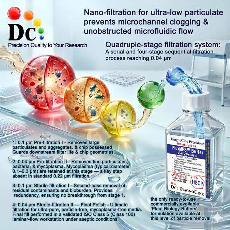
Quadruple-stage filtration system
A serial four-stage sequential filtration process reaching 0.04 µm—the only commercially available buffer at this level of particle removal.
-
1
0.1 µm Pre-filtration I
Removes large particulates and aggregates. Primary guard to extend downstream filter lifetime.
-
2
0.04 µm Pre-filtration II
Removes fine particulates, bacteria, and mycoplasma (0.1–0.3 µm)—a key step absent in standard 0.22 µm filtration.
-
3
0.1 µm Sterile-filtration I
Second-pass removal of residual contaminants and bioburden with full redundancy.
-
4
0.04 µm Sterile-filtration II — Final Polish
Ultimate filtration for particle-free, mycoplasma-free product. Final fill in validated ISO Class 5 laminar-flow workstation.
Performance vs. conventional reagents
Standard 0.22 µm single-pass filtration leaves particles in the 0.05–0.22 µm range. FluxMPS™ removes these contaminants for cleaner results and higher reproducibility.
Validated applications
- Detection of oxidative stress responses in detached leaves after abiotic or biotic treatments.
- Comparison of ROS accumulation between genotypes, treatments, or time points in plant physiology workflows.
- Visualization of hydrogen peroxide localization in leaves, roots, seedlings, and other soft tissues suitable for immersion staining.
- Immerse fresh plant tissue in the ready-to-use DAB Staining Solution and incubate in the dark.
- Replace the stain with Bleaching/Clearing Solution to remove chlorophyll and improve visibility of the brown precipitate.
Quality assurance & logistics
ISO 13485:2016 Certified
Manufactured under certified QMS with full traceability and lot documentation.
Made in USA
All manufacturing, QA testing, and final assembly at DiagnoCine Precision, Totowa, NJ.
Supporting literature
- Daudi A, O'Brien JA. Detection of hydrogen peroxide by DAB staining in Arabidopsis leaves. Bio-protocol. 2012;2(18):e263.
- Thordal-Christensen H, Zhang Z, Wei Y, Collinge DB. Subcellular localization of H₂O₂ in plants: H₂O₂ accumulation in papillae and hypersensitive response during the barley-powdery mildew interaction. Plant J. 1997;11(6):1187-1194.
- Kumar D, Yusuf MA, Singh P, et al. Histochemical detection of superoxide and H₂O₂ accumulation in Brassica juncea seedlings. Bio-protocol. 2014;4(8):e1108.