


Easy-WESTERN-II Quick & Super full set
Product#: FNK-BCL-EZ23
$424.80
Availability:
Ships in 1-2 Weeks
Easy-WESTERN-II Quick & Super full set
Cat. No.: FNK-BCL-EZ23
Size: 1 set
Storage: -20℃ (MAD reagent, marker detection reagent, and mouse IgG enhancer reagent) 4℃ (other reagents)
Principle
Easy-WESTERN (EZW) is a primary antibody detection reagent kit for Western blots. The kit is based on Multi-Antibody Detection (MAD) technology. The MAD reagent consists of nanosize protein particles with high affinity for antibodies. Each particle is composed of about 100 antibody-binding proteins and is labeled with 50 HRP molecules. Due to these properties, MAD reagent enables high sensitivity and rapid detection of primary antibodies.
The Easy-WESTERN kit is ideal for high sensitivity, signal enhancement, and simultaneous detection of multiple antigens, which is not possible with standard Western blot techniques.
<Advantages>
1. Easy to use: Primary antibodies from most animal species1) can be detected by simply using MAD in place of a secondary antibody.
2. Higher signal than conventional methods2): It is possible to detect low-expression antigens. In addition, the amount of primary antibody can be reduced, which saves on expensive primary antibodies.
3. Enhancement of signals detected by conventional methods: If the signal detected by a secondary antibody is weak, a stronger signal can be obtained by reprobing with MAD reagent.
4. Rapid detection: By mixing the MAD reagent with the primary antibody, detection can be performed in a one-step reaction.
Note:
1) Some antibody species (mainly mouse IgG1 and goat IgG) are difficult to detect using MAD reagent. However, mouse IgG1 can be detected with high sensitivity using a mouse IgG enhancer reagent.
2) The performance of this reagent may vary depending on the combination of antigen and antibody.
3) When using samples containing serum, please note that IgG in the serum will be detected.
Product Characteristics
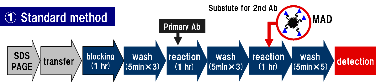
The basic components of Easy-Western are MAD regents and MAD dilution buffer. MAD is used as a substitute of 2nd antibody. MAD dilution buffer is used to dilute MAD to eliminate non-specific binding. When using Easy-Western, there are many kinds of ways depending on the purpose. Some of examples are shown below.
1. High sensitivity detection without secondary antibody
With ordinary protocol just change primary antibody to MAD reagents.
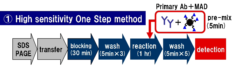
2. One step detection with high sensitivity
Mix primary antibody with MAD reagents and react membrane with the mixture.
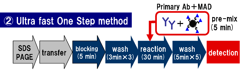
3. Save time by using One-Step detection
Mix primary antibody with MAD reagents and react membrane with the mixture using short time protocol.
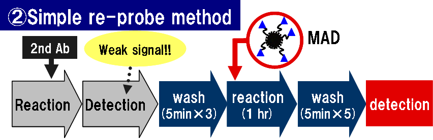
4. Signal enhancement of weak signal using ordinary western
Membrane with weak signal by ordinary secondary antibody method can be enhanced second reaction using MAD reagent.
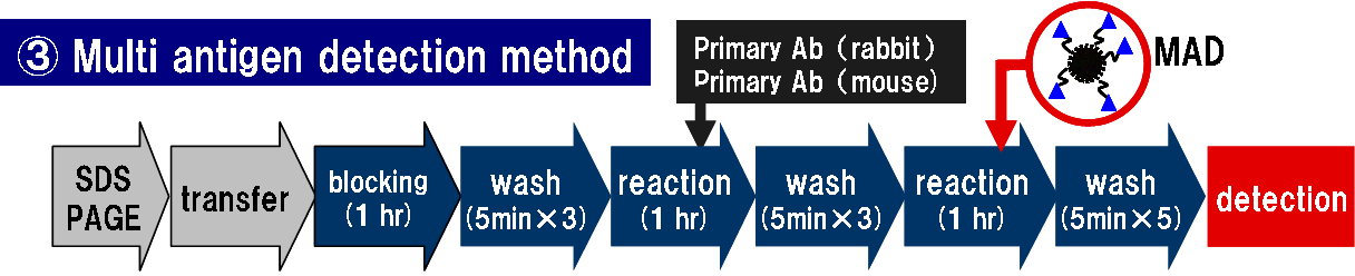
5. Multiple antigen detection
By using multiple primary antibodies regardless animal species, multiple antigen detection is possible by a single procedure with MAD reagents.
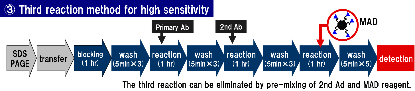
6. Ultra high sensitivity by using MAD as third reaction
Using third reaction with MAD after 2nd antibody reaction.
Preparation and use of supplementary reagents
1. Prepare TBS-T (150 mM NaCl, 10 mM Tris-HCl, 0.1% Tween-20, pH 7.6).
2. Dilute the 10x dilution buffer provided with the kit 10-fold with purified water to prepare 1x dilution buffer. This is used to dilute the MAD reagent.
3. Dilute the 1x dilution buffer 10-fold with TBS-T to make 1/10x dilution buffer. This is used to dilute the primary antibody.
4. A PVDF membrane with a pore size of 0.2 µm is recommended. If you use a pore size of 0.45 µm, the background may be slightly higher.
5. Please prepare a blocking solution. If using skim milk, ensure it is of reagent grade.
6. Mouse IgG Enhancer Reagent is used when detection is weak with mouse IgG. It is especially essential when using IgG1 as the primary antibody. When using this reagent, add 1/2000 vol. of it to the MAD reagent diluent and mix before the reaction.
7. Marker detector is a reagent that enables detection of molecular weight markers that are normally detected with secondary antibodies, such as MagicMark XP (Invitrogen). When using, add 1/10,000 vol. of it to the primary antibody diluent and mix before the reaction.
Standard Protocol
This is the most common method used to obtain higher sensitivity.
1. Perform SDS-PAGE using a suitable gel.
2. Transfer the protein to a PVDF membrane from the SDS-PAGE gel using standard methods.
3. Block the PVDF membrane with blocking solution by incubating it for 1 hour at room temperature.
4. Wash the PVDF membrane with TBS-T (5 min x 3 times).
5. Dilute the primary antibody with 1/10x dilution buffer and incubate it with the PVDF membrane for 1 hour at room temperature. Please follow the dilution ratio recommended by the manufacturer. Depending on the antibody, sufficient bands may be detectable even if the antibody is further diluted up to about 5 times. Signal detection can be further enhanced by using Signal Booster solution A (code: BCL-125A) as the dilution solution.
6. Wash the PVDF membrane with TBS-T (5 min x 3 times).
7. Dilute the MAD reagent 2000-fold (recommended range: 1000-4000-fold) with 1x dilution buffer and incubate it with the PVDF membrane for 1 hour at room temperature.
8. Wash the PVDF membrane with TBS-T (5 min x 5 times).
9. Detect signals using commercially available HRP detection reagents.
2. Transfer the protein to a PVDF membrane from the SDS-PAGE gel using standard methods.
3. Block the PVDF membrane with blocking solution by incubating it for 1 hour at room temperature.
4. Wash the PVDF membrane with TBS-T (5 min x 3 times).
5. Dilute the primary antibody with 1/10x dilution buffer and incubate it with the PVDF membrane for 1 hour at room temperature. Please follow the dilution ratio recommended by the manufacturer. Depending on the antibody, sufficient bands may be detectable even if the antibody is further diluted up to about 5 times. Signal detection can be further enhanced by using Signal Booster solution A (code: BCL-125A) as the dilution solution.
6. Wash the PVDF membrane with TBS-T (5 min x 3 times).
7. Dilute the MAD reagent 2000-fold (recommended range: 1000-4000-fold) with 1x dilution buffer and incubate it with the PVDF membrane for 1 hour at room temperature.
8. Wash the PVDF membrane with TBS-T (5 min x 5 times).
9. Detect signals using commercially available HRP detection reagents.
Note: If using the mouse IgG enhancer reagent or marker detector reagent, refer to steps 6 and 7 in Preparation and use of supplementary reagents.
One-step quick Protocol
This is a method for rapid detection in 65 minutes. By adjusting the conditions, highsensitivity detection is also possible.
1. Perform steps 1 and 2 in Standard Protocol.
2. Block the PVDF membrane with blocking solution by incubating it for 5 minutes at room temperature.
3. Dilute and mix the primary antibody and MAD reagent with 1x dilution buffer. Follow the primary antibody dilution ratio recommended by the manufacturer. A 2,000-fold dilution of MAD reagent is recommended. The mixture can be used 5 minutes after mixing. If you are using a primary antibody solution containing NaN3, mix the MAD reagent after diluting the primary antibody so that the final concentration of NaN3 is 0.001% or less.
4. Incubate the PVDF membrane in the solution prepared in step 3 for 30 minutes at room temperature.
5. Wash the PVDF membrane with TBS-T (5 min x 5 times).
2. Block the PVDF membrane with blocking solution by incubating it for 5 minutes at room temperature.
3. Dilute and mix the primary antibody and MAD reagent with 1x dilution buffer. Follow the primary antibody dilution ratio recommended by the manufacturer. A 2,000-fold dilution of MAD reagent is recommended. The mixture can be used 5 minutes after mixing. If you are using a primary antibody solution containing NaN3, mix the MAD reagent after diluting the primary antibody so that the final concentration of NaN3 is 0.001% or less.
4. Incubate the PVDF membrane in the solution prepared in step 3 for 30 minutes at room temperature.
5. Wash the PVDF membrane with TBS-T (5 min x 5 times).
6. Detect signals using commercially available HRP detection reagents.
Important: For high-sensitivity detection using this method, extend the blocking time to 30 minutes and the incubation time in step 4 to a maximum of 1 hour.
Note: If using the mouse IgG enhancer reagent or marker detector reagent, refer to steps 6 and 7 in Preparation and use of supplementary reagents.
Enhanced signal using 2nd antibody Protocol
This is a method to enhance signals by adding to standard secondary antibody reactions.
1. Perform SDS-PAGE, transfer to a PVDF membrane, block, and perform primary and secondary antibody reactions.
2. Wash the PVDF membrane with TBS-T (5 min x 3 times).
3. Dilute the MAD reagent 2000-fold with 1x dilution buffer and incubate it with the PVDF membrane for 1 hour at room temperature.
4. Wash the PVDF membrane with TBS-T (5 min x 5 times).
5. Detect signals using commercially available HRP detection reagents.
2. Wash the PVDF membrane with TBS-T (5 min x 3 times).
3. Dilute the MAD reagent 2000-fold with 1x dilution buffer and incubate it with the PVDF membrane for 1 hour at room temperature.
4. Wash the PVDF membrane with TBS-T (5 min x 5 times).
5. Detect signals using commercially available HRP detection reagents.
Note: If using the mouse IgG enhancer reagent or marker detector reagent, refer to steps 6 and 7 in Preparation and use of supplementary reagents.
Multi-antigen detection Protocol
This method allows for the detection of multiple antigens on a single membrane
simultaneously.
1. Perform steps 1 to 4 in Standard Protocol.
2. Incubate two or more primary antibodies simultaneously with the PVDF membrane for 1 hour at room temperature. (Follow the dilution ratio recommended by the manufacturer. Use 1/10x dilution buffer for antibody dilution.)
3. Wash the PVDF membrane with TBS-T (5 min x 3 times).
4. Incubate the PVDF membrane with MAD reagent diluent. (refer to step 7 in Standard Protocol)
5. Wash the PVDF membrane with TBS-T (5 min x 5 times).
6. Detect signals using commercially available HRP detection reagents.
Note: If using the mouse IgG enhancer reagent or marker detector reagent, refer to steps 6 and 7 in Preparation and use of supplementary reagents.2. Incubate two or more primary antibodies simultaneously with the PVDF membrane for 1 hour at room temperature. (Follow the dilution ratio recommended by the manufacturer. Use 1/10x dilution buffer for antibody dilution.)
3. Wash the PVDF membrane with TBS-T (5 min x 3 times).
4. Incubate the PVDF membrane with MAD reagent diluent. (refer to step 7 in Standard Protocol)
5. Wash the PVDF membrane with TBS-T (5 min x 5 times).
6. Detect signals using commercially available HRP detection reagents.
Reprobing Protocol
This method increases signal intensity on membranes with weak signals using conventional
methods and allows for re-detection.
1. Membranes with weak detection signals from conventional methods (e.g., secondary antibodies) can be used. Dried membranes cannot be used.
2. Wash the PVDF membrane with TBS-T (5 min x 3 times).
3. Incubate the PVDF membrane with MAD reagent diluent. (Refer to steps 7 in Standard Protocol)
4. Wash the PVDF membrane with TBS-T (5 min x 5 times).
5. Detect signals using commercially available HRP detection reagents.
Note: If using the mouse IgG enhancer reagent or marker detector reagent, refer to steps 6 and 7 in Preparation and use of supplementary reagents.2. Wash the PVDF membrane with TBS-T (5 min x 3 times).
3. Incubate the PVDF membrane with MAD reagent diluent. (Refer to steps 7 in Standard Protocol)
4. Wash the PVDF membrane with TBS-T (5 min x 5 times).
5. Detect signals using commercially available HRP detection reagents.
Troubleshooting
| Problem | Possible Solutions |
| Weak signal | 1. Increase antigen concentration. |
| 2. Increase primary antibody concentration. | |
| 3. Increase the electric current or extend the transfer time to improve protein transfer to the membrane. | |
| 4. Over-blocking can reduce signal intensity. Reduce the blocking time or lower the concentration of blocking agents. | |
| 5. The primary antibody is an antibody species (mouse IgG1, goat IgG) that is difficult to detect with Easy-WESTERN. In the case of mouse antibodies, please use "Mouse IgG Enhancer Reagent". Additionally, if using a primary antibody type other than mouse, please consider using Easy WESTERN Multi. | |
| 6. When diluting MAD Reagent in buffer without blocking agents, use low protein-binding tubes. | |
| White out of luminescent signal | 7. Excessive antigen or antibody. Too much signal inhibits luminescence. Reduce the concentration of antigen or antibody used. |
| Too many extra bands | 8. Non-specific binding of the primary antibody. Reduce the antibody concentration to an appropriate level. |
| 9. Excessive protein. Reduce the amount of protein in electrophoresis. | |
| 10. Insufficient blocking. Block the membrane with 5% skim milk in TBS-T for over 1 hour | |
| 11. MAD reagent may become inactivated due to improper storage. MAD Reagent should be stored at -20°C. Inactivated MAD may produce nonspecific signals. Replace the MAD Reagent if necessary. | |
| 12. When using a serum-containing sample, an extra band appears around 50 kDa. This band represents the heavy chain of IgG in the serum. Remove IgG using a protein A column or similar method. | |
| High background | 13. Insufficient washing. Increase the number and the duration of washes |
| 14. If there is adequate signal but with high background, decrease the primary antibody concentration and/or shorten the incubation time. | |
| 15. Reduce the concentration of MAD Reagent. | |
| 16. When using antigen-antibody reaction enhancers (such as Signal Booster Solution A, code: BCL-125A), insufficient washing can cause high background. Increase the number and the duration of washes. | |
| Weak signal of 1 antigen when using the Multi-Antigen Detection Protocol | 17. One primary antibody may weakly bind to the antigen or MAD Reagent. Increase the concentration of the primary antibody with the weak signal. |
| 18. Insufficient washing of the primary antibody. Increase the number and the duration of washes. | |
| No signal with Quick Protocol | 19. If using a non-purified antibody such as serum, the Quick Protocol may not work well due to contaminating IgGs. Use purified antibodies or follow the Standard Protocol. |
| Weak Marker band | 20. Increase the concentration of the marker detection reagent, or dilute the marker detection reagent with TBS-T and incubate it with the PVDF membrane for about 15 minutes before the antibody/MAD reagent reaction. |