

DynaMarker, RNA High
DynaMarker, RNA High
Cat. No. FNK-DM160
Size 50 μg (56 μl), 0.9 mg/ml
Storage -80°C
Range 200-8,000 base of RNA
Storage buffer 10 mM Tris-HCl (pH 8.0) buffer containing 1 mM EDTA
Storage conditionStore at -80 o C. Repeated freeze/thaw cycles should be avoided.
Description
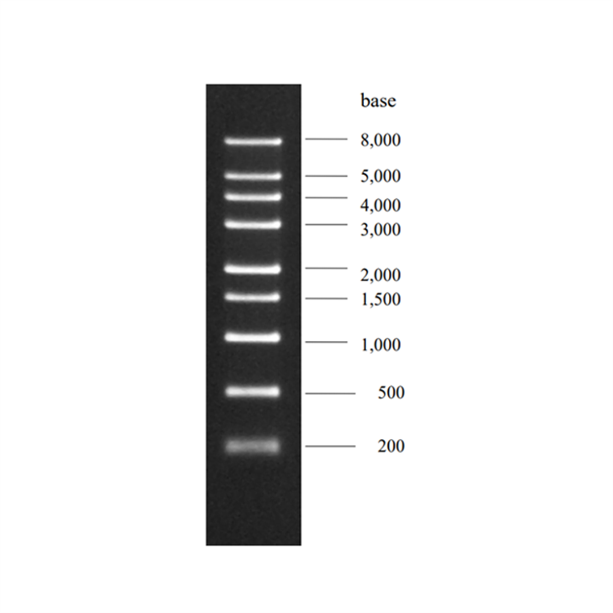
DynaMarker RNA High consists of nine single-stranded RNAs, 200, 500, 1,000, 1,500, 2,000, 3,000, 4,000, 5,000 and 8,000 bases.
The DynaMarker RNA High consists of nine single-stranded RNAs, 200, 500, 1,000, 1,500, 2,000, 3,000, 4,000, 5,000 and 8,000 bases, which are synthesized by in vitro transcription. The DynaMarker RNA High is suitable for determinating size of single-stranded RNAs in denaturing agarose gel electrophoresis. The concentration of each RNA (200-8,000 base) in the marker is approximately 0.1 μg/μl. It is useful for estimating of RNA amount. The DynaMarker RNA High can be visualized by UV light after ethidium bromide staining or exposure to film with end labeling.

Electrophoresis profile of DynaMarker RNA High (0.9 μg) on formaldehyde-agarose (1%) gel
Solutions and reagents to mix the marker should be high grade and nuclease-free. To use, thaw the DynaMarker RNA
High on ice and keep it on ice while using.
Recommended usage
Procedure
20 mM Sodium acetate
10 mM EDTA (pH 8.0)
2. Denaturation of RNA
Prepare denaturated DynaMarker RNA High and RNA to be analysed in a small tube as below.
10 × MOPS buffer 2 μl
37 % formaldehyde solution 4 μl
formamide 10 μl
200 μg/ml ethidum brimode 1 μl
After mixing, heat the RNA solution at 75 °C for 3 min, then quickly transfer the tube on ice.
* Required RNA amount depends on experiments. For northern analysis, up to 15 μg of RNA is loaded. For detection of DynaMarker RNA High by ethidum bromide staining, load 0.5-4 μg of the marker.
3. Loading and electrophoresis
Add 2 μl of 10 × formaldehyde gel-loading buffer* to each RNA solution and return the tube on ice. Set up the prepared agarose gel containing formaldehyde in a horizontal electrophoresis apparatus submerged in 1 × MOPS buffer. Load the denatured RNA solution to a well. and start electrophoresis. After the tracking dyes have migrated an appropriate distance through gel, stop the electrophoresis. RNA bands can be seen under UV illumination.
10 mM EDTA (pH 8.0)
0.025 % (w/v) bromophenol blue
0.025 % (w/v) xylene cyanol FF
- Sambrook, J. and Russell, D.W. (2001) Molecular Cloning: A Laboratory Manual, 3rd ed., Cold Spring Harbor Laboratory Press, Cold Spring Harbor, NY.
